An Introduction to Cellular and Molecular Neuroscience
John H. Byrne,Ruth Heidelberger,M. Neal Waxham
This is a test
This is a test
Buch teilen
694 Seiten
English
ePUB (handyfreundlich)
Über iOS und Android verfügbar
eBook - ePub
From Molecules to Networks
An Introduction to Cellular and Molecular Neuroscience
John H. Byrne,Ruth Heidelberger,M. Neal Waxham
Angaben zum Buch
Buchvorschau
Inhaltsverzeichnis
Quellenangaben
Über dieses Buch
An understanding of the nervous system at virtually any level of analysis requires an understanding of its basic building block, the neuron. The third edition of From Molecules to Networks provides the solid foundation of the morphological, biochemical, and biophysical properties of nerve cells. In keeping with previous editions, the unique content focus on cellular and molecular neurobiology and related computational neuroscience is maintained and enhanced.
All chapters have been thoroughly revised for this third edition to reflect the significant advances of the past five years. The new edition expands on the network aspects of cellular neurobiology by adding new coverage of specific research methods (e.g., patch-clamp electrophysiology, including applications for ion channel function and transmitter release; ligand binding; structural methods such as x-ray crystallography).
Written and edited by leading experts in the field, the third edition completely and comprehensively updates all chapters of this unique textbook and insures that all references to primary research represent the latest results.
The first treatment of cellular and molecular neuroscience that includes an introduction to mathematical modeling and simulation approaches
80% updated and new content
New Chapter on "Biophysics of Voltage-Gated Ion Channels"
New Chapter on "Synaptic Plasticity"
Includes a chapter on the Neurobiology of Disease
Highly referenced, comprehensive and quantitative
Full color, professional graphics throughout
All graphics are available in electronic version for teaching purposes
Häufig gestellte Fragen
Wie kann ich mein Abo kündigen?
Gehe einfach zum Kontobereich in den Einstellungen und klicke auf „Abo kündigen“ – ganz einfach. Nachdem du gekündigt hast, bleibt deine Mitgliedschaft für den verbleibenden Abozeitraum, den du bereits bezahlt hast, aktiv. Mehr Informationen hier.
(Wie) Kann ich Bücher herunterladen?
Derzeit stehen all unsere auf Mobilgeräte reagierenden ePub-Bücher zum Download über die App zur Verfügung. Die meisten unserer PDFs stehen ebenfalls zum Download bereit; wir arbeiten daran, auch die übrigen PDFs zum Download anzubieten, bei denen dies aktuell noch nicht möglich ist. Weitere Informationen hier.
Welcher Unterschied besteht bei den Preisen zwischen den Aboplänen?
Mit beiden Aboplänen erhältst du vollen Zugang zur Bibliothek und allen Funktionen von Perlego. Die einzigen Unterschiede bestehen im Preis und dem Abozeitraum: Mit dem Jahresabo sparst du auf 12 Monate gerechnet im Vergleich zum Monatsabo rund 30 %.
Was ist Perlego?
Wir sind ein Online-Abodienst für Lehrbücher, bei dem du für weniger als den Preis eines einzelnen Buches pro Monat Zugang zu einer ganzen Online-Bibliothek erhältst. Mit über 1 Million Büchern zu über 1.000 verschiedenen Themen haben wir bestimmt alles, was du brauchst! Weitere Informationen hier.
Unterstützt Perlego Text-zu-Sprache?
Achte auf das Symbol zum Vorlesen in deinem nächsten Buch, um zu sehen, ob du es dir auch anhören kannst. Bei diesem Tool wird dir Text laut vorgelesen, wobei der Text beim Vorlesen auch grafisch hervorgehoben wird. Du kannst das Vorlesen jederzeit anhalten, beschleunigen und verlangsamen. Weitere Informationen hier.
Ist From Molecules to Networks als Online-PDF/ePub verfügbar?
Ja, du hast Zugang zu From Molecules to Networks von John H. Byrne,Ruth Heidelberger,M. Neal Waxham im PDF- und/oder ePub-Format sowie zu anderen beliebten Büchern aus Biological Sciences & Neuroscience. Aus unserem Katalog stehen dir über 1 Million Bücher zur Verfügung.
Patrick R. Hof, Grahame Kidd, Javier DeFelipe, Jean de Vellis, Miguel A. Gama Sosa, Gregory A. Elder and Bruce D. Trapp
This chapter provides general information on the various types of cells that compose nervous tissues and serves as an introduction to cellular neuroscience. Different classes of neurons are presented in terms of their function, morphology, neurochemistry, and place in neural circuits. Cortical pyramidal neurons and inhibitory interneuron subtypes are discussed together with examples of specialized neurons from subcortical regions. The structure of excitatory and inhibitory synapses is also reviewed. Each type of glial cell (astrocytes, oligodendrocytes, as well as microglia) is introduced in separate sections that review their particular function, structure, and interactions with neurons. A final section presents brain endothelial cells and their role in maintenance of the blood-brain barrier.
Several types of cellular elements are integrated to constitute normally functioning brain tissue. The neuron is the communicating cell, and many neuronal subtypes are connected to one another via complex circuitries, usually involving multiple synaptic connections. Neuronal physiology is supported and maintained by neuroglial cells, which have highly diverse functions. These include myelination, secretion of trophic factors, maintenance of the extracellular milieu, and scavenging of molecular and cellular debris. Neuroglial cells also participate in the formation and maintenance of the blood–brain barrier, a multicomponent structure that is interposed between the circulatory system and the brain substance and that serves as the molecular gateway to brain tissue.
Neurons
The neuron is a highly specialized cell type and is the essential cellular element in the CNS (central nervous system). All neurological processes are dependent on complex cell–cell interactions among single neurons as well as groups of related neurons. Neurons can be categorized according to their size, shape, neurochemical characteristics, location, and connectivity, which determine their particular functional role in the brain. More importantly, neurons form circuits, and these circuits constitute the structural basis for brain function. Macrocircuits involve a population of neurons projecting from one brain region to another region, and microcircuits reflect the local cell–cell interactions within a brain region. The detailed analysis of these macro- and microcircuits is an essential step in understanding the neuronal basis of a given cortical function in the healthy and the diseased brain. Thus, these cellular characteristics allow us to appreciate the special structural and biochemical qualities of a neuron in relation to its neighbors and to place it in the context of a specific neuronal subset, circuit, or function.
Broadly speaking, therefore, there are five general categories of neurons: inhibitory neurons that make local contacts (e.g., GABAergic interneurons in the cerebral and cerebellar cortex), inhibitory neurons that make distant contacts (e.g., medium spiny neurons of the basal ganglia or Purkinje cells of the cerebellar cortex), excitatory neurons that make local contacts (e.g., spiny stellate cells of the cerebral cortex), excitatory neurons that make distant contacts (e.g., pyramidal neurons in the cerebral cortex), and neuromodulatory neurons that influence neurotransmission, often at large distances. Within these general classes, careful analyses of the structural variation of the anatomic features of neurons have led to various categorizations and to the development of the concept of cell type. The grouping of neurons into descriptive cell types (such as chandelier, double bouquet, or bipolar cells) allows the analysis of populations of neurons and the linking of specified cellular characteristics with certain functional roles.
General Features of Neuronal Morphology
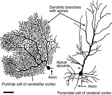
Neurons are highly polarized cells, meaning that they develop distinct subcellular domains that subserve different functions. Morphologically, in a typical neuron, three major regions can be defined: (1) the cell body (soma or perikaryon), which contains the nucleus and the major cytoplasmic organelles; (2) a variable number of dendrites, which emanate from the perikaryon and ramify over a certain volume of gray matter and which differ in size and shape, depending on the neuronal type; and (3) a single axon, which extends, in most cases, much farther from the cell body than the dendritic arbor (Fig. 1.1). Dendrites may be spiny (as in pyramidal cells) or non-spiny (as in most interneurons), whereas the axon is generally smooth and emits a variable number of branches (collaterals). In vertebrates, many axons are surrounded by an insulating myelin sheath, which facilitates rapid impulse conduction. The axon terminal region, where contacts with other cells are made, displays a wide range of morphological specializations, depending on its target area in the central or peripheral nervous system.
Figure 1.1Typical morphology of projection neurons. (Left) A Purkinje cell of the cerebellar cortex and (right) a pyramidal neuron of the neocortex. These neurons are highly polarized. Each has an extensively branched, spiny apical dendrite, shorter basal dendrites, and a single axon emerging from the basal pole of the cell.
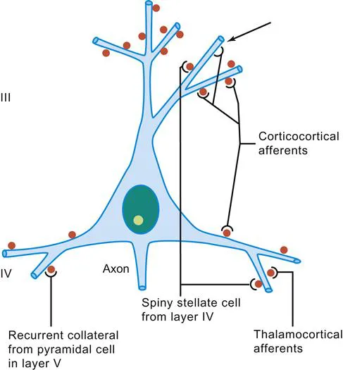
The cell body and dendrites are the two major domains of the cell that receive inputs, and dendrites play a critically important role in providing a massive receptive area on the neuronal surface (see also Chapters 16 and 17). In addition, there is a characteristic shape for each dendritic arbor, which can be used to classify neurons into morphological types. Both the structure of the dendritic arbor and the distribution of axonal terminal ramifications confer a high level of subcellular specificity in the localization of particular synaptic contacts on a given neuron. The three-dimensional distribution of dendritic arborization is also important with respect to the type of information transferred to the neuron. A neuron with a dendritic tree restricted to a particular cortical layer typically receives a very limited pool of afferents, whereas the widely expanded dendritic arborization of a large pyramidal neuron receives highly diversified inputs (Fig. 1.2) (Mountcastle, 1978). The structure of the dendritic tree is maintained by surface interactions between adhesion molecules and, intracellularly, by an array of cytoskeletal components (microtubules, neurofilaments, and associated proteins), which also take part in the movement of organelles within the dendritic cytoplasm.
Figure 1.2Schematic representation of the spatial distribution of four major excitatory inputs to pyramidal neurons. A pyramidal neuron in layer III is shown as an example. Note the preferential distribution of synaptic contacts on spines. Spines are indicated in red. Arrow shows a contact directly on the dendritic shaft.
An important specialization of the dendritic arbor of certain neurons is the presence of large numbers of dendritic spines, which are membranous protrusions. They are abundant in large pyramidal neurons and are much sparser on the dendrites of interneurons (see below).
The perikaryon contains the nucleus and a variety of cytoplasmic organelles. Stacks of rough endoplasmic reticulum are conspicuous in large neurons and, when interposed with arrays of free polyribosomes, are referred to as Nissl substance. Another feature of the perikaryal cytoplasm is the presence of a rich cytoskeleton composed primarily of neurofilaments and microtubules. These cytoskeletal elements are dispersed in bundles that extend from the soma into the axon and dendrites.
Whereas dendrites and the cell body are the domains of the neuron that receive afferents, the axon, at the other pole of the neuron, is responsible for transmitting neural information. This information may be primary, in the case of a sensory receptor, or processed information that has already been modified through a series of integrative steps. The morphology of the axon and its course through the nervous system are correlated with the type of information processed by the particular neuron and by its connectivity patterns with other neurons. The axon leaves the cell body from a small swelling called the axon hillock. This structure is particularly apparent in large pyramidal neurons; in other cell types, the axon sometimes emerges from one of the main dendrites. At the axon hillock, microtubules are packed into bundles that enter the axon as parallel fascicles. The axon hillock is the part of the neuron where the action potential is generated (see Chapter 12). The axon is generally unmyelinated in local circuit neurons (such as inhibitory interneurons), but it is myelinated in neurons that furnish connections between different parts of the nervous system. Axons usually have higher numbers of neurofilaments than dendrites, although this distinction can be difficult to make in small elements that contain fewer neurofilaments. In addition, the axon may show extensive, spatially constrained ramifications, as in certain local circuit neurons; it may give out a large number of recurrent collaterals, as in neurons connecting different cortical regions; or it may be relatively straight in the case of projections to subcortical centers, as in cortical motor neurons that send their very long axons to the ventral horn of the spinal cord. At the interface of axon terminals with target cells are the synapses, which represent specialized zones of contact consisting of a presynaptic (axonal) element, a narrow synaptic cleft, and a postsynaptic element on a dendrite or perikaryon.
Synapses and Spines
Synapses
Each synapse is a complex of several components: (1) a presynaptic element, (2) a cleft, and (3) a postsynaptic e...