Introduces readers to the enlightening world of the modern light microscope
There have been rapid advances in science and technology over the last decade, and the light microscope, together with the information that it gives about the image, has changed too. Yet the fundamental principles of setting up and using a microscope rests upon unchanging physical principles that have been understood for years. This informative, practical, full-colour guide fills the gap between specialised edited texts on detailed research topics, and introductory books, which concentrate on an optical approach to the light microscope. It also provides comprehensive coverage of confocal microscopy, which has revolutionised light microscopy over the last few decades.
Written to help the reader understand, set up, and use the often very expensive and complex modern research light microscope properly, Understanding Light Microscopy keeps mathematical formulae to a minimum—containing and explaining them within boxes in the text. Chapters provide in-depth coverage of basic microscope optics and design; ergonomics; illumination; diffraction and image formation; reflected-light, polarised-light, and fluorescence microscopy; deconvolution; TIRF microscopy; FRAP & FRET; super-resolution techniques; biological and materials specimen preparation; and more.
Gives a didactic introduction to the light microscope
Encourages readers to use advanced fluorescence and confocal microscopes within a research institute or core microscopy facility
Features full-colour illustrations and workable practical protocols
Understanding Light Microscopy is intended for any scientist who wishes to understand and use a modern light microscope. It is also ideal as supporting material for a formal taught course, or for individual students to learn the key aspects of light microscopy through their own study.
Häufig gestellte Fragen
Wie kann ich mein Abo kündigen?
Gehe einfach zum Kontobereich in den Einstellungen und klicke auf „Abo kündigen“ – ganz einfach. Nachdem du gekündigt hast, bleibt deine Mitgliedschaft für den verbleibenden Abozeitraum, den du bereits bezahlt hast, aktiv. Mehr Informationen hier.
(Wie) Kann ich Bücher herunterladen?
Derzeit stehen all unsere auf Mobilgeräte reagierenden ePub-Bücher zum Download über die App zur Verfügung. Die meisten unserer PDFs stehen ebenfalls zum Download bereit; wir arbeiten daran, auch die übrigen PDFs zum Download anzubieten, bei denen dies aktuell noch nicht möglich ist. Weitere Informationen hier.
Welcher Unterschied besteht bei den Preisen zwischen den Aboplänen?
Mit beiden Aboplänen erhältst du vollen Zugang zur Bibliothek und allen Funktionen von Perlego. Die einzigen Unterschiede bestehen im Preis und dem Abozeitraum: Mit dem Jahresabo sparst du auf 12 Monate gerechnet im Vergleich zum Monatsabo rund 30 %.
Was ist Perlego?
Wir sind ein Online-Abodienst für Lehrbücher, bei dem du für weniger als den Preis eines einzelnen Buches pro Monat Zugang zu einer ganzen Online-Bibliothek erhältst. Mit über 1 Million Büchern zu über 1.000 verschiedenen Themen haben wir bestimmt alles, was du brauchst! Weitere Informationen hier.
Unterstützt Perlego Text-zu-Sprache?
Achte auf das Symbol zum Vorlesen in deinem nächsten Buch, um zu sehen, ob du es dir auch anhören kannst. Bei diesem Tool wird dir Text laut vorgelesen, wobei der Text beim Vorlesen auch grafisch hervorgehoben wird. Du kannst das Vorlesen jederzeit anhalten, beschleunigen und verlangsamen. Weitere Informationen hier.
Ist Understanding Light Microscopy als Online-PDF/ePub verfügbar?
Ja, du hast Zugang zu Understanding Light Microscopy von Jeremy Sanderson im PDF- und/oder ePub-Format sowie zu anderen beliebten Büchern aus Physical Sciences & Science History & Practice. Aus unserem Katalog stehen dir über 1 Million Bücher zur Verfügung.
‘Ah, I see!’ demonstrates how dependent we are on our sight, and how it is linked inextricably with understanding. We are a visually oriented species, and our two eyes are our ‘window on the world’. It is easy to forget that not all animals depend upon sight to the extent that humans do. Bats use echolocating sonar to navigate, and bees exploit ultraviolet (UV) light to discriminate foliage. At the other end of the visible spectrum, vipers sense infrared radiation to detect warm blooded prey. For those creatures whose habitat is underground (e.g. moles), vision necessarily cedes priority to other senses such as touch and smell. Other animals, chiefly birds, are more dependent upon their sight than humans, with a much greater proportion of their head devoted to large eyes.
Via the optic nerve, the eyes are a direct extension of the brain. The human eye is a wonderful device. If it were a camera, it might boast autofocus, wide-angle lens, auto-exposure, high sensitivity to light, automatic colour balancing, one-hundred megapixel resolution (each retina contains around 100 million sensitive cells) and 3D imaging (when used in pairs). We can see in bright daylight or discriminate a single light in the dark several miles away. The eye has naturally evolved to suit its principal function: helping its owner navigate the world.
Many modern microscopes are designed to be operated from a computer screen, rather than be used by viewing the image directly down the eyepieces. Nevertheless, all images must eventually be seen by our eyes, so in this chapter we shall discuss how our eyes function. We shall consider how well our eyes are adapted for their purpose but also the limitations and aberrations that they suffer from. These limitations are relevant to microscopy, affecting how we acquire and interpret scientific images (i.e. data) using the light microscope. We shall see how a magnifying glass works and why the microscope was developed to assist our vision.
1.2 How Our Eyes Work
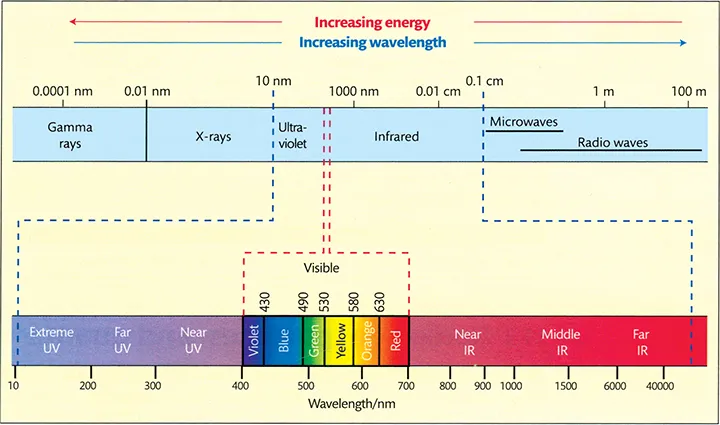
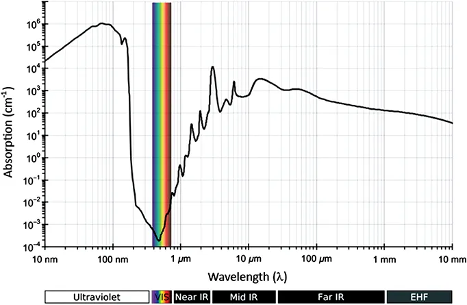
Any image must ultimately be seen by the eye. Light is electromagnetic radiation that stimulates the eye. It is merely a fraction of the entire electromagnetic spectrum (Figure 1.1). Only a small proportion of solar radiation reaches the earth’s surface. We depend on the ozone layer for protection; atmospheric dust, smoke, air molecules and water vapour also absorb a significant proportion of insolation, or incident solar radiation. Our eyes evolved from aquatic animals and contain a significant amount of water. Human sensitivity to electromagnetic radiation (Figure 1.2) corresponds closely to the wavelengths of minimum water absorbance, located away from harmful UV radiation and towards the infrared end of the spectrum.
Figure 1.1The Electromagnetic spectrum The electromagnetic spectrum extends from beyond low frequencies used for modern radio to very high-frequency gamma radiation at the short-wavelength end of the spectrum, encompassing wavelengths from thousands of kilometres down to a fraction of a nanometre. Humans are able to discriminate the visible wavelengths from long wavelength, low energy, red at around 710 nm to short wavelength, high-energy violet radiation at around 380 nm. A very small number of people can see into the infrared, and people suffering from cataracts who have their lens removed (i.e. who are aphakic) can see into the UV, even if they can’t focus well.
Source: Glencross et al. 2011. Reproduced with permission from Oxford University Press.
Figure 1.2Human sensitivity to light Visible light corresponds closely to the wavelengths of minimum water absorbance on the electromagnetic spectrum. See also Chapter 2, Section 2.5. Since our eyes are composed largely of water, it is advisable to use a heat filter (e.g. Schott KG5) in the light path to cut out any absorption of infrared light.
Source: Adapted from Kebes at English Wikipedia under the terms of the Creative Commons Attribution licence, CC BY-SA 3.0 (http://creativecommons.org/licenses/by-sa/3.0).
Our eyes respond to the visible part of the electromagnetic spectrum from near UV at a wavelength of 380 nm to deep red at 710 nm. This stimulation depends on both the energy (frequency, expressed as wavelength) and the quantity (number of photons) of light. The wavelength of light is perceived as colour, and the quantity of light (expressed as the amplitude of the light wave) is seen as intensity1 (see also Appendix A1.1.3 and Table A1.1, pages 45–46 in: Tilley, 2010). Suppose we have four LEDs: blue emitting at 490 nm; green at 555 nm; far red at 670 nm and infrared at 940 nm (commonly used for remote controls) all emitting the same radiant flux of 5 mW absolute power, measured in radiometric units. If we measure the respective light output of each of these LEDs in photometric units, the green LED will be the brightest (3.4 lm); the blue will be the second one (0.75 lm), and the far red will be the third (0.1 lm). The infrared LED will have a recorded emission of zero lumens (this example is taken from Tilley, 2010).
Further details about the electromagnetic spectrum and the nature of light are discussed in Chapter 2, Section 2.2 onwards. Light itself has no inherent colour; our perception of different hues is fundamentally a complex judgment experienced as a sensation by our brains. We discriminate colour very well, although brightness less so. This is why we choose paint in a range of colours, rather than different intensities.
The retina is the photo-sensitive tissue of the eye (Figure 1.3), withi...