![]()
1
INTRODUCTION
Andreas Filippi
The tongue is by far the largest organ in the oral cavity. With its excellent innervation and mobility, it performs important functions for humans, such as touching, tasting, speaking, whistling, sucking, and cleaning the mouth. The tongue also fulfills an important role in mastication: It moves the food around in the mouth so that it can easily be chewed and lubricated with saliva. If one or more of these functions is restricted, quality of life is often greatly impaired. Many people suffering from advanced dysfunction or even complete loss of individual functions can also suffer from depression as a result. Articulation as well as the sense of touch and taste are so important to quality of life that patients who undergo radiotherapy to the head and neck area repeatedly report complaints. In addition to mucositis, they suffer severe impairment of their sense of taste (eg, things taste differently or blander than usual). In some cases, people develop aversions to particular foods and stop enjoying things that they used to like eating. Fortunately, these sensory disturbances usually go away after oncology treatment is finished.
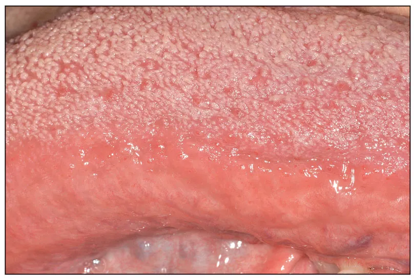
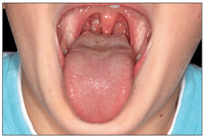
The dorsal surface (ie, top) of the tongue is the only oral mucosa to have a microrough surface. This harbors more than half of all oral microorganisms, which live there in a highly organized biofilm that protects them against chemical and mechanical influences. Aerobes are more likely to be found on the surface and anaerobes in the depth of the tongue. The latter reside in the fissures of the strikingly rugged filiform papillae, which are only present on the dorsal surface of the tongue (Figs 1-1 and 1-2).
Fig 1-1 (a) The tongue surface is covered with filiform papillae. (b and c) As magnification increases, it becomes clear that the papillae are very rugged.
Fig 1-2 Difference between upper and lower surfaces of the tongue.
Treatment of the consequences of microbiologic diseases in the oral cavity (eg, caries, marginal periodontitis, apical periodontitis) is the most common type of work done in dental practices. Some causal microorganisms may reside on the teeth or in the sulcus or periodontal pockets, but a great many reside on the tongue. Even if great skill and effort is put into cleaning individual periodontal areas, this may not actually have a sustained impact on reinfection of the periodontium. Consider the current debate about the benefits of dental floss or the fact that the market introduces new toothbrushes each year that supposedly clean better than past toothbrushes. When it comes to caries prevention, clean teeth are only one consideration. In light of this growth of knowledge in prevention and therapy, modern medicine is fortunately focusing more and more on the largest site of oral microorganisms: the tongue. This is illustrated by examples such as full-mouth disinfection, modern halitosis treatment, and the idea of caries prevention by means of tongue cleaning. Furthermore, attempts are repeatedly being made to alter the oral biofilm (the largest of
which is located on the dorsal surface of the tongue) with the aid of probiotic medicines or probiotic foods to benefit oral health (Fig 1-3). There has been some success in relation to gut flora with certain changes or diseases, although attempts have not yet been successful in the oral cavity. However, in recent years there has been growing awareness of diagnostics of the tongue among dental practitioners and especially dental hygienists. This started with the extensive professional tongue diagnostics performed on halitosis patients, which is beyond the scope of this book but covered in other textbooks.3 Fundamentally, dentistry should not concentrate solely on the teeth. It is not without reason that universities in many countries have departments such as Oral Medicine, Oral Diagnostic Sciences, or Oral Health—a trend that should spread across the globe.
Fig 1-3 The largest oral biofilm is found on the tongue.
During a thorough dental examination, the borders, underside, and base of the tongue as well as the floor of the mouth should be inspected as a basic principle. If there are any visible or merely palpable changes, further diagnostic investigation should be discussed and—depending on the results—suitable treatment initiated. As well as visible and/or palpable changes, subjective complaints are playing a growing role in the aging population, who often require drug treatment in the general medical practice. The decrease in the saliva flow rate is a common problem that can lead to redness, inflammation, fungal infections, and a burning sensation of the tongue. This frequently demands an interdisciplinary approach to provide satisfactory help to patients whose quality of life is often impaired.
![]()
2
ANATOMY AND PHYSIOLOGY OF THE TONGUE
Ralf J. Radlanski
In its resting state when the mouth is closed, the tongue completely fills the oral cavity palatal to the rows of teeth (ie, the oral cavity proper). Because of its muscular core, the tongue is so variably mobile that the tip of the tongue is able to reach nearly every point in the mouth (Fig 2-1).1 Only a shortened lingual frenulum (ie, ankyloglossia) would limit this mobility.
Fig 2-1 The tongue in lateral view (a) and ventral view (b). (Reprinted with permission from Radlanski.1)
PARTS OF THE TONGUE
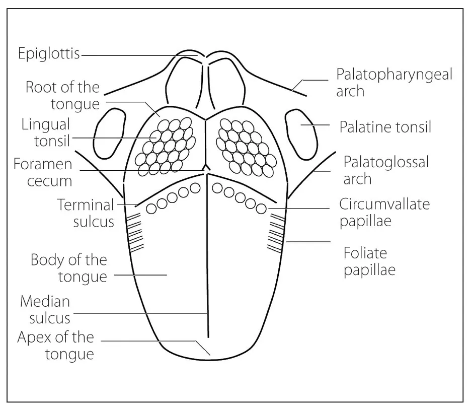
The median sulcus (ie, midline groove) divides the left and right half of the body of the tongue in the sagittal direction. The terminal sulcus runs in the transverse direction as a slightly V-shaped line (Fig 2-2) and divides the tongue into two parts. One part is the root or base, which lies in the pharynx. The second part comprises the body and tip, which lie within the oral cavity (Fig 2-3). The root accounts for roughly one-third of tongue volume, while the two anterior parts make up two thirds. The circumvallate papillae are on the body of the tongue, and the foramen cecum is located dorsally to the terminal sulcus (ie, on the root of the tongue).2–4
Fig 2-2 Macroscopic anatomy of the tongue in schematic diagram, cranial view.
Fig 2-3 Schematic diagram of the tongue in sagittal section.
DEVELOPMENT OF THE TONGUE
The pharyngeal arches have a major influence on the development of the face, including the tongue. The apex and body of the tongue develop from the first pharyngeal arch, and the root of the tongue develops from the third and fourth pharyngeal arches. The foramen cecum marks the endpoint of the thyroglossal duct and is evidence of the descent of the thyroid gland during the embryonic period of development.5
INTERNAL STRUCTURE OF THE TONGUE
The body of the tongue is permeated by intrinsic muscles that run in the sagittal, transverse, and vertical directions, partly interwoven with each other (Figs 2-4 to 2-6). The superior longitudinal muscle, the inferior longitudinal muscle, the genioglossus, and the geniohyoid muscles run sagittally; the transverse muscle runs transversally; and the vertical muscle runs vertically. Fasciae running in these directions appropriately compartmentalize the muscles. The lingual septum runs sagittally-vertically in the middle, and the lingual aponeurosis covers the dorsum of the tongue.3,4
Fig 2-4 Schematic diagram of the tongue in frontal section.
Fig 2-5 (a) Sagittal section through the tongue. Hematoxylin-eosin stain. (b) Tongue musculature cut longitudinally and transversally. (Reprinted with permission from Radlanski.1)
Fig 2-6 Sagittal section through the floor of the mouth region of the tongue and its intrinsic musculature. The epithelium on the underside of the tongue is free of papillae, smooth and thin, like the epithelium on the floor of the mouth. In the submucosa of the floor of the mouth, the sublingual gland and the excretory duct of the submandibular gland are cut. Hematoxylin-eosin stain. (Reprinted with permission from Radlanski.1)
The genioglossus, hyoglossus, palatoglossus, and styloglossus muscles radiate from outside into the tongue. Mediated by insertions of individual muscles onto the hyoid bone, these muscles afford support...