Bringing together material scattered across many disciplines, Semiconductor Radiation Detectors providesreaders with aconsolidated source of information on the properties of a wide range of semiconductors; their growth, characterization and the fabrication of radiation sensors with emphasis on the X- and gamma-ray regimes. It explores the promise and limitations of both the traditional and new generation of semiconductors and discusses where the future in semiconductor development and radiation detection may lie.
The purpose of this book is two-fold; firstly to serve as a text book for those new to the field of semiconductors and radiation detection and measurement, and secondly as a reference book for established researchers working in related disciplines within physics and engineering.
Features:
The only comprehensive book covering this topic
Fully up-to-date with new developments in the field
Provides a wide-ranging source of further reference material
Reproduction of the cartoon on the front cover of P.J. van Heerden’s seminal Doctoral thesis, entitled “The Crystal Counter: A New Instrument in Nuclear Physics” (Rijksuniversiteit Utrecht, July 1945). The left side shows a stylistic depiction of the “crystal counter”, which was essentially a solid state ionization chamber – the precursor of all modern semiconductor energy resolving radiation detectors. The right side of the image shows the “deflections” (pulse heights) of individual events when the crystal was exposed to an external γ-ray source.
CONTENTS
1.1 The Discovery of Radiation
1.1.1 Understanding the Atom and Its Structure
1.2 Radiation Detection
1.2.1 Early Monitoring Devices
1.2.2 Early Recording Devices
1.2.3 Electro-Optical Approaches
1.3 Early Work with Semiconductors
1.3.1 Photoconduction Detectors
1.3.2 Do Semiconductors Exist?
1.3.3 Theoretical Stagnation and Salvation
1.3.4 Crystal Counters
1.4 Post-1960 Evolution
1.4.1 The Current Situation
1.4.1.1 Other Technologies
1.5 Future Directions
1.5.1 Exploring the Nano-Scale Properties of Materials
1.5.2 Exploiting New Degrees of Freedom
1.5.3 Biological Based Detection Systems
References
1.1 The Discovery of Radiation
The study of radiation and radiation detection really begins with the discovery of X-rays by Röntgen in 1895 [1], who while investigating cathode rays using a Hittorf-Crookes tube, observed that when the rays hit the glass wall, a mysterious radiation was given off which could fog photographic plates and cause various materials to fluoresce. The rays became known as Röntgen rays. The first corroborative reports of radiation detection took place almost immediately afterwards – which is curious since, apart from photographic plates, radiation detectors had not yet been developed. In 1896, Brandes [2] reported seeing an “effect” which he described as a faint “blue-gray” glow that seemed to originate from the eye itself when standing close to an X-ray tube. In his first communication, Röntgen [1], had stated that the eye was insensitive to the new rays, but later in his third communication [3] reported seeing “a feeble sensation of light that spread over the whole field of vision”. Whilst the mechanism was not understood, the observed effects (known by the grandiose but vacuous title of “radiation phosphenes”1), were assumed to be due to the direct action of the X-rays on the photoreceptors of the retina [4]. Much later, when radiotherapy became a standard medical modality, the visual effects of X-rays were immediately apparent and were attributed to Cherenkov radiation generated in the ocular media by secondary electrons [5].
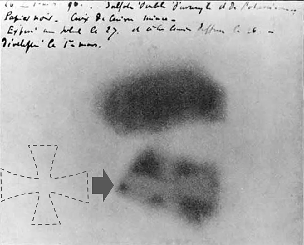
FIGURE 1.1 An exposed photographic plate made by Henri Becquerel [6] showing the fogging effects of “invisible rays” emitted by two crystals of uranium salt. The shadow of a metal Maltese cross sandwiched between the plate and the lower crystal is visible.
While investigating Röntgen’s work on X-rays, Becquerel [6] decided to test Poincare’s hypothesis [7] that the emission of X-rays could be related to phosphorescence, essentially the delayed emission of light by a substance after its exposure to light. To do this, he placed crystals of potassium uranyl sulfate (K2UO2(SO4)2.2H2O) on top of a copper Maltese cross and a photographic plate wrapped in black paper. He had originally planned to expose the uranium salts to sunlight before placing them on the cross and plate, believing that the uranium would absorb the sun’s energy and then emit it as X-rays. However, the sky was overcast, so he placed the entire assembly into a darkened bureau draw and waited for the weather to improve. It did not, and so after several days he decided to develop the plate anyway and was surprised to see a distinct image of the cross (see Fig. 1.1). Since the plate had not been exposed to light and the crystals were non-luminous, the only conclusion that could be drawn was that the crystals were emitting a previously unknown energetic radiation which became known as Becquerel rays or uranium rays. At first the relationship between uranium rays and Röntgen rays was not clear. Becquerel rays seemed to have intermediate properties between light and Röntgen rays [8]. Of significance, he also observed that an electroscope loses its charge under the effect of the radiation, meaning that the radiation produces charges in the air. In 1898, Marie Curie [9] discovered that thorium minerals also behaved like uranium and suggested that a new radioactive element may be found in pitchblende based on the fact that it was more active than metallic uranium itself. In the same year, Pierre and Marie Currie announced the discovery of two new elements, polonium and radium [10],[11] and concluded that uranium, thorium, polonium and radium all emit uranium rays. They coined the word “radioactivity”, although at the time the meaning of the word was subtly different from today’s. Today, we understand radioactivity to be the property exhibited by certain types of matter to emit energy and subatomic particles spontaneously. At the time, it was more an expression of how active these elements were with respect to metallic uranium. Thus, radium was more “active” than polonium which was more “active” than thorium which was more “active” than uranium. The true nature of the radiation was not revealed until 1899 when Rutherford [12] showed by absorption and conduction measurements that the emanations from uranium consisted of two components. He called the less penetrating component “alpha radiation” and the more penetrating one “beta radiation”. Magnetic deflection measurements showed both to be particular in nature and to be of high energy. In the same year, Becquerel measured the mass-to-charge ratio (e/m) for beta particles by the method J. J. Thomson used to study cathode rays, which led to the identification of the electron [13]. He found that e/m for a beta particle is the same as for Thomson’s electron and therefore suggested that the beta particle is, in fact, an electron. Later work by Rutherford showed that alpha particles were bare helium nuclei [14]. Also in 1900, Villard [15] demonstrated the presence of an even more penetrating ray emitted by radium. Later experiments showed that they frequently accompanied alpha and beta emission. In 1903, Rutherford renamed Villard’s rays “gamma-rays” following the prosaic naming convention he had used for the hard and soft components of Becquerel’s uranium rays.
The discovery of radium and polonium filled two empty places in the Periodic Table. However later studies showed that some radioactive elements had the same chemical properties as known stable elements but differed in the amount of radioactivity. Since this appeared to contract the Daltonian model of the elements, (i.e, that two elements could not occupy the same place in the Periodic Table) these new “elements” (now known as isotopes) were referred to as radioelements, identified by adding letters to the original parent element (for example UrX, ThA, ThB, ThC, ThX, RaA, RaB, RaC, etc.). In 1903, Rutherford and Soddy concluded that radioactive elements were undergoing a spontaneous transformation from one radioelement to another and that the emanations they were detecting were the signature of that transition [16].
FIGURE 1.2 Flow diagram illustration of the key events following the discovery of radioactive emanations, their effects and the subsequent unravelling of their various components. Most of the initial discoveries took place within a relatively short period of 15 years and can be attributed to a handful of people. By 1932, all of the “standard” types of radiation as we understand them today had been discovered and quantified and a workable theoretical model of the atom and nuclear structure realized.
1.1.1 Understanding the Atom and Its Structure
In 1911, Rutherford and co-workers observed that while a beam of alpha particles passed through a thin gold foil undeflected, a few were elastically scattered through very large angles [17]. This was completely unexpected since theoretical models of the atom at the time, assumed that atoms consisted of spheres of positive charge in which the electrons were uniformly embedded – the so-called “plum-pudding” model. As such, impinging alpha particles should pass through attenuated but with minimal scattering. Rutherford concluded that the bulk of the mass contained in the gold atoms must be concentrated in a tiny, central region, which we now know as the nucleus [17] and led directly to the more familiar sun and planet type model in which the atom is mostly empty space with the positive charge confined in a tiny compact core, surrounded by an orbiting cloud of electrons. As Rutherford described it at the time “The mobile electrons constitute, so to speak, the bricks of the atomic structure, while the positive electricity acts as the necessary mortar to bind them together”. The vexed problem of why the electrons did not radiate energy according to classical electromagnetic theory and as a consequence spiral into the nucleus was explained by Niels Bohr in 1913, who assumed that the orbits of the electrons were quantized [18]. An atomic system, he claimed, “can only exist in certain stationary states in which revolving electrons do not emit energy. Only when the system changes abruptly from a higher state E2 to a lower state E1 will the energy difference appear as radiation”. By 1916, the nuclear atom, which now had become the Bohr-Rutherford model of the atom, was generally accepted in the physics community. Later experiments in which light materials were bombarded by alpha particles were able to show that elements could be transmuted into other elements and by studying the reaction products, the exist...
Table of contents
Cover
Half Title
Series Page
Title Page
Copyright Page
Dedication
Table of Contents
List of Acronyms
Preface
About the Author
1. Introduction to Radiation and Its Detection: An Historical Perspective
2. Semiconductors
3. Crystal Structure
4. Growth Techniques
5. Contacting Systems
6. Detector Fabrication
7. Detector Characterization
8. Radiation Detection and Measurement
9. Materials Used for General Radiation Detection
10. Current Materials Used for Neutron Detection
11. Performance Limiting Factors
12. Improving Performance
13. Future Directions in Radiation Detection
Appendix A: Supplementary Reference Material and Further Reading List
Appendix B: Table of Physical Constants
Appendix C: Units and Conversions
Appendix D: Periodic Table of the Elements
Appendix E: Properties of the Elements
Appendix F: General Properties of Semiconducting Materials
Appendix G: Radiation Environments
Appendix H: Table of Radioactive Calibration Sources
Index
Frequently asked questions
Yes, you can cancel anytime from the Subscription tab in your account settings on the Perlego website. Your subscription will stay active until the end of your current billing period. Learn how to cancel your subscription
No, books cannot be downloaded as external files, such as PDFs, for use outside of Perlego. However, you can download books within the Perlego app for offline reading on mobile or tablet. Learn how to download books offline
Perlego offers two plans: Essential and Complete
Essential is ideal for learners and professionals who enjoy exploring a wide range of subjects. Access the Essential Library with 800,000+ trusted titles and best-sellers across business, personal growth, and the humanities. Includes unlimited reading time and Standard Read Aloud voice.
Complete: Perfect for advanced learners and researchers needing full, unrestricted access. Unlock 1.4M+ books across hundreds of subjects, including academic and specialized titles. The Complete Plan also includes advanced features like Premium Read Aloud and Research Assistant.
Both plans are available with monthly, semester, or annual billing cycles.
We are an online textbook subscription service, where you can get access to an entire online library for less than the price of a single book per month. With over 1 million books across 990+ topics, we’ve got you covered! Learn about our mission
Look out for the read-aloud symbol on your next book to see if you can listen to it. The read-aloud tool reads text aloud for you, highlighting the text as it is being read. You can pause it, speed it up and slow it down. Learn more about Read Aloud
Yes! You can use the Perlego app on both iOS and Android devices to read anytime, anywhere — even offline. Perfect for commutes or when you’re on the go. Please note we cannot support devices running on iOS 13 and Android 7 or earlier. Learn more about using the app
Yes, you can access Semiconductor Radiation Detectors by Alan Owens in PDF and/or ePUB format, as well as other popular books in Physical Sciences & Physics. We have over one million books available in our catalogue for you to explore.