eBook - ePub
Dermatoscopy of Non-Pigmented Skin Tumors
Pink - Think - Blink
- 158 pages
- English
- ePUB (mobile friendly)
- Available on iOS & Android
eBook - ePub
Dermatoscopy of Non-Pigmented Skin Tumors
Pink - Think - Blink
About this book
Although many skin lesions are pigmented, Dermatoscopy of Non-pigmented Skin Tumors: Pink - Think - Blink addresses non-pigmented lesions, which may be more difficult to diagnose. It discusses dermatoscopy not only as a reliable tool for diagnosis, but also for the monitoring of treatment outcomes following topical therapy.The clinical diagnosis of
Trusted by 375,005 students
Access to over 1.5 million titles for a fair monthly price.
Study more efficiently using our study tools.
Information
Topic
MedicineSubtopic
DermatologySECTION III
Specific Dermatoscopic Patterns and Their Diagnostic Significance
12 | Intradermal nevi (including Unna and Miescher types) |
KEY FEATURE
Monomorphous comma vessels are the hallmark of intradermal nevi.
KEY MANAGEMENT
No further treatment is required only if the diagnosis of intradermal nevus can be made with high confidence.
The very common papillomatous nevus was described first by Paul Gerson Unna in 1896.1 The nevus, which was defined by Unna as “sessile, button-shaped, raspberry-like, molluscoid or fibromatous soft nevus usually seen in adults” is predominately located on the trunk. A special variant of an intradermal nevus,2 most common seen in the face, is the Miescher nevus. Histology of both nevi shows intradermal melanocytes in nests and cords. The main difference is that melanocytes involve mainly the papillary dermis in Unna nevi, whereas they widely penetrate the reticular dermis in Miescher nevi.3,4
CLINICAL CHARACTERISTICS
Intradermal nevi are common in the general population. About 30% of attendees of a recent screening campaign showed at least one intradermal nevus.5 Unna nevi typically present clinically as a soft exophytic papule with a diameter of about 5 mm. The color varies from skin colored to dark brown. Characteristic is the papillomatous surface (Figure 12.1).
The Miescher nevus is normally located on the face. This nevus displays a dome-shaped contour with a smooth surface. The color is in most of the cases skin colored or faint brown (Figure 12.2).
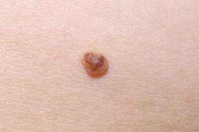
Figure 12.1 Typical papillomatous nevus (Unna nevus) with a diameter of 5 mm on the trunk.
DERMOSCOPIC FEATURES
The predominant pattern of Unna nevi is the globular pattern. Due to the papillomatous surface sometimes a cobblestone pattern is formed. The homogenous pattern is second frequent seen in Unna nevi. Hypo/hyperpigmentation is characteristic for Unna nevi and lead sometimes to an asymmetric distribution of colors. A specific finding are the comma vessels (linear curved vessels), which are usually found at the periphery of Unna nevi (Figure 12.3).
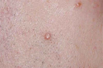
Figure 12.2 Miescher nevi on the face: pink domeshaped papules with a smooth surface.
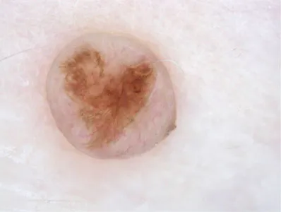
Figure 12.3 Dermoscopic image of the Unna nevus of Figure 12.1: irregular pigmentation in the center and comma-like vessels at the periphery.
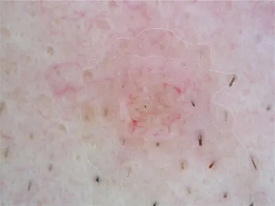
Figure 12.4 Dermoscopic image of the Miescher nevus of Figure 12.2: remnants of pigmentation and comma-like vessels.
Miescher nevi display usually a homogeneous pattern. They are symmetric and the color is in most of the cases pink to brownish. Comma vessels are also typical for Miescher nevi (Figure 12.4). An additional dermoscopic feature seen in some Miescher nevi is a golden brown halo.6
CLINICAL MANAGEMENT
Miescher and Unna nevi are benign melanocytic nevi and do not need special treatment or follow-up if the diagnosis can be made without any doubt. Differential diagnosis encompasses basal cell carcinoma, amelanotic melanoma, and benign tumors such as fibrous papule of the nose, fibroma, neurofibroma, and skin tags. If there is a change in color, outline, or contour, one should consider a melanoma arising in an intradermal nevus and histology is obligatory.
REFERENCES
1. Unna PG. Histopathology of Diseases of the Skin. New York: MacMillan; 1896: 1129–44.
2. Miescher G, von Albertini A. Histologie de 100 cas de nevi pigmentaires d’apres les methodes de Masson. Bull Soc Franc Deratol Syph 1935; 42: 1265–73.
3. Fernandez-Flores A, Sanchez-Velicia L, Manjon JA, Alija A, Soto F. A hypothesis on the morphologic differences between Unna and Miescher nevi on the head and neck, based on embryologic bases. Am J Dermatopathol 2012; 34: 602–6.
4. Ackerman AB, Magana-Garcia M. Naming acquired melanocytic nevi: Unna’s, Miescher’s, Spitz’s Clark’s. Am J Dermatopathol 1990; 12(2): 193–209.
5. Niederkorn A, Ahlgrimm-Siess V, Fink-Puches R et al. Frequency, clinical and dermoscopic features of benign papillomatous melanocytic naevi (Unna type). Br J Dermatol 2009; 161: 510–4.
6. Grazzini M, Marghoob AA. The “golden brown halo” of the Miescher nevus. Arch Dermatol 2012: 148: 870.
13 | Clark nevi in fair skin types |
KEY FEATURES
Fair-skinned individuals have a high proportion of poorly pigmented nevi. A clinical selection of nevi for dermoscopy by size and intensity of pigmentation carries a bias toward large and pigmented lesions.
KEY MANAGEMENT
Clinical atypia does not always correspond to histopathologic dysplasia and vice versa.
It is well known that nevi in people with skin type IV–VI mostly are very dark in appearance. In the fair-skinned population large, asymmetric, and dark nevi attract attention and are frequently excised under suspicion of melanoma. If inspected by dermoscopy, many of these nevi display rather orderly patterns and architecture, e.g., a regular network or cobblestone pattern. In contrast other nevi may appear quite normal clinically. However, if viewed dermoscopically one may come across features not encountered in the neighboring nevi of the same individual. Even nevi of little clinical atypicality may appear highly irregular on dermoscopy and cannot be distinguished reliably from melanoma. Especially in hypopigmented nevi, features of pigmentation may be faint or even absent. Consequently, vascular findings are becoming more important and may be the most important clues to diagnosis.
If nevi are selected for dermoscopic inspection just by their atypical appearance on inspection with the naked eye, other ones—small, symmetric, or poorly pigmented nevi—may escape dermoscopic analysis. The latter may in fact reveal significantly atypical features requiring immediate action. Dermoscopy of nevi should therefore be performed in a nonselective manner not to miss the nevi inconspicuous for the naked eye but highly suspicious under the microscope. Comparison with neighboring nevi is helpful to identify the “ugly duckling” requiring excision.
When Clark et al.1 described a type of nevus assumed to be a melanoma precursor they used several clinical criteria many of which were typical for melanoma as well. The so-called “dysplastic nevus” was clinically similar to melanoma but did not fulfill all histological criteria for malignancy. Many investigators analyzed the correlation of clinical and histopathological features. Some believe that clinical atypicality characterizes nevi with histopathological signs of dysplasia, whereas others would not confirm that correlation.2 Furthermore it could be demonstrated that mechanical irritation or exposure to ultraviolet light would induce histopathological features of dysplasia or even of early melanoma. However, these alterations were reversible within a few weeks. Today, the “dysplastic” nevus is not considered to be an obligatory melanoma precursor.
Dermoscopy helps to identify nevi which are severely dysplastic histopathologically and cannot be distinguished from melanoma with certainty. For many n...
Table of contents
- Cover
- Half Title
- Title Page
- Copyright Page
- Dedication
- Table of Contents
- Foreword
- Preface
- Contributors
- SECTION I TECHNICAL ASPECTS
- SECTION II DERMATOSCOPY OF NON-PIGMENTED LESIONS
- SECTION III SPECIFIC DERMATOSCOPIC PATTERNS AND THEIR DIAGNOSTIC SIGNIFICANCE
Frequently asked questions
Yes, you can cancel anytime from the Subscription tab in your account settings on the Perlego website. Your subscription will stay active until the end of your current billing period. Learn how to cancel your subscription
No, books cannot be downloaded as external files, such as PDFs, for use outside of Perlego. However, you can download books within the Perlego app for offline reading on mobile or tablet. Learn how to download books offline
Perlego offers two plans: Essential and Complete
- Essential is ideal for learners and professionals who enjoy exploring a wide range of subjects. Access the Essential Library with 800,000+ trusted titles and best-sellers across business, personal growth, and the humanities. Includes unlimited reading time and Standard Read Aloud voice.
- Complete: Perfect for advanced learners and researchers needing full, unrestricted access. Unlock 1.5M+ books across hundreds of subjects, including academic and specialized titles. The Complete Plan also includes advanced features like Premium Read Aloud and Research Assistant.
We are an online textbook subscription service, where you can get access to an entire online library for less than the price of a single book per month. With over 1.5 million books across 990+ topics, we’ve got you covered! Learn about our mission
Look out for the read-aloud symbol on your next book to see if you can listen to it. The read-aloud tool reads text aloud for you, highlighting the text as it is being read. You can pause it, speed it up and slow it down. Learn more about Read Aloud
Yes! You can use the Perlego app on both iOS and Android devices to read anytime, anywhere — even offline. Perfect for commutes or when you’re on the go.
Please note we cannot support devices running on iOS 13 and Android 7 or earlier. Learn more about using the app
Please note we cannot support devices running on iOS 13 and Android 7 or earlier. Learn more about using the app
Yes, you can access Dermatoscopy of Non-Pigmented Skin Tumors by Iris Zalaudek, Giuseppe Argenziano, Jason Giacomel, Iris Zalaudek,Giuseppe Argenziano,Jason Giacomel in PDF and/or ePUB format, as well as other popular books in Medicine & Dermatology. We have over 1.5 million books available in our catalogue for you to explore.