eBook - ePub
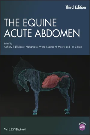
The Equine Acute Abdomen
- English
- ePUB (mobile friendly)
- Available on iOS & Android
eBook - ePub
The Equine Acute Abdomen
About this book
Written and edited by leading experts on equine digestive diseases, The Equine Acute Abdomen, Third Edition is the preeminent text on diagnosing and treating acute abdominal diseases in horses, donkeys, and mules.
- The definitive guide to acute abdominal disorders in equine patients, fully updated and revised to reflect the latest developments in the field
- Lavishly illustrated with more than 450 color illustrations, photographs, line drawings, and figures
- A companion website features video clips and images from the book available for download
- Provides an invaluable resource to equine surgery and internal medicine specialists, researchers, practitioners, and students who deal with colic
Tools to learn more effectively

Saving Books

Keyword Search

Annotating Text

Listen to it instead
Information
Part I
Normal Anatomy and Physiology
1
Gross and Microscopic Anatomy of the Equine Gastrointestinal Tract
Thomas M. Krunkosky1, Carla L. Jarrett1, and James N. Moore2
1 Department of Veterinary Biosciences and Diagnostic Imaging, College of Veterinary Medicine, University of Georgia, Athens, Georgia, USA
2 College of Veterinary Medicine, University of Georgia, Athens, Georgia, USA
Introduction
Gaining a working knowledge of the equine gastrointestinal tract and associated intra‐abdominal organs can be challenging, especially for inexperienced individuals. Experienced veterinarians who examine and treat horses with conditions characterized by acute abdominal pain (colic) know that the key to the diagnosis often lies in recognizing changes in anatomic structures or relationships among different organs. With this in mind, the focus of this chapter is the gross and microscopic structure of the horse’s alimentary tract (Figure 1.1A, B, C, and D), starting with the esophagus. Because some conditions characterized by colic involve other organs within the abdomen, we have reviewed the relevant structural aspects of the liver, spleen, and pancreas. In compiling this information, our goal is to provide veterinary students and veterinarians with the foundational materials needed to understand clinical conditions that result in colic.

Figure 1.1 (A) The abdominal organs from the left side of the horse. (B) A view from the cranial‐most aspect of the abdomen. (C) The abdominal organs visible from the caudal‐most aspect. (D) The abdominal organs visible from the horse’s right side.
Source: Courtesy of The Glass Horse, Science In 3D.
Esophagus
Gross Anatomic Features
The esophagus is the long muscular tube that connects the pharynx to the stomach. It is regionally subdivided into cervical, thoracic, and abdominal parts. Individual and breed variations exist, but in general the esophagus is positioned on the dorsal aspect of the trachea at the level of the 1st cervical vertebra, inclines to the left lateral surface of the trachea at the level of the 4th cervical vertebra, and is positioned ventrolateral to the trachea from the level of the 6th cervical vertebra up to and during passage through the thoracic inlet. The thoracic portion of the esophagus travels within the mediastinum and is positioned dorsal to the trachea to the level of the tracheal bifurcation. The esophagus passes dorsal to the base of the heart and continues caudally until it penetrates the diaphragm at the esophageal hiatus, accompanied by the dorsal and ventral vagal trunks. The abdominal portion of the esophagus is short and travels over the dorsal border of the liver, creating an esophageal impression, before joining the cardia of the stomach at an acute angle.
The esophagus is more superficial and therefore more accessible for surgery in the mid‐ to caudal‐third of the left side of the neck ventromedial to the jugular groove. Deep cervical fascia ensheathes the esophagus as it passes along the neck and also forms the left carotid sheath enclosing the left common carotid artery, the left vagosympathetic trunk, and the left internal jugular vein (when present). These structures, along with the neighboring left recurrent laryngeal nerve and the left tracheal lymphatic trunk (embedded within the deep cervical fascia that ensheathes the trachea), are to be avoided during surgical approaches to the esophagus.
Microscopic Features
The esophagus is designed to facilitate the delivery of ingesta to the stomach. Longitudinally oriented folds occur along the length of the mucosa of the esophagus to allow for expansion of its lumen during the passage of a food bolus. The mucosa of the esophagus is considerably mobile upon the underlying submucosa. The tunica mucosa is composed of three layers, or laminae (Figure 1.2). The lamina epithelialis is nonkeratinized stratified squamous epithelium (Figure 1.3); mild to moderate keratinization of the epithelium may occur, depending on the nature of the swallowed material. The lamina propria varies from loose to dense irregular connective tissue. The lamina muscularis mucosa consists of isolated bundles of longitudinally oriented smooth muscle in the cranial esophagus. The muscle bundles increase in density and coalesce into a distinct layer towards the caudal esophagus. Because the lamina muscularis mucosa serves as a demarcation between the mucosa and the submucosa, it is difficult to distinguish these layers where the muscularis is sparse or absent. The tunica submucosa is dense irregular connective tissue that contains prominent vasculature and the submucosal nerve plexus. Simple branching tubuloalveolar mucus‐secreting submucosal glands are present at the pharyngoesophageal junction (Figure 1.4). The tunica muscularis is skeletal muscle in the cranial two‐thirds of the esophagus and transitions into smooth muscle in the caudal third of the esophagus. There are two muscle layers in the tunica muscularis; however, the layers are not always distinguishable due to spiraling and interlacing of the muscle bundles. The cervical region of the esophagus has a tunica adventitia of dense irregular connective tissue that blends with the surrounding tissues. The thoracic and abdominal regions of the esophagus have a tunica serosa, which is mediastinal pleura and visceral peritoneum, respectively.

Figure 1.2 Full‐thickness section of the thoracic esophagus. H&E stain.

Figure 1.3 The lamina epithelialis of the esophagus. The epithelium is nonkeratinized with retention of nuclei throughout the most superficial layer (the stratum superficiale). The lamina propria is dense irregular connective tissue. The lamina propria and lamina epithelialis interdigitate via finger‐like projections of the epidermis (epidermal pegs) and dermis (dermal papillae). H&E stain.

Figure 1.4 Esophageal submucosal glands. The mucous secretory products of the submucosal glands are ducted into the esophageal lumen. The larger clear spaces are sections of ducts. H&E stain.
Esophagus–Stomach Junction
The true gastroesophageal junction in the equine is microscopically similar to the caudal esophagus with the addition of a thickening in the inner circular layer of the tunica muscularis that functions as a sphincter between the two organs. The combination of the muscular cardiac sphincter and the oblique angle at which the distal end of the esophagus joins the cardia of the stomach makes it exceptionally difficult for horses to vomit.
Stomach
Gross Anatomic Features
The stomach is enclosed within the ribcage between the 9th and 15th ribs and is positioned in the left half of the abdomen, caudal to the diaphragm and liver and cranial to the spleen. It has four compartments, the cardia, fundus (saccus cecus), body, and pyloric regions (Figure 1.5). The cardia is the most cranial region and is firmly fixed to the diaphragm near the dorsal surface of the 11th rib. The fundus is dorsal to the cardia and is large and lined by a nonglandular mucosa. The body is the largest portion of the stomach and spans between the nonglandular region ventral to the cardia to the acute angle of the lesser curvature (the angular incisure). The pyloric region spans between the angular incisure to the duodenum and is subdivided into the pyloric antrum, canal, and the strong muscular sphincter, the pylorus. The pylorus is the only portion of the stomach located to the right of the median plane. The cardiac and pyloric regions are in close proximity due to the acute angle of the concave cranial s...
Table of contents
- Cover
- Title Page
- Table of Contents
- Editors
- List of Contributors
- Preface
- About the Companion Website
- Part I: Normal Anatomy and Physiology
- Part II: Pathophysiology of Gastrointestinal Diseases
- Part III: Intestinal Parasitism
- Part IV: Epidemiology of Colic
- Part V: Diagnosis of Gastrointestinal Disease
- Part VI: Medical Management
- Part VII: Colic in the Foal
- Part VIII: Colic in the Donkey
- Part IX: Nutritional Management
- Part X: Anesthesia for Abdominal Surgery
- Part XI: Surgery for Acute Abdominal Disease
- Part XII: Intensive Care and Postoperative Care
- Part XIII: Specific Diseases of Horses
- Index
- End User License Agreement
Frequently asked questions
Yes, you can cancel anytime from the Subscription tab in your account settings on the Perlego website. Your subscription will stay active until the end of your current billing period. Learn how to cancel your subscription
No, books cannot be downloaded as external files, such as PDFs, for use outside of Perlego. However, you can download books within the Perlego app for offline reading on mobile or tablet. Learn how to download books offline
Perlego offers two plans: Essential and Complete
- Essential is ideal for learners and professionals who enjoy exploring a wide range of subjects. Access the Essential Library with 800,000+ trusted titles and best-sellers across business, personal growth, and the humanities. Includes unlimited reading time and Standard Read Aloud voice.
- Complete: Perfect for advanced learners and researchers needing full, unrestricted access. Unlock 1.4M+ books across hundreds of subjects, including academic and specialized titles. The Complete Plan also includes advanced features like Premium Read Aloud and Research Assistant.
We are an online textbook subscription service, where you can get access to an entire online library for less than the price of a single book per month. With over 1 million books across 990+ topics, we’ve got you covered! Learn about our mission
Look out for the read-aloud symbol on your next book to see if you can listen to it. The read-aloud tool reads text aloud for you, highlighting the text as it is being read. You can pause it, speed it up and slow it down. Learn more about Read Aloud
Yes! You can use the Perlego app on both iOS and Android devices to read anytime, anywhere — even offline. Perfect for commutes or when you’re on the go.
Please note we cannot support devices running on iOS 13 and Android 7 or earlier. Learn more about using the app
Please note we cannot support devices running on iOS 13 and Android 7 or earlier. Learn more about using the app
Yes, you can access The Equine Acute Abdomen by Anthony T. Blikslager, Nathaniel A. White, James N. Moore, Tim S. Mair, Anthony T. Blikslager,Nathaniel A. White, II,James N. Moore,Tim S. Mair in PDF and/or ePUB format, as well as other popular books in Medicine & Dentistry. We have over one million books available in our catalogue for you to explore.