- English
- ePUB (mobile friendly)
- Available on iOS & Android
eBook - ePub
The Chemistry of Molecular Imaging
About this book
Molecular imaging is primarily about the chemistry of novel biological probes, yet the vast majority of practitioners are not chemists or biochemists. This is the first book, written from a chemist's point of view, to address the nature of the chemical interaction between probe and environment to help elucidate biochemical detail instead of bulk anatomy.
- Covers all of the fundamentals of modern imaging methodologies, including their techniques and application within medicine and industry
- Focuses primarily on the chemistry of probes and imaging agents, and chemical methodology for labelling and bioconjugation
- First book to investigate the chemistry of molecular imaging
- Aimed at students as well as researchers involved in the area of molecular imaging
Tools to learn more effectively

Saving Books

Keyword Search

Annotating Text

Listen to it instead
Information
1
An Introduction to Molecular Imaging
Ga-Lai Law and Wing-Tak Wong
Department of Applied Biology and Chemical Technology, Hong Kong Polytechnic University, Hung Hom, Kowloon, Hong Kong SAR, China
1.1 Introduction
The aim of this book is to introduce the concepts of different imaging techniques that are employed for diagnostics and therapy and the role that chemistry has played in their evolution. The book provides a general introduction to the area of molecular imaging, giving an account of the role of molecular design and its importance in modern-day techniques, with an in-depth introduction of some of the probes and methodologies employed. This first chapter introduces the different types of imaging modalities currently at the forefront of imaging and illustrates some basic concepts underlying these techniques. It acts as a simplified background to set the scene for the following chapters, which will discuss the chemical properties of molecules and the role they play in different imaging modalities. For the interested readers, other textbooks are referenced that will provide more detailed information regarding the different techniques reviewed.
In life everything is incessantly changing. There is constant evolution in life sciences, evolution in the way problems arise, and evolution in the way they are solved. Diagnostics and therapy are both important, but as Einstein said, “intellectuals solve problems, geniuses prevent them.” The key challenge still remains to unravel the hidden knowledge within life sciences, which constantly challenges us with new diseases and mechanistic mutation of biological systems and pathways [1]. Again, as stated by Einstein, “once we accept our limits, we go beyond them.”
Molecular imaging aims to detect and monitor mechanistic processes in cells, tissues, or living organisms with the use of instruments and contrast mechanisms without perturbing their living system. Ultimately, it is a field that utilises molecular building blocks to bring solutions to problems by specialised imaging techniques that have matured into a large integrated field enveloped within various branches of science (Figure 1.1) [2]. In the area of modern-day imaging where technology is at its pinnacle, molecular design still holds a dominant role in the forefront of molecular imaging.
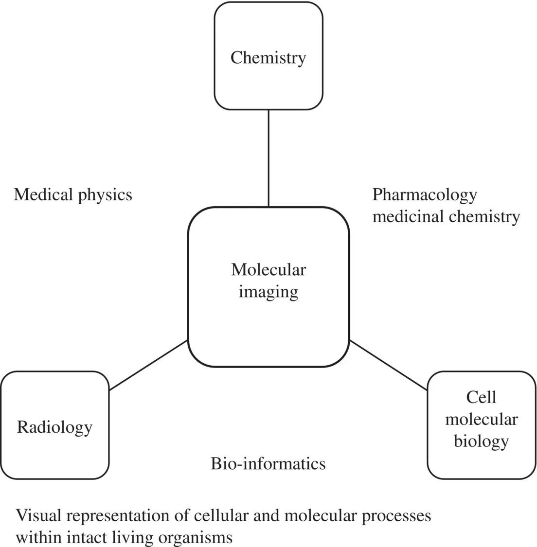
Figure 1.1 Types of multidisciplinary fields related to molecular imaging.
In the past, developments in contrast agents, probes, and dyes have brought about an era of creativity where new techniques, materials, and designs have flourished to form a concrete foundation resulting in today’s achievements in diagnosis and therapy (Figure 1.2). The construction of better chemical molecules will continue to help us develop a more comprehensive picture of learning about life science. Figure 1.3depicts a timeline in the development of the field [1–3].

Figure 1.2 Diagram showing the links in the design rationale of imaging agents.

Figure 1.3 An approximate timeline showing the development of the different imaging modalities [1–3].
1.2 What Is Positron Emission Tomography (PET)?
Positron Emission Tomography (PET) is a nuclear medicine tomographic modality and one of the most sensitive methods for quantitative measurement of physiologic processes in vivo [4]. This technique utilises positron-emitting radionuclides and requires the use of radiotracers that decay and produce two 511 keV γ-rays resulting from the annihilation of a positron and an electron. One of the most commonly used molecules is 18 F-labelled fluorodeoxyglucose (18FDG), which has radioactive fluorine and is readily taken up by tumours (Figure 1.4) [5].
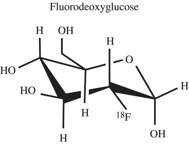
Figure 1.4 18FDG, a typical contrast agent used in PET.
1.2.1 Basic Principles
In PET, a neutron-deficient isotope causes positron annihilation to produce two 511 keV γ-rays, which are simultaneously emitted when a positron from a nuclear disintegration annihilates in tissue. PET imaging, unlike MRI, ultrasound, and optical imaging, does not require any external sources for probing or excitation; instead, the source is generated from radioisotopes and emitted from but not transmitted through an object/patient, as in CT imaging [4–7]. Radionuclides are incorporated as part of a small metabolically active molecule to generate radiotracers such as 18FDG, which are then intravenously injected into patients at trace dosage for PET imaging. 18FDG is a favourable radiotracer because it is inhibited from metabolic degradation before it decays due to the fluorine at the 2' position in the molecule. Upon decay, the fluorine is converted into 18O. There is generally a short period of time before accumulation of radiotracers into the targeted organs or tissues that are being examined, so it is important for radiotracers to have a suitable half-life—some commonly used radionuclei have very short half-lives. Some common radionuclides used in PET are 11-C (half-life ~20 min), 13-N (~10 min), 15-O (~2 min) and 18-F (~110 min). These are produced by a cyclotron, whereas 82-Rb (76 s), which is used in clinical cardiac PET, is produced by a generator [8–9].
When a radioisotope undergoes positron emission decay (positive β-decay), it emits a positron that travels through the tissue for a short distance (~ < 2 mm) whilst decelerating by the loss of its kinetic energy until it collides with an electron. This results in back-to-back annihilation of γ-ray photons, which move in opposite directions and are emitted nearly 180 degrees apart before being detected by scintillators an...
Table of contents
- Cover
- Title page
- Copyright page
- Preface
- List of Contributors
- 1 An Introduction to Molecular Imaging
- 2 Chemical Methodology for Labelling and Bioconjugation
- 3 Recent Developments in the Chemistry of [18F]Fluoride for PET
- 4 Carbon-11, Nitrogen-13, and Oxygen-15 Chemistry: An Introduction to Chemistry with Short-Lived Radioisotopes
- 5 The Chemistry of Inorganic Nuclides (86Y, 68Ga, 64Cu, 89Zr, 124I)
- 6 The Radiopharmaceutical Chemistry of Technetium and Rhenium
- 7 The Radiopharmaceutical Chemistry of Gallium(III) and Indium(III) for SPECT Imaging
- 8 The Chemistry of Lanthanide MRI Contrast Agents
- 9 Nanoparticulate MRI Contrast Agents
- 10 CEST and PARACEST Agents for Molecular Imaging
- 11 Organic Molecules for Optical Imaging
- 12 Application of d- and f-Block Fluorescent Cell Imaging Agents
- 13 Lanthanide-Based Upconversion Nanophosphors for Bioimaging
- 14 Microbubbles: Contrast Agents for Ultrasound and MRI
- 15 Non-Nanoparticle-Based Dual-Modality Imaging Agents
- 16 Chemical Strategies for the Development of Multimodal Imaging Probes Using Nanoparticles
- Index
- End User License Agreement
Frequently asked questions
Yes, you can cancel anytime from the Subscription tab in your account settings on the Perlego website. Your subscription will stay active until the end of your current billing period. Learn how to cancel your subscription
No, books cannot be downloaded as external files, such as PDFs, for use outside of Perlego. However, you can download books within the Perlego app for offline reading on mobile or tablet. Learn how to download books offline
Perlego offers two plans: Essential and Complete
- Essential is ideal for learners and professionals who enjoy exploring a wide range of subjects. Access the Essential Library with 800,000+ trusted titles and best-sellers across business, personal growth, and the humanities. Includes unlimited reading time and Standard Read Aloud voice.
- Complete: Perfect for advanced learners and researchers needing full, unrestricted access. Unlock 1.4M+ books across hundreds of subjects, including academic and specialized titles. The Complete Plan also includes advanced features like Premium Read Aloud and Research Assistant.
We are an online textbook subscription service, where you can get access to an entire online library for less than the price of a single book per month. With over 1 million books across 990+ topics, we’ve got you covered! Learn about our mission
Look out for the read-aloud symbol on your next book to see if you can listen to it. The read-aloud tool reads text aloud for you, highlighting the text as it is being read. You can pause it, speed it up and slow it down. Learn more about Read Aloud
Yes! You can use the Perlego app on both iOS and Android devices to read anytime, anywhere — even offline. Perfect for commutes or when you’re on the go.
Please note we cannot support devices running on iOS 13 and Android 7 or earlier. Learn more about using the app
Please note we cannot support devices running on iOS 13 and Android 7 or earlier. Learn more about using the app
Yes, you can access The Chemistry of Molecular Imaging by Nicholas Long,Wing-Tak Wong in PDF and/or ePUB format, as well as other popular books in Technology & Engineering & Diagnostics Imaging. We have over one million books available in our catalogue for you to explore.