The seeming simplicity of day-to-day movement can belie the complexity of the structures that facilitate motion. More than just a framework around which the body develops, the human skeleton has evolved over time to allow humans to walk and stand upright. Muscles likewise perform a range of functions, without which the body could not manage. This comprehensive book details the anatomy and mechanisms that allow bones and muscles to operate naturally and examines the consequences of disease and injury on these fundamental components of the human body.
Häufig gestellte Fragen
Wie kann ich mein Abo kündigen?
Gehe einfach zum Kontobereich in den Einstellungen und klicke auf „Abo kündigen“ – ganz einfach. Nachdem du gekündigt hast, bleibt deine Mitgliedschaft für den verbleibenden Abozeitraum, den du bereits bezahlt hast, aktiv. Mehr Informationen hier.
(Wie) Kann ich Bücher herunterladen?
Derzeit stehen all unsere auf Mobilgeräte reagierenden ePub-Bücher zum Download über die App zur Verfügung. Die meisten unserer PDFs stehen ebenfalls zum Download bereit; wir arbeiten daran, auch die übrigen PDFs zum Download anzubieten, bei denen dies aktuell noch nicht möglich ist. Weitere Informationen hier.
Welcher Unterschied besteht bei den Preisen zwischen den Aboplänen?
Mit beiden Aboplänen erhältst du vollen Zugang zur Bibliothek und allen Funktionen von Perlego. Die einzigen Unterschiede bestehen im Preis und dem Abozeitraum: Mit dem Jahresabo sparst du auf 12 Monate gerechnet im Vergleich zum Monatsabo rund 30 %.
Was ist Perlego?
Wir sind ein Online-Abodienst für Lehrbücher, bei dem du für weniger als den Preis eines einzelnen Buches pro Monat Zugang zu einer ganzen Online-Bibliothek erhältst. Mit über 1 Million Büchern zu über 1.000 verschiedenen Themen haben wir bestimmt alles, was du brauchst! Weitere Informationen hier.
Unterstützt Perlego Text-zu-Sprache?
Achte auf das Symbol zum Vorlesen in deinem nächsten Buch, um zu sehen, ob du es dir auch anhören kannst. Bei diesem Tool wird dir Text laut vorgelesen, wobei der Text beim Vorlesen auch grafisch hervorgehoben wird. Du kannst das Vorlesen jederzeit anhalten, beschleunigen und verlangsamen. Weitere Informationen hier.
Ist Bone and Muscle als Online-PDF/ePub verfügbar?
Ja, du hast Zugang zu Bone and Muscle von Britannica Educational Publishing, Kara Rogers im PDF- und/oder ePub-Format sowie zu anderen beliebten Büchern aus Ciencias biológicas & Fisiología. Aus unserem Katalog stehen dir über 1 Million Bücher zur Verfügung.
The skeleton is the most fundamental component of human anatomy. It serves not only as a framework for the body, providing places for the attachment of muscles and other tissues, but also serves as a protective barrier for vital organs, such as the brain and heart. The skeleton consists of many individual bones and cartilages. There also are bands of fibrous connective tissue—the ligaments and the tendons—in intimate relationship with the parts of the skeleton.
The human skeleton, like that of other vertebrates, consists of two principal subdivisions, each with origins distinct from the others and each presenting certain individual features. These are (1) the axial, comprising the vertebral column—the spine—and much of the skull, and (2) the appendicular, to which the pelvic (hip) and pectoral (shoulder) girdles and the bones and cartilages of the limbs belong.
When one considers the relation of these subdivisions of the skeleton to the soft parts of the human body—such as the nervous system, the digestive system, the respiratory system, the cardiovascular system, and the voluntary muscles of the muscle system—it is clear that the functions of the skeleton are of three different types: support, protection, and motion. Of these functions, support is the most primitive and the oldest; likewise, the axial part of the skeleton was the first to evolve. The vertebral column, corresponding to the notochord in lower organisms, is the main support of the trunk.
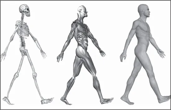
3-D side view of a walking male’s skeleton and muscles. Shutterstock.com
A distinctive characteristic of humans as compared with other mammals is erect posture. The human body is to some extent like a walking tower that moves on pillars, represented by the legs. Tremendous advantages have been gained from this erect posture, the chief among which has been the freeing of the arms for a great variety of uses. Nevertheless, erect posture has created a number of mechanical problems—in particular, weight bearing. These problems have had to be met by adaptations of the skeletal system.
Protection of the heart, lungs, and other organs and structures in the chest requires a flexible and elastic covering that can move with the organs as they expand and contract. Such a covering is provided by the bony thoracic basket, or rib cage, which forms the skeleton of the wall of the chest, or thorax. The connection of the ribs to the breastbone—the sternum—is in all cases a secondary one, brought about by the relatively pliable rib (costal) cartilages. The small joints between the ribs and the vertebrae permit a gliding motion of the ribs on the vertebrae during breathing and other activities. The motion is limited by the ligamentous attachments between ribs and vertebrae.
The third general function of the skeleton is that of motion. The great majority of the skeletal muscles are firmly anchored to the skeleton, usually to at least two bones and in some cases to many bones. Thus, the motions of the body and its parts, all the way from the lunge of the football player to the delicate manipulations of a handicraft artist or of the use of complicated instruments by a scientist, are made possible by separate and individual engineering arrangements between muscle and bone.
AXIAL AND VISCERAL SKELETON
THE CRANIUM
The cranium—the part of the skull that encloses the brain—is sometimes called the braincase. The primary function of the cranium is to protect the brain; however, it also serves as an important role in providing a connective medium for the muscles of the face and for the tissues of the brain.
Development of Cranial Bones
The cranium is formed of bones of two different types of developmental origin—the cartilaginous, or substitution, bones, which replace cartilages preformed in the general shape of the bone; and membrane bones, which are laid down within layers of connective tissue. For the most part, the substitution bones form the floor of the cranium, while membrane bones form the sides and roof.
The range in the capacity of the cranial cavity is wide but is not directly proportional to the size of the skull, because there are variations also in the thickness of the bones and in the size of the air pockets, or sinuses. The cranial cavity has a rough, uneven floor, but its landmarks and details of structure generally are consistent from one skull to another.
The cranium forms all the upper portion of the skull, with the bones of the face situated beneath its forward part. It consists of a relatively few large bones, the frontal bone, the sphenoid bone, two temporal bones, two parietal bones, and the occipital bone. The frontal bone underlies the forehead region and extends back to the coronal suture, an arching line that separates the frontal bone from the two parietal bones, on the sides of the cranium. In front, the frontal bone forms a joint with the two small bones of the bridge of the nose and with the zygomatic bone (which forms part of the cheekbone), the sphenoid, and the maxillary bones. Between the nasal and zygomatic bones, the horizontal portion of the frontal bone extends back to form a part of the roof of the eye socket, or orbit; it thus serves an important protective function for the eye and its accessory structures.
Each parietal bone has a generally four-sided outline. Together they form a large portion of the side walls of the cranium. Each adjoins the frontal, the sphenoid, the temporal, and the occipital bones and its fellow of the opposite side. They are almost exclusively cranial bones, having less relation to other structures than the other bones that help to form the cranium.
Interior of the Cranium
The interior of the cranium shows a multitude of details, reflecting the shapes of the softer structures that are in contact with the bones. In addition the base of the cranium is divided into three major depressions, or fossae, which are divided strictly according to the borders of the bones of the cranium but are related to major portions of the brain. The anterior cranial fossa serves as the bed in which rest the frontal lobes of the cerebrum, the large forward part of the brain. The middle cranial fossa, sharply divided into two lateral halves by a central eminence of bone, contains the temporal lobes of the cerebrum. The posterior cranial fossa serves as a bed for the hemispheres of the cerebellum (a mass of brain tissue behind the brain stem and beneath the rear portion of the cerebrum) and for the front and middle portion of the brain stem. Major portions of the brain are thus partially enfolded by the bones of the cranial wall.
There are openings in the three fossae for the passage of nerves and blood vessels, and the markings on the internal surface of the bones are from the attachments of the brain coverings—the meninges—and venous sinuses and other blood vessels.
The anterior cranial fossa shows a crestlike projection in the midline, the crista galli (“crest of the cock”). This is a place of firm attachment for the falx cerebri, a subdivision of dura mater that separates the right and left cerebral hemispheres. On either side of the crest is the cribriform (pierced with small holes) plate of the ethmoid bone, a midline bone important as a part both of the cranium and of the nose. At the sides of the cribriform plate are the orbital plates of the frontal bone, which form the roofs of the eye sockets.
The rear part of the anterior cranial fossa is formed by those portions of the sphenoid bone called its body and lesser wings. Projections from the lesser wings, the anterior clinoid (bedlike) processes, extend back to a point beside each optic foramen, an opening through which important optic nerves, or tracts, enter into the protection of the cranial cavity after a relatively short course within the eye socket.
The central eminence of the middle cranial fossa is specialized as a saddlelike seat for the pituitary gland. The posterior portion of this seat, or sella turcica (“Turk’s saddle”), is actually wall-like and is called the dorsum sellae. The pituitary gland is thus situated in almost the centre of the cranial cavity.
The deep lateral portions of the middle cranial fossa contain the temporal lobes of the cerebrum. Also in the middle fossa is the jagged opening called the foramen lacerum. The lower part of the foramen lacerum is blocked by fibrocartilage, but through its upper part passes the internal carotid artery, surrounded by a network of nerves, as it makes its way to the interior of the cranial cavity.
The posterior cranial fossa is above the vertebral column and the muscles of the back of the neck. The foramen magnum, the opening through which the brain and the spinal cord make connection, is in the lowest part of the fossa. Through other openings in the posterior cranial fossa, including the jugular foramina, pass the large blood channels called the sigmoid sinuses and also the 9th (glossopharyngeal), 10th (vagus), and 11th (spinal accessory) cranial nerves as they leave the cranial cavity.
THE HYOID BONE
The primary function of the hyoid bone is to serve as an anchoring structure for the tongue. The bone is situated at the root of the tongue in the front of the neck and between the lower jaw and the largest cartilage of the larynx, or voice box. It has no articulation with other bones and thus has a purely anchoring function, and it is more or less in the shape of a U, with the body forming the central part, or base, of the letter.
The hyoid bone has certain muscles of the tongue attached to it. Through these muscle attachments, the hyoid plays an important role in chewing (mastication), in swallowing, and in voice production. At the beginning of a swallowing motion, the geniohyoid and mylohyoid muscles elevate the bone and the floor of the mouth simultaneously. These muscles are assisted by the stylohyoid and digastric muscles. The tongue is pressed upward against the palate, and the food is forced backward.
THE FACIAL BONES AND THEIR COMPLEX FUNCTIONS
The Upper Jaws
The larger part of the skeleton of the face is formed by the maxillae. Though they are called the upper jaws, the extent and functions of the maxillae include much more than serving as complements to the lower jaw, or mandible. They form the middle and lower portion of the eye socket. They have the opening for the nose between them, and they form the sharp projection known as the anterior nasal spine.
The infraorbital foramen, an opening into the floor of the eye socket, is the forward end of a canal through which passes the infraorbital branch of the maxillary nerve, the second division of the fifth cranial nerve. In addition the alveolar margin, containing the alveoli, or sockets, in which all the upper teeth are set, forms the lower part of each maxilla, and lateral projections form the zygomatic process, creating a joint with the zygomatic, or malar, bone (cheekbone).
The Lower Jaw
The left and right halves of the lower jaw, or mandible, begin originally as two distinct bones, but in the second year of life the two bones fuse at the midline. The horizontal central part on each side is the body of the mandible. The projecting chin, at the lower part of the body in the midline, is said to be a distinctive characteristic of the human skull.
The ascending parts of the mandible at the side are called rami (branches). The joints by means of which the lower jaw is able to make all its varied movements are between a rounded knob, or condyle, at the upper back corner of each ramus and a depression, called a glenoid fossa, in each temporal bone. Another, rather sharp projection at the top of each ramus and in front, called a coronoid process, attaches to the temporalis muscle, which serves with other muscles in shutting the jaws. On the inner side of the ramus of either side is a large, obliquely placed opening into a channel, the mandibular canal, for nerves, arteries, and veins.
The zygomatic arch, forming the cheekbone, consists of portions of three bones: the maxilla, in front; the zygomatic bone, centrally in the arch; and a projection from the temporal bone to form the rear part. The zygomatic arch actually serves as a firm bony origin for the powerful masseter muscle, which descends from it to insert on the outer side of the mandible. The masseter muscle shares with the temporalis muscle and lateral and medial pterygoid muscles the function of elevating the mandible in order to bring the lower against the upper teeth, thus achieving a bite.
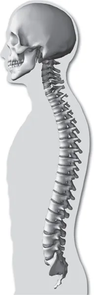
THE SPINE
The assumption of erect posture during the development of the human species has led to a need for adaptation and changes in the human skeletal system. The very form of the human vertebral column is due to such adaptations and changes.
The Vertebral Column
Viewed from the side, the vertebral column is not actually a column but rather a sort of spiral spring in the form of the letter S. The newborn child has a relatively straight backbone. The development of the curvatures occurs as the supporting functions of the vertebral column in humans—i.e., holding up the trunk, keeping the head erect, serving as an anchor for the extremities—are developed.
The S-curvature enables the vertebral column to absorb the shocks of walking on hard surfaces; a straight column would conduct the jarring shocks directly from the pelvic girdle to the head. The curvature meets the problem of the weight of the viscera. In an erect animal with a straight column, the column would be pulled forward by the viscera. Additional space for the viscera is provided by the concavities of the thoracic and pelvic regions.
The Spinal Cord
The space between the spinal cord and the vertebrae is occupied by the meninges, by the cerebrospinal fluid, and by a certain amount of fat and connective tissue. In front are the heavy centrums, or bodies, of the vertebrae and the intervertebral disks—the tough, resilient pads between the vertebral bodies. The portion of each vertebra called the neural arch encloses and protects the back and sides of the spinal cord. Between the neural arches are sheets of elastic connective tissue, the interlaminar ligaments, or ligamenta flava. Here some protective function has to be sacrificed for the sake of motion, because a forward bending of part of the column leads to separation between the laminae and between the spines of the neural arches of adjoining vertebrae.
Besides its role in support and protection, the vertebral column is important in the anchoring of muscles. Many of the muscles attached to it act to move either the column itself or various segments of it. Some are relatively superficial, and others are deep-lying. The large and important erector spinae, as the name implies, holds the spine erect. It begins on the sacrum (the large triangular bone at the base of the spinal column) and passes upward, forming a mass of muscle on either side of the spines of the lumbar vertebrae. It then divides into three columns, ascending over the back of the chest.
Small muscles run between the transverse processes (projections from the sides of the neural rings) of adjacent vertebrae, between the vertebral spines (projections from the centres of the rings), and from transverse process to spine, giving great mobility to the segmented bony column.
The anchoring function of the spinal column is of great importance for the muscles that arise on the trunk, in whole or in part from the column or from ligaments attached to it, and that are inserted on the bones of the arms and legs. Of these muscles, the most important for the arms are the latissimus dorsi (drawing the arm backward and downward and rotating it inward), the trapezius (rotating the shoulder blade), the rhomboideus, and the levator scapulae (raising and lowering the shoulder blade); for the legs, the psoas (loin) muscles.
THE RIB CAGE
The rib cage, or thoracic basket, consists of the 12 thoracic (chest) vertebrae, the 24 ribs, and t...
Inhaltsverzeichnis
Cover Page
Title Page
Copyright Page
Contents
Introduction
Chapter 1: Human Skeletal and Muscle Systems
Chapter 2: The Nature of Bone
Chapter 3: Bones of the Human Anatomy
Chapter 4: The Nature of Muscle
Chapter 5: The Human Body in Motion
Chapter 6: Diseases and Injuries of Bone
Chapter 7: Diseases and Injuries of Muscle
Chapter 8: Diseases and Injuries of Joints
Conclusion
Glossary
Bibliography
Index
Zitierstile für Bone and Muscle
APA 6 Citation
Educational, B. (2010). Bone and Muscle ([edition unavailable]). Britannica Educational Publishing. Retrieved from https://www.perlego.com/book/1638341/bone-and-muscle-structure-force-and-motion-pdf (Original work published 2010)
Chicago Citation
Educational, Britannica. (2010) 2010. Bone and Muscle. [Edition unavailable]. Britannica Educational Publishing. https://www.perlego.com/book/1638341/bone-and-muscle-structure-force-and-motion-pdf.
Harvard Citation
Educational, B. (2010) Bone and Muscle. [edition unavailable]. Britannica Educational Publishing. Available at: https://www.perlego.com/book/1638341/bone-and-muscle-structure-force-and-motion-pdf (Accessed: 14 October 2022).
MLA 7 Citation
Educational, Britannica. Bone and Muscle. [edition unavailable]. Britannica Educational Publishing, 2010. Web. 14 Oct. 2022.