Handbook of Medical Image Processing and Analysis
eBook - ePub

Handbook of Medical Image Processing and Analysis

Isaac Bankman

  1. 1,000 Seiten
  2. English
  3. ePUB (handyfreundlich)
  4. Über iOS und Android verfügbar
eBook - ePub

Handbook of Medical Image Processing and Analysis

Isaac Bankman

Angaben zum Buch
Buchvorschau
Inhaltsverzeichnis
Quellenangaben

Über dieses Buch

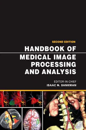
The Handbook of Medical Image Processing and Analysis is a comprehensive compilation of concepts and techniques used for processing and analyzing medical images after they have been generated or digitized. The Handbook is organized into six sections that relate to the main functions: enhancement, segmentation, quantification, registration, visualization, and compression, storage and communication.The second edition is extensively revised and updated throughout, reflecting new technology and research, and includes new chapters on: higher order statistics for tissue segmentation; tumor growth modeling in oncological image analysis; analysis of cell nuclear features in fluorescence microscopy images; imaging and communication in medical and public health informatics; and dynamic mammogram retrieval from web-based image libraries.For those looking to explore advanced concepts and access essential information, this second edition of Handbook of Medical Image Processing and Analysis is an invaluable resource. It remains the most complete single volume reference for biomedical engineers, researchers, professionals and those working in medical imaging and medical image processing. Dr. Isaac N. Bankman is the supervisor of a group that specializes on imaging, laser and sensor systems, modeling, algorithms and testing at the Johns Hopkins University Applied Physics Laboratory. He received his BSc degree in Electrical Engineering from Bogazici University, Turkey, in 1977, the MSc degree in Electronics from University of Wales, Britain, in 1979, and a PhD in Biomedical Engineering from the Israel Institute of Technology, Israel, in 1985. He is a member of SPIE.

  • Includes contributions from internationally renowned authors from leading institutions
  • NEW! 35 of 56 chapters have been revised and updated. Additionally, five new chapters have been added on important topics incluling Nonlinear 3D Boundary Detection, Adaptive Algorithms for Cancer Cytological Diagnosis, Dynamic Mammogram Retrieval from Web-Based Image Libraries, Imaging and Communication in Health Informatics and Tumor Growth Modeling in Oncological Image Analysis.
  • Provides a complete collection of algorithms in computer processing of medical images
  • Contains over 60 pages of stunning, four-color images

Häufig gestellte Fragen

Wie kann ich mein Abo kündigen?
Gehe einfach zum Kontobereich in den Einstellungen und klicke auf „Abo kündigen“ – ganz einfach. Nachdem du gekündigt hast, bleibt deine Mitgliedschaft für den verbleibenden Abozeitraum, den du bereits bezahlt hast, aktiv. Mehr Informationen hier.
(Wie) Kann ich Bücher herunterladen?
Derzeit stehen all unsere auf Mobilgeräte reagierenden ePub-Bücher zum Download über die App zur Verfügung. Die meisten unserer PDFs stehen ebenfalls zum Download bereit; wir arbeiten daran, auch die übrigen PDFs zum Download anzubieten, bei denen dies aktuell noch nicht möglich ist. Weitere Informationen hier.
Welcher Unterschied besteht bei den Preisen zwischen den Aboplänen?
Mit beiden Aboplänen erhältst du vollen Zugang zur Bibliothek und allen Funktionen von Perlego. Die einzigen Unterschiede bestehen im Preis und dem Abozeitraum: Mit dem Jahresabo sparst du auf 12 Monate gerechnet im Vergleich zum Monatsabo rund 30 %.
Was ist Perlego?
Wir sind ein Online-Abodienst für Lehrbücher, bei dem du für weniger als den Preis eines einzelnen Buches pro Monat Zugang zu einer ganzen Online-Bibliothek erhältst. Mit über 1 Million Büchern zu über 1.000 verschiedenen Themen haben wir bestimmt alles, was du brauchst! Weitere Informationen hier.
Unterstützt Perlego Text-zu-Sprache?
Achte auf das Symbol zum Vorlesen in deinem nächsten Buch, um zu sehen, ob du es dir auch anhören kannst. Bei diesem Tool wird dir Text laut vorgelesen, wobei der Text beim Vorlesen auch grafisch hervorgehoben wird. Du kannst das Vorlesen jederzeit anhalten, beschleunigen und verlangsamen. Weitere Informationen hier.
Ist Handbook of Medical Image Processing and Analysis als Online-PDF/ePub verfügbar?
Ja, du hast Zugang zu Handbook of Medical Image Processing and Analysis von Isaac Bankman im PDF- und/oder ePub-Format sowie zu anderen beliebten Büchern aus Computer Science & Digital Media. Aus unserem Katalog stehen dir über 1 Million Bücher zur Verfügung.

Information

Jahr
2008
ISBN
9780080559148
Part I
Enhancement
Chapter 1 Fundamental Enhancement Techniques
Raman B. Paranjape
University of Regina
1.1 Introduction
1.2 Preliminaries and Definitions
1.3 Pixel Operations
1.3.1 Compensation for Nonlinear Characteristics of Display or Print Media
1.3.2 Intensity Scaling
1.3.3 Histogram Equalization
1.4 Local Operators
1.4.1 Noise Suppression by Mean Filtering
1.4.2 Noise Suppression by Median Filtering
1.4.3 Edge Enhancement
1.4.4 Local-Area Histogram Equalization
1.5 Operations with Multiple Images
1.5.1 Noise Suppression by Image Averaging
1.5.2 Change Enhancement by Image Subtraction
1.6 Frequency Domain Techniques
1.7 Concluding Remarks
1.8 References

1.1 Introduction

Image enhancement techniques are used to refine a given image so that desired image features become easier to perceive for the human visual system or more likely to be detected by automated image analysis systems [1, 13]. Image enhancement allows the observer to see details in images that may not be immediately observable in the original image. This may be the case, for example, when the dynamic range of the data and that of the display are not commensurate, when the image has a high level of noise, or when contrast is insufficient [4, 5, 8, 9].
Fundamentally, image enhancement is the transformation or mapping of one image to another [10, 14]. This transformation is not necessarily one-to-one, so two different input images may transform into the same or similar output images after enhancement. More commonly, one may want to generate multiple enhanced versions of a given image. This aspect also means that enhancement techniques may be irreversible.
Often the enhancement of certain features in images is accompanied by undesirable effects. Valuable image information may be lost, or the enhanced image may be a poor representation of the original. Furthermore, enhancement algorithms cannot be expected to provide information that is not present in the original image. If the image does not contain the feature to be enhanced, noise or other unwanted image components may be inadvertently enhanced without any benefit to the user.
In this chapter we present established image enhancement algorithms commonly used for medical images. Initial concepts and definitions are presented in Section 1.2. Pixel-based enhancement techniques described in Section 1.3 are transformations applied to each pixel without utilizing specifically the information in the neighborhood of the pixel. Section 1.4 presents enhancement with local operators that modify the value of each pixel using the pixels in a local neighborhood. Enhancement that can be achieved with multiple images of the same scene is outlined in Section 1.5. Spectral domain filters that can be used for enhancement are presented in Section 1.6. The techniques described in this chapter are applicable to dental and medical images.

1.2 Preliminaries and Definitions

We define a digital image as a two-dimensional array of numbers that represent the real, continuous intensity distribution of a spatial signal. The continuous spatial signal is sampled at regular intervals, and the intensity is quantized to a finite number of levels. Each element of the array is referred to as a picture element or pixel. The digital image is defined as a spatially distributed intensity signal f (m, n), where f is the intensity of the pixel, and m and n define the position of the pixel along a pair of orthogonal axes usually defined as horizontal and vertical. We shall assume that the image has M rows and N columns and that the digital image has P quantized levels of intensity (gray levels) with values ranging from 0 to P − 1.
The histogram of an image, commonly used in image enhancement and image characterization, is defined as a vector that contains the count of the number of pixels in the image at each gray level. The histogram, h(i), can be defined as

image

where

image

A useful image enhancement operation is convolution using local operators, also known as kernels. Considering a kernel w(k, l) to be an array of (2K + 1 × 2L + 1) coefficients where the point (k, l) = (0, 0) is t...

Inhaltsverzeichnis

  1. Cover Image
  2. Title page
  3. Copyright
  4. Foreword
  5. Contributors
  6. Preface
  7. Dedication
  8. Acknowledgments
  9. Table of Contents
  10. Part I: Enhancement
  11. Part II: Segmentation
  12. Part III: Quantification
  13. Part IV: Registration
  14. Part V: Visualization
  15. Part VI: Compression, Storage, and Communication
  16. Index
Zitierstile für Handbook of Medical Image Processing and Analysis

APA 6 Citation

Bankman, I. (2008). Handbook of Medical Image Processing and Analysis (2nd ed.). Elsevier Science. Retrieved from https://www.perlego.com/book/1834654/handbook-of-medical-image-processing-and-analysis-pdf (Original work published 2008)

Chicago Citation

Bankman, Isaac. (2008) 2008. Handbook of Medical Image Processing and Analysis. 2nd ed. Elsevier Science. https://www.perlego.com/book/1834654/handbook-of-medical-image-processing-and-analysis-pdf.

Harvard Citation

Bankman, I. (2008) Handbook of Medical Image Processing and Analysis. 2nd edn. Elsevier Science. Available at: https://www.perlego.com/book/1834654/handbook-of-medical-image-processing-and-analysis-pdf (Accessed: 15 October 2022).

MLA 7 Citation

Bankman, Isaac. Handbook of Medical Image Processing and Analysis. 2nd ed. Elsevier Science, 2008. Web. 15 Oct. 2022.