Paediatric Radiology for MRCPCH and FRCR, Second Edition
eBook - ePub

Paediatric Radiology for MRCPCH and FRCR, Second Edition

Christopher Schelvan, Annabel Copeman, Jacqueline Davis, Annmarie Jeanes, Jane Young, Christopher Schelvan, Annabel Copeman, Jacqueline Davis, Annmarie Jeanes, Jane Young, Christopher Schelvan, Annabel Copeman, Jacqueline Davis, Annmarie Jeanes, Jane Young

  1. 310 Seiten
  2. English
  3. ePUB (handyfreundlich)
  4. Über iOS und Android verfügbar
eBook - ePub

Paediatric Radiology for MRCPCH and FRCR, Second Edition

Christopher Schelvan, Annabel Copeman, Jacqueline Davis, Annmarie Jeanes, Jane Young, Christopher Schelvan, Annabel Copeman, Jacqueline Davis, Annmarie Jeanes, Jane Young, Christopher Schelvan, Annabel Copeman, Jacqueline Davis, Annmarie Jeanes, Jane Young

Angaben zum Buch
Buchvorschau
Inhaltsverzeichnis
Quellenangaben

Über dieses Buch

Radiology plays a fundamental role in the diagnosis and management of childhood diseases. This is reflected in both paediatric and radiology post graduate exams, where candidates are expected to have a working knowledge of paediatric pathology, clinical manifestations and appropriate radiological investigations. Building on the great success of the first edition, Paediatric Radiology for MRCPCH and FRCR retains the popular preexisting structure of the book, but presents an improved variety of clinical cases as well as updated text in-keeping with advances in medical practice and technology. There is more emphasis on cross-sectional imaging, as candidates are increasingly encountering these sophisticated imaging tests in postgraduate exams. Images have been updated, and all the clinical information has been reviewed and revised accordingly.

  • Contains over 100 clinical cases, presented in exam format, with answers overleaf
  • Includes a wide range of common and rare paediatric conditions with supplementary images to illustrate additional points
  • Uses classic examination images, with salient radiological and clinical summaries of each condition - the "hot lists"
  • Carries specific information for paediatricians and radiologists for each case
  • An introductory chapter on the basic concepts of imaging aims to provide the reader with an approach to radiological imaging and an awareness of the different modalities available, with new sections on non-accidental injury and radiation protection.

Häufig gestellte Fragen

Wie kann ich mein Abo kündigen?
Gehe einfach zum Kontobereich in den Einstellungen und klicke auf „Abo kündigen“ – ganz einfach. Nachdem du gekündigt hast, bleibt deine Mitgliedschaft für den verbleibenden Abozeitraum, den du bereits bezahlt hast, aktiv. Mehr Informationen hier.
(Wie) Kann ich Bücher herunterladen?
Derzeit stehen all unsere auf Mobilgeräte reagierenden ePub-Bücher zum Download über die App zur Verfügung. Die meisten unserer PDFs stehen ebenfalls zum Download bereit; wir arbeiten daran, auch die übrigen PDFs zum Download anzubieten, bei denen dies aktuell noch nicht möglich ist. Weitere Informationen hier.
Welcher Unterschied besteht bei den Preisen zwischen den Aboplänen?
Mit beiden Aboplänen erhältst du vollen Zugang zur Bibliothek und allen Funktionen von Perlego. Die einzigen Unterschiede bestehen im Preis und dem Abozeitraum: Mit dem Jahresabo sparst du auf 12 Monate gerechnet im Vergleich zum Monatsabo rund 30 %.
Was ist Perlego?
Wir sind ein Online-Abodienst für Lehrbücher, bei dem du für weniger als den Preis eines einzelnen Buches pro Monat Zugang zu einer ganzen Online-Bibliothek erhältst. Mit über 1 Million Büchern zu über 1.000 verschiedenen Themen haben wir bestimmt alles, was du brauchst! Weitere Informationen hier.
Unterstützt Perlego Text-zu-Sprache?
Achte auf das Symbol zum Vorlesen in deinem nächsten Buch, um zu sehen, ob du es dir auch anhören kannst. Bei diesem Tool wird dir Text laut vorgelesen, wobei der Text beim Vorlesen auch grafisch hervorgehoben wird. Du kannst das Vorlesen jederzeit anhalten, beschleunigen und verlangsamen. Weitere Informationen hier.
Ist Paediatric Radiology for MRCPCH and FRCR, Second Edition als Online-PDF/ePub verfügbar?
Ja, du hast Zugang zu Paediatric Radiology for MRCPCH and FRCR, Second Edition von Christopher Schelvan, Annabel Copeman, Jacqueline Davis, Annmarie Jeanes, Jane Young, Christopher Schelvan, Annabel Copeman, Jacqueline Davis, Annmarie Jeanes, Jane Young, Christopher Schelvan, Annabel Copeman, Jacqueline Davis, Annmarie Jeanes, Jane Young im PDF- und/oder ePub-Format sowie zu anderen beliebten Büchern aus Medicine & Medical Theory, Practice & Reference. Aus unserem Katalog stehen dir über 1 Million Bücher zur Verfügung.

Information

Verlag
CRC Press
Jahr
2020
ISBN
9781000153156

The cases

Case 1

Image
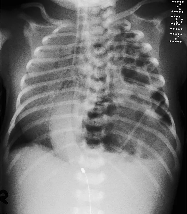
This term baby was admitted to the neonatal unit with respiratory distress.
1 What abnormalities are seen on the frontal chest radiograph?
2 What is the diagnosis?

ANSWERS

1 There are multiple lucencies in the left hemithorax, mediastinal shift to the right and there is little aeration of the right lung. The gastric bubble is not seen in the normal position and there is no bowel gas seen below the diaphragm. The tip of the nasogastric tube is in the left chest.
2 Left congenital diaphragmatic hernia, with multiple loops of bowel within the left hemithorax.

RADIOLOGY HOT LIST

• Initial radiographs may show an opaque hemithorax, as the bowel loops are fluid filled. As the baby swallows air, the characteristic gas-filled bowel loops appear in the thorax.
• Placement of a nasogastric tube verifies that the stomach is in the chest.
• The differential diagnosis of cystic intrathoracic lesions is congenital cystic adenomatoid malformation, where there is a normal abdominal bowel gas pattern and the NG tube is normally sited.
• Ultrasound can be helpful to confirm the presence of bowel loops within the chest and demonstrate the diaphragmatic defect. In doubtful cases, an upper GI contrast study can be performed.

CLINICAL HOT LIST

• Incidence 1 : 2500 live births, most diagnosed antenatally.
• 80% of herniations are left posterolateral (Bochdalek).
• Embryology: the pleuroperitoneal opening (foramen of Bochdalek) fails to close. Abdominal viscera herniate into the thorax, compromising pulmonary development.
• Morbidity depends on the degree of pulmonary hypoplasia and associated pulmonary hypertension: the presence of aerated lung on both sides is a good prognostic indicator.

Case 2

Image
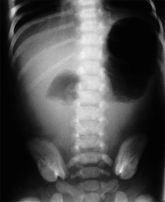
This baby was reviewed on the postnatal ward for poor feeding and bilious vomiting.
1 What abnormality is seen on the plain abdominal radiograph?
2 What is the most likely diagnosis?
3 With what condition is this associated?

ANSWERS

1 There is gas in the stomach and proximal duodenum but none elsewhere, giving the ‘double-bubble’ sign.
2 Duodenal atresia.
3 Down’s syndrome.

RADIOLOGY HOT LIST

• Duodenal atresia represents a complete obstruction, with no gas seen beyond the duodenum. The abdominal X-ray is diagnostic and a contrast study is not required.
• If the abdominal radiograph suggests incomplete obstruction (with a small amount of gas in the distal bowel), a careful upper gastrointestinal (GI) contrast study should be performed to assess the site of obstruction, and exclude a malrotation/midgut volvulus, which is a surgical emergency.
• Other causes of neonatal duodenal obstruction include duodenal stenosis, duodenal web, annular pancreas, Ladd’s bands and midgut volvulus, which all show gas in the distal bowel.

CLINICAL HOT LIST

• Incidence 1 : 1000 live births.
• The cause appears to be a failure in canalization of the fetal duodenum due to early developmental insult. There is a strong association with other abnormalities of the GI and biliary tracts, e.g. malrotation, oesophageal atresia and anal anomalies. Cardiac and renal abnormalities are sometimes seen.
• Up to 30% have Down’s syndrome.
• Most atresias occur distal to the ampulla of Vater, presenting with bilious vomiting in the first few hours of life. An incomplete obstruction may present later.
• Antenatal diagnosis is possible; 40% will have maternal polyhydramnios.

Case 3

Image
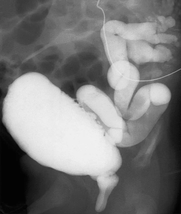
This 4-day-old boy is in renal failure. Antenatal ultrasound had shown bilateral hydronephrosis.
1 What investigation is this?
2 What does it show?
3 What is the diagnosis?

ANSWERS

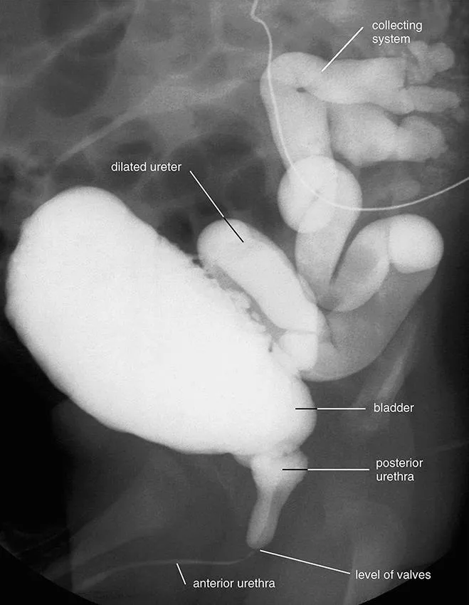
1 A micturating cysto-urethrogram (MCUG).
2 There is an abrupt change in calibre of the urethra, with dilatation of the posterior urethra. The bladder wall is trabeculated. There is bilateral vesicoureteric reflux into dilated and tortuous ureters.
3 Posterior urethral valves.
Image

RADIOLOGY HOT LIST

• The diagnosis of posterior urethral valves is usually made on an MCUG. This will show dilatation of the posterior urethra, a transverse filling defect (valves) and reduction of the urethral calibre distal to the obstruction.
• The bladder may be large or small volume, with trabeculation of the bladder wall.
• Vesicoureteric reflux is common and associated with a worse prognosis. It occurs in approximately 50% and is bilateral in 15%.
• Antenatal ultrasound may suggest the diagnosis, showing dilatation of the ureters and pelvicalyceal systems and a thick-walled bladder. There may be oligohydraminios, which is associated with a worse prognosis.
• All children should receive antibiotic cover at the time of the MCUG. The investigation should not be performed in the presence of a urinary tract infection (UTI).

CLINICAL HOT LIST

• Valves are mucosal folds that close on voiding, leading to obstruction.
• It is rare, but remains the commonest obstructive uropathy in boys.
• There is a spectrum of severity from mild to severe. The...

Inhaltsverzeichnis

  1. Cover
  2. Half Title
  3. Dedication
  4. Title Page
  5. Copyright Page
  6. Contents
  7. Foreword
  8. Foreword from first edition
  9. Preface
  10. Acknowledgement
  11. Rules and tools
  12. Cases
  13. Bibliography
  14. Index
Zitierstile für Paediatric Radiology for MRCPCH and FRCR, Second Edition

APA 6 Citation

[author missing]. (2020). Paediatric Radiology for MRCPCH and FRCR, Second Edition (2nd ed.). CRC Press. Retrieved from https://www.perlego.com/book/1900016/paediatric-radiology-for-mrcpch-and-frcr-second-edition-pdf (Original work published 2020)

Chicago Citation

[author missing]. (2020) 2020. Paediatric Radiology for MRCPCH and FRCR, Second Edition. 2nd ed. CRC Press. https://www.perlego.com/book/1900016/paediatric-radiology-for-mrcpch-and-frcr-second-edition-pdf.

Harvard Citation

[author missing] (2020) Paediatric Radiology for MRCPCH and FRCR, Second Edition. 2nd edn. CRC Press. Available at: https://www.perlego.com/book/1900016/paediatric-radiology-for-mrcpch-and-frcr-second-edition-pdf (Accessed: 15 October 2022).

MLA 7 Citation

[author missing]. Paediatric Radiology for MRCPCH and FRCR, Second Edition. 2nd ed. CRC Press, 2020. Web. 15 Oct. 2022.