Equine Hematology, Cytology, and Clinical Chemistry
eBook - ePub

Equine Hematology, Cytology, and Clinical Chemistry

Raquel M. Walton, Rick L. Cowell, Amy C. Valenciano, Raquel M. Walton, Rick L. Cowell, Amy C. Valenciano

  1. English
  2. ePUB (handyfreundlich)
  3. Über iOS und Android verfügbar
eBook - ePub

Equine Hematology, Cytology, and Clinical Chemistry

Raquel M. Walton, Rick L. Cowell, Amy C. Valenciano, Raquel M. Walton, Rick L. Cowell, Amy C. Valenciano

Angaben zum Buch
Buchvorschau
Inhaltsverzeichnis
Quellenangaben

Über dieses Buch

The all-new Equine Hematology, Cytology, and Clinical Chemistry draws on hematology and clinical chemistry information featured in the first edition of Equine Clinical Pathology and adds valuable cytopathology material from Diagnostic Cytology and Hematology of the Horse, making it a truly definitive reference to clinical pathology in equids. Thoroughly updated and expanded throughout, this Second Edition offers more images, more information, and new knowledge for previous chapters and entirely new chapters on bone marrow evaluation and cytopathology.

Designed to present clear, concise, and clinically relevant information, the book is logically organized for easy reference. Numerous figures, tables and images support the text, together with summarized information for ease of use.

  • Offers a focus on clinical pathology in the horse, with in-depth information on hematology, clinical chemistry, and cytopathology in equids
  • Presents equine disease from a systems-based, clinicopathological perspective
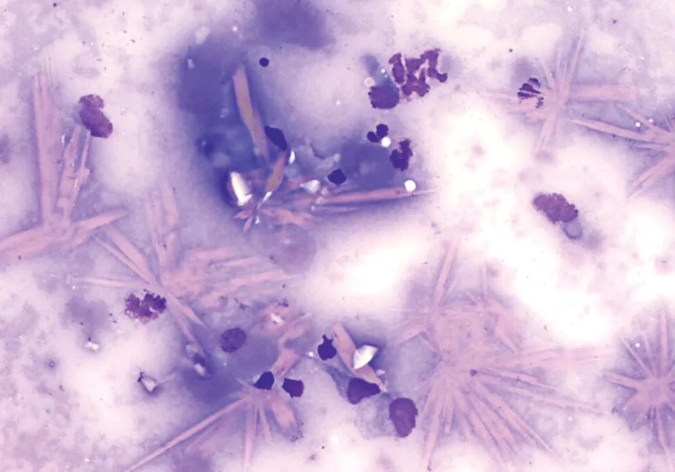
  • Features hundreds of high-quality images
  • Includes contributions from veterinary specialists with expert knowledge of clinical pathology

A must-have purchase for anyone using hematology, clinical chemistry, and cytology in equine patients, Equine Hematology, Cytology, and Clinical Chemistry, 2nd Edition is a valuable resource for equine practitioners, clinical pathologists and residents, and veterinary students.

Häufig gestellte Fragen

Wie kann ich mein Abo kündigen?
Gehe einfach zum Kontobereich in den Einstellungen und klicke auf „Abo kündigen“ – ganz einfach. Nachdem du gekündigt hast, bleibt deine Mitgliedschaft für den verbleibenden Abozeitraum, den du bereits bezahlt hast, aktiv. Mehr Informationen hier.
(Wie) Kann ich Bücher herunterladen?
Derzeit stehen all unsere auf Mobilgeräte reagierenden ePub-Bücher zum Download über die App zur Verfügung. Die meisten unserer PDFs stehen ebenfalls zum Download bereit; wir arbeiten daran, auch die übrigen PDFs zum Download anzubieten, bei denen dies aktuell noch nicht möglich ist. Weitere Informationen hier.
Welcher Unterschied besteht bei den Preisen zwischen den Aboplänen?
Mit beiden Aboplänen erhältst du vollen Zugang zur Bibliothek und allen Funktionen von Perlego. Die einzigen Unterschiede bestehen im Preis und dem Abozeitraum: Mit dem Jahresabo sparst du auf 12 Monate gerechnet im Vergleich zum Monatsabo rund 30 %.
Was ist Perlego?
Wir sind ein Online-Abodienst für Lehrbücher, bei dem du für weniger als den Preis eines einzelnen Buches pro Monat Zugang zu einer ganzen Online-Bibliothek erhältst. Mit über 1 Million Büchern zu über 1.000 verschiedenen Themen haben wir bestimmt alles, was du brauchst! Weitere Informationen hier.
Unterstützt Perlego Text-zu-Sprache?
Achte auf das Symbol zum Vorlesen in deinem nächsten Buch, um zu sehen, ob du es dir auch anhören kannst. Bei diesem Tool wird dir Text laut vorgelesen, wobei der Text beim Vorlesen auch grafisch hervorgehoben wird. Du kannst das Vorlesen jederzeit anhalten, beschleunigen und verlangsamen. Weitere Informationen hier.
Ist Equine Hematology, Cytology, and Clinical Chemistry als Online-PDF/ePub verfügbar?
Ja, du hast Zugang zu Equine Hematology, Cytology, and Clinical Chemistry von Raquel M. Walton, Rick L. Cowell, Amy C. Valenciano, Raquel M. Walton, Rick L. Cowell, Amy C. Valenciano im PDF- und/oder ePub-Format sowie zu anderen beliebten Büchern aus Medicine & Equine Veterinary Science. Aus unserem Katalog stehen dir über 1 Million Bücher zur Verfügung.

Information

Jahr
2020
ISBN
9781119500193

1
General Laboratory Medicine

Raquel M. Walton
IDEXX Laboratories, Inc., Langhorne, PA, USA
Acronyms and abbreviations that appear in this chapter include: Hb, hemoglobin; MCH, mean cell Hb; MCHC, mean cell Hb concentration; MCV, mean cell volume; PCV, packed cell volume; POC, point of care; POCT, point‐of‐care testing; RBC, red blood cells; TP, total protein; TPRef, refractometer total protein; TS, total solids.

1.1 Introduction to Laboratory Medicine

Laboratory medicine, more commonly referred to as clinical pathology (or bioanalytical pathology), is a distinct specialty that overlaps other medicine specialties such as internal medicine and oncology in the area of diagnostics. In contrast to internists, clinical pathologists practice a systems‐based rather than problem‐based approach when interpreting hematological and biochemical results. However, in addition to recognizing disease‐associated changes, two other phenomena contribute to test interpretation: how test results are generated and how “normal” is defined. Artifacts due to sample preparation, sample condition or disease processes need to be identified and distinguished from true disease‐associated changes. Similarly, test interpretation is always performed in context – the context of health. The accuracy and sensitivity of tests and the use of appropriately established reference intervals are essential to the ability to diagnose disease.
This chapter will provide selected information on hematological and biochemical test methodologies and validation, and will discuss the basic knowledge needed for generating and/or using reference intervals. The remainder of the book will address test interpretation using a systems‐based approach.

1.2 Preanalytical Factors

Preanalytical factors that may affect test results should be minimized in order to ensure result accuracy [1]. Specimens should be collected according to standard practices and transported to the laboratory in a timely manner under conditions appropriate for the type of specimen and its stability. The minimum information on a specimen label for laboratory evaluation should include the full name of the patient (animal and owner), the patient signalment, and the specimen type (e.g., whole blood, serum or plasma). Especially for hematological evaluation, it is important that the patient's signalment be correct as analyzer settings vary with respect to species.
Anticoagulated specimens for hematology that have visible macroclots in the tube will produce variably erroneous results. Because the degree of inaccuracy cannot be predicted, clotted specimens are unsuitable for analyzis and these specimens should not be analyzed or submitted for analysis.
Blood films and cytology smears should not be refrigerated and should be protected from condensation and freezing during transport to the laboratory to avoid condensation artifact (Figure 1.1). Failure to fully dry blood films or cytology preparations before placing them into slide holders can also result in moisture artefact.
Image described by caption.
Figure 1.1 Condensation artifact caused by exposure of unfixed slides to moisture. The nucleated cells are lyzed and many hemoglobin crystals are present, formed from erythrocytes.

1.3 Basic Hematological Techniques

1.3.1 Packed Cell Volume and Plasma Evaluation: Disease and Artifacts

Measurement of the percentage of red blood cells in whole blood can provide more information than simply the packed cell volume (PCV). In addition to the packed erythrocytes at the bottom of a microhematocrit tube, there is the white buffy coat layer and a plasma layer. The size of the buffy coat is related to the white blood cell (WBC) (and platelet) count; a thick buffy coat would indicate a high leukocyte (and/or platelet) count, whereas a scant buffy coat suggests leukopenia. The character of the plasma can also yield valuable information pertaining to a disease process, as well as contributing to spurious results. The plasma can appear hemolyzed, icteric or lipemic (Figure 1.2).
Hemolysis in samples from horses usually indicates an in vivo phenomenon due to toxins or immune‐mediated disease (see Chapter 4). However, hemolysis can also occur during blood collection if excessive force or too small needle gauge is used in phlebotomy. Whether in vivo or in vitro, hemolysis produces a color change that can make refractometer readings difficult or interfere with spectrophotometric tests.
Icterus indicates hyperbilirubinemia that usually exceeds 1.5 mg/dL (see Chapter 5). However, in herbivorous animals yellow‐colored plasma is not a reliable indicator of hyperbilirubinemia due to the presence of diet‐associated carotene pigments, which impart a yellow color to plasma. Icterus has not been demonstrated to interfere with refractometer readings [2]. Depending upon the chemistry analyzer, icterus can cause interference with some serum chemistry tests.
Image described by caption.
Figure 1.2 Evaluation of plasma. From left to right: normal plasma color and consistency; lipemic and slightly hemolyzed plasma; hemolyzed plasma; icteric plasma.
Lipemia is visible to the eye as increased turbidity in plasma or serum at triglyceride concentrations >300 mg/dL. Whether physiological (postprandial) or pathological (see Chapter 9), lipemia can cause spuriously high refractometer readings and will interfere with many chemistry tests.

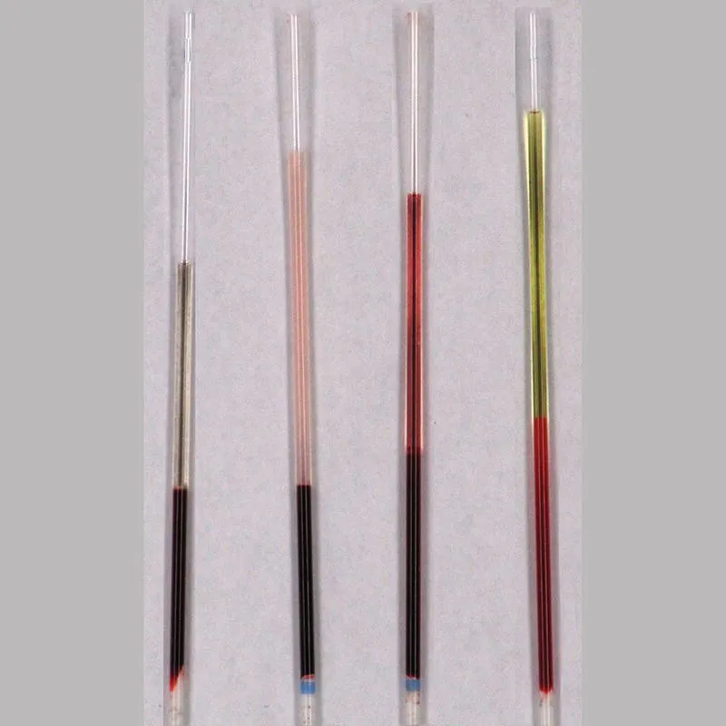
1.3.2 Protein Measurement by Refractometer

Protein can be rapidly and accurately measured by hand‐held refractometers. Because refractometers measure protein via a total solids‐based technique, the total dissolved solids in the sample affect light refraction. In addition to protein, total solids include electrolytes, glucose, urea, and lipids. The term “total solids” has caused much confusion in the reporting of refractometric protein results. Total protein (TP) and total solids (TS) are not synonymous. Currently, the vast majority of refractometers incorporate a conversion factor in their design so that the scales report TP and not TS. Contributing to the confusion is the fact that at least one refractometer is named the “TS meter” (AO Corporation) when it is in fact calibrated to report TP. While the altered refraction of plasma is mostly due to protein content, increases in lipid, glucose or urea content interfere with refractometric protein measurements. However, marked increases in urea or glucose (273 and 649 mg/dL, respectively) are needed to increase protein measurement by 0.4–0.5 g/dL. Increases in plasma cholesterol of 39 mg/dL are shown to increase the refractometer TP (TPRef) by 0.14 g/dL [2].
Another potential cause of erroneous refractometer readings is the addition of EDTA from K3EDTA anticoagulant tubes. At the standard concentration of EDTA (5 μmol/mL), K3EDTA...

Inhaltsverzeichnis