Handbook of Equine Parasite Control, Second Edition offers a thorough revision to this practical manual of parasitology in the horse. Incorporating new information and diagnostic knowledge throughout, it adds five new sections, new information on computer simulation methods, and new maps to show the spread of anthelmintic resistance. The book also features 30 new high-quality figures and expanded information on parasite occurrence and epidemiology, new diagnostics, treatment strategies, clinical significance of infections, anthelmintic resistance, and environmental persistence.
This second edition of Handbook of Equine Parasite Control brings together all the details needed to appropriately manage parasites in equine patients and support discussions between horse owners and their veterinarians. It offers comprehensive coverage of internal parasites and factors affecting their transmission; principles of equine parasite control; and diagnosis and assessment of parasitologic information. Additionally, the book provides numerous new case histories, covering egg count results from yearlings, peritonitis and parasites, confinement and deworming, quarantine advice, abdominal distress in a foal, and more.
A clear and concise user-friendly guide to equine parasite control for veterinary practitioners and students
Fully updated with new knowledge and diagnostic methods throughout
Features brand new case studies
Presents 30 new high-quality figures, including new life-cycle charts
Provides maps to show the spread of anthelmintic resistance
Handbook of Equine Parasite Control is an essential guide for equine practitioners, veterinary students, and veterinary technicians dealing with parasites in the horse.
Häufig gestellte Fragen
Wie kann ich mein Abo kündigen?
Gehe einfach zum Kontobereich in den Einstellungen und klicke auf „Abo kündigen“ – ganz einfach. Nachdem du gekündigt hast, bleibt deine Mitgliedschaft für den verbleibenden Abozeitraum, den du bereits bezahlt hast, aktiv. Mehr Informationen hier.
(Wie) Kann ich Bücher herunterladen?
Derzeit stehen all unsere auf Mobilgeräte reagierenden ePub-Bücher zum Download über die App zur Verfügung. Die meisten unserer PDFs stehen ebenfalls zum Download bereit; wir arbeiten daran, auch die übrigen PDFs zum Download anzubieten, bei denen dies aktuell noch nicht möglich ist. Weitere Informationen hier.
Welcher Unterschied besteht bei den Preisen zwischen den Aboplänen?
Mit beiden Aboplänen erhältst du vollen Zugang zur Bibliothek und allen Funktionen von Perlego. Die einzigen Unterschiede bestehen im Preis und dem Abozeitraum: Mit dem Jahresabo sparst du auf 12 Monate gerechnet im Vergleich zum Monatsabo rund 30 %.
Was ist Perlego?
Wir sind ein Online-Abodienst für Lehrbücher, bei dem du für weniger als den Preis eines einzelnen Buches pro Monat Zugang zu einer ganzen Online-Bibliothek erhältst. Mit über 1 Million Büchern zu über 1.000 verschiedenen Themen haben wir bestimmt alles, was du brauchst! Weitere Informationen hier.
Unterstützt Perlego Text-zu-Sprache?
Achte auf das Symbol zum Vorlesen in deinem nächsten Buch, um zu sehen, ob du es dir auch anhören kannst. Bei diesem Tool wird dir Text laut vorgelesen, wobei der Text beim Vorlesen auch grafisch hervorgehoben wird. Du kannst das Vorlesen jederzeit anhalten, beschleunigen und verlangsamen. Weitere Informationen hier.
Ist Handbook of Equine Parasite Control als Online-PDF/ePub verfügbar?
Ja, du hast Zugang zu Handbook of Equine Parasite Control von Martin K. Nielsen, Craig R. Reinemeyer im PDF- und/oder ePub-Format sowie zu anderen beliebten Büchern aus Medicine & Equine Veterinary Science. Aus unserem Katalog stehen dir über 1 Million Bücher zur Verfügung.
Section I Internal Parasites and Factors Affecting Their Transmission
1 Biology and Life Cycles of Equine Parasites
Life cycles are the road maps that guide parasites to their ultimate goal – propagating a subsequent generation. Some parasites follow a single, direct path to grandma’s house, while yet others may travel by convoluted routes, sojourn for protracted periods at some wayside convenience, or even pick up a passenger or two. These differences represent alternate strategies for coping with the vagaries of the environment and of their eventual hosts.
A thorough knowledge of life cycles is not emphasized merely to torment veterinary students. Rather, life cycle details reveal opportunities to control parasites through chemical or management interventions, to exploit unfavorable environmental conditions, or to promote natural enemies that might act as agents of biological control. Taking advantage of these potential control opportunities will be emphasized in individual chapters in this volume.
At the root of all life cycles is a fundamental principle that distinguishes helminth parasites from other infectious agents such as viruses, bacteria, fungi, and protozoa. Through various types of clonal expansion, the latter can all amplify their numbers within a host animal. Literally millions of individual organisms may arise from infective burdens that are orders of magnitude smaller. The reproductive products of nearly all helminths, however, are required to leave the host and undergo essential change in a different location. Defecation is the most common means by which reproductive products exit the host, but a notable exception includes immature parasitic stages that are ingested by blood‐sucking arthropods (e.g., Onchocerca, Setaria). Most parasitic products can become infective in the environment, whereas others require intermediate hosts or vectors. Regardless, all of these essential transformations occur “outside the definitive host”. Indeed, dramatic biological change is mandatory before a parasitic organism is capable of infecting a new host animal or of reinfecting the original host.
Compared to those organisms that amplify their numbers through clonal expansion, helminth disease is a numbers game. Simply put, as the number of invading parasites increases, greater tissue damage or nutrient loss results, and the range and severity of clinical signs become more extensive.
In this chapter, we propose to describe the basic life cycles of the major helminth parasites of equids. Specific control opportunities may be mentioned in this overview, but these will be discussed more fully elsewhere in the volume.
Nematodes
Superfamily Strongyloidea
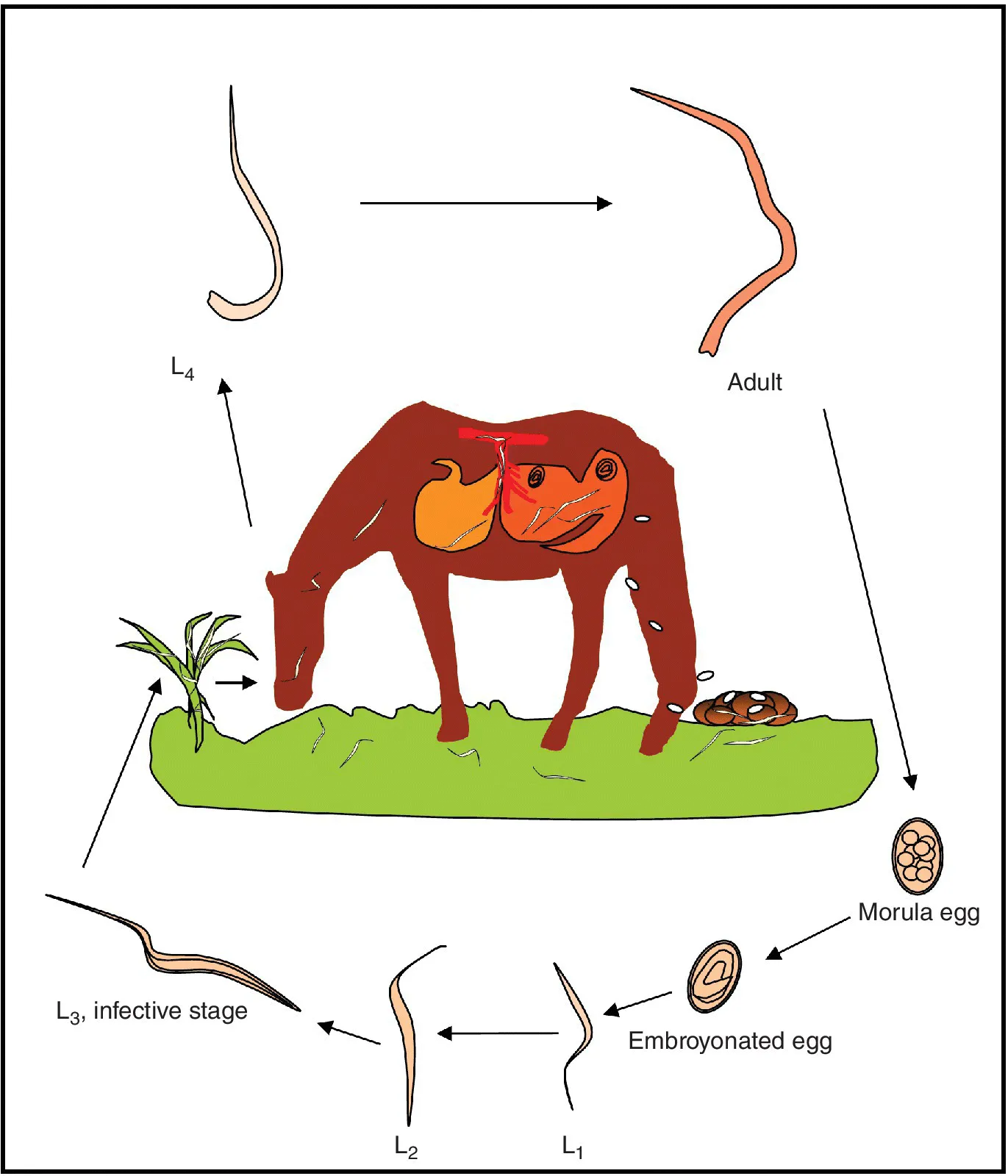
The members of the Strongyloidea (“strongyles”) are moderately sized, stout worms with substantial buccal capsules. The males have a copulatory bursa at the posterior end and females of all species produce eggs that are similar in appearance. Eggs of small strongyles cannot be differentiated microscopically from those of large strongyles, and the only practical method of differentiation (other than molecular approaches) is through coproculture. The strongyloids of horses all have direct life cycles; intermediate or paratenic hosts are never used (Figure 1.1).
Figure 1.1 Strongyle life cycle. The life cycle of strongyle parasites. Parasitic stages can be seen above the horse and preparasitic stages below it. Fertilized eggs are shed by adult females in the cecum and colon, and excreted to the environment in the feces. Here, the eggs hatch and a first‐stage larva (L1) emerges. The L1 then molts to L2 in the feces. Another molt gives rise to the L3, which retains its L2 cuticle and thus has a double‐layered sheath. The L3 leaves the fecal pat and migrates on to forage, where it is ingested by a horse. Inside the horse, the L3 exsheathes and invades the mucosa of the large intestine. Large strongyles (Strongylus spp.) undergo extensive migration in various organs of the horse, while cyathostomins encyst in the mucosal lining of the large intestine. After returning to the large intestinal lumen, the worms reach sexual maturity and start shedding eggs.
Strongyloid eggs pass in feces and hatch in favorable environmental conditions of moisture, temperature, and oxygenation. All species exhibit three sequential larval stages, first (L1), second (L2), and third (L3). The L1 and L2 stages feed on organic material in the environment, but the third stage develops within the sheath of the L2. This protective covering helps L3s to resistant environmental conditions, but it has no oral opening, so third stage larvae are unable to ingest nutrients. The L3 is the infective stage for all strongyloid nematodes of equids. Infection invariably occurs through inadvertent ingestion, whether while grazing or via oral contact with elements of the environment.
Apparently, horses never develop absolute immunity to strongyloids, so these are often the sole nematode parasites recovered from well‐managed, mature equids. The Strongyloidea of horses are comprised of two distinct subfamilies, the Strongylinae and the Cyathostominae.
Strongylinae (large strongyles)
Members of the subclass Strongylinae tend to be larger, on average, than most genera that comprise the Cyathostominae. In addition, Strongylinae have large buccal capsules, adapted for attachment to, and even ingestion of, the gut mucosa. The larval stages of at least one strongylin genus undergo extensive, albeit stereotypic, migration within the host prior to returning to the gut to mature and begin reproduction.
Strongylus vulgaris
Strongylus vulgaris is widely acknowledged as the single most pathogenic nematode parasite of horses. Adult worms measure about 1.5–2.5 cm in length and the females are larger than the males. Adults are usually found attached to the mucosa of the cecum and the ventral colon (Figure 1.2). After ingestion from the environment, third stage larvae invade the mucosa of the distal small intestine, cecum, and colon. Here, they molt to the fourth stage (L4) before penetrating local arterioles and migrating proximally beneath the intimal layer of local blood vessels. Migrating S. vulgaris L4s leave subintimal tracts in their wake and congregate near the root of the cranial mesenteric artery. A portion of the infecting larvae may continue to migrate, even t...