
Understanding Light Microscopy
Jeremy Sanderson
- English
- ePUB (handyfreundlich)
- Über iOS und Android verfügbar
Understanding Light Microscopy
Jeremy Sanderson
Über dieses Buch
Introduces readers to the enlightening world of the modern light microscope
There have been rapid advances in science and technology over the last decade, and the light microscope, together with the information that it gives about the image, has changed too. Yet the fundamental principles of setting up and using a microscope rests upon unchanging physical principles that have been understood for years. This informative, practical, full-colour guide fills the gap between specialised edited texts on detailed research topics, and introductory books, which concentrate on an optical approach to the light microscope. It also provides comprehensive coverage of confocal microscopy, which has revolutionised light microscopy over the last few decades.
Written to help the reader understand, set up, and use the often very expensive and complex modern research light microscope properly, Understanding Light Microscopy keeps mathematical formulae to a minimum—containing and explaining them within boxes in the text. Chapters provide in-depth coverage of basic microscope optics and design; ergonomics; illumination; diffraction and image formation; reflected-light, polarised-light, and fluorescence microscopy; deconvolution; TIRF microscopy; FRAP & FRET; super-resolution techniques; biological and materials specimen preparation; and more.
- Gives a didactic introduction to the light microscope
- Encourages readers to use advanced fluorescence and confocal microscopes within a research institute or core microscopy facility
- Features full-colour illustrations and workable practical protocols
Understanding Light Microscopy is intended for any scientist who wishes to understand and use a modern light microscope. It is also ideal as supporting material for a formal taught course, or for individual students to learn the key aspects of light microscopy through their own study.
Häufig gestellte Fragen
Information
1
Our Eyes and the Microscope
Outline
- The eye-brain system
- The features of the eye
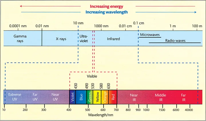
- Response to visible electromagnetic radiation
- Rods and cones
- Basis of colour vision
- Eye sensitivity to brightness
- Dark vision
- Ability to see shades of grey
- Anatomy of the eye
- Floaters
- Lens focusing
- Macula and fovea
- Visual acuity
- Eye dominance
- Spherical aberration
- Chromatic aberration
- Astigmatism
- Saccades – tiling our visual field
- Stereoscopic vision
- Parallax
- Near point
- Accommodation
- Nearest distance of distinct vision
- The magnifying glass
- Magnification of a simple lens
- Myopia
- Emetropia
- Hyperopia
- Presbyopia
- Filling in and bias
- Images as scientific data sets
- The requirement for measurement
- BioNumbers database
- Nomenclature
- 8 points
1.1 Introduction
1.2 How Our Eyes Work
Inhaltsverzeichnis
- Cover
- Understanding Light Microscopy
- About the Author
- Acknowledgements
- Look-Up Guide to Feature Boxes by Theme
- Glossary and Definitions
- Notes
- Introduction
- 1 Our Eyes and the Microscope
- 2 Light
- 3 Basic Microscope Optics
- 4 Microscope Anatomy and Design
- 5 Ergonomics
- 6 Optical Aberrations of the Microscope
- 7 The Microscope Objective
- 8 Condensers and Eyepieces
- 9 Illumination in the Microscope
- 10 Diffraction and Image Formation in Microscopy
- 11 Contrast Generation and Enhancement
- 12 Reflected-Light Microscopy
- 13 Polarised-Light Microscopy: Part 1 – Theory
- 14 Polarised-Light Microscopy: Part 2 – Applied
- 15 Fluorescence Microscopy
- 16 Fluorophores and Fluorescent Proteins
- 17 Optical Sectioning and Confocal Microscopy
- 18 Operating the Confocal Microscope
- 19 Light-Sheet Microscopy
- 20 Bleed-Through and Spectral Unmixing
- 21 Deconvolution
- 22 Multi-Photon Microscopy
- 23 Total Internal Reflection Fluorescence Microscopy
- 24 FRAP and FRET
- 25 Colocalisation
- 26 Super-Resolution Fluorescence Microscopy
- 27 Choosing a Microscope Platform and Core Imaging Facilities
- 28 Biological Specimen Preparation
- 29 Materials Specimen Preparation
- 30 Recording the Image: Part 1 – Theory
- 31 Recording the Image: Part 2 – Applied
- Appendix 1 Buying, and Tendering for, a Light Microscope
- Appendix 2 Troubleshooting Poor Image Quality
- Appendix 3 The Michel-Lévy Interference Colour Chart
- Appendix 4 Cleaning and Maintenance of the Light Microscope
- Appendix 5 Selected Suppliers
- Appendix 6 Historical Background
- Appendix 7 Timeline of Key Events
- Index
- Foldout
- WILEY END USER LICENSE AGREEMENT