
eBook - ePub
Surface Electromyography
Physiology, Engineering, and Applications
Roberto Merletti, Dario Farina, Roberto Merletti, Dario Farina
This is a test
- English
- ePUB (handyfreundlich)
- Über iOS und Android verfügbar
eBook - ePub
Surface Electromyography
Physiology, Engineering, and Applications
Roberto Merletti, Dario Farina, Roberto Merletti, Dario Farina
Angaben zum Buch
Buchvorschau
Inhaltsverzeichnis
Quellenangaben
Über dieses Buch
Reflects on developments in noninvasive electromyography, and includes advances and applications in signal detection, processing and interpretation
- Addresses EMG imaging technology together with the issue of decomposition of surface EMG
- Includes advanced single and multi-channel techniques for information extraction from surface EMG signals
- Presents the analysis and information extraction of surface EMG at various scales, from motor units to the concept of muscle synergies.
Häufig gestellte Fragen
Wie kann ich mein Abo kündigen?
Gehe einfach zum Kontobereich in den Einstellungen und klicke auf „Abo kündigen“ – ganz einfach. Nachdem du gekündigt hast, bleibt deine Mitgliedschaft für den verbleibenden Abozeitraum, den du bereits bezahlt hast, aktiv. Mehr Informationen hier.
(Wie) Kann ich Bücher herunterladen?
Derzeit stehen all unsere auf Mobilgeräte reagierenden ePub-Bücher zum Download über die App zur Verfügung. Die meisten unserer PDFs stehen ebenfalls zum Download bereit; wir arbeiten daran, auch die übrigen PDFs zum Download anzubieten, bei denen dies aktuell noch nicht möglich ist. Weitere Informationen hier.
Welcher Unterschied besteht bei den Preisen zwischen den Aboplänen?
Mit beiden Aboplänen erhältst du vollen Zugang zur Bibliothek und allen Funktionen von Perlego. Die einzigen Unterschiede bestehen im Preis und dem Abozeitraum: Mit dem Jahresabo sparst du auf 12 Monate gerechnet im Vergleich zum Monatsabo rund 30 %.
Was ist Perlego?
Wir sind ein Online-Abodienst für Lehrbücher, bei dem du für weniger als den Preis eines einzelnen Buches pro Monat Zugang zu einer ganzen Online-Bibliothek erhältst. Mit über 1 Million Büchern zu über 1.000 verschiedenen Themen haben wir bestimmt alles, was du brauchst! Weitere Informationen hier.
Unterstützt Perlego Text-zu-Sprache?
Achte auf das Symbol zum Vorlesen in deinem nächsten Buch, um zu sehen, ob du es dir auch anhören kannst. Bei diesem Tool wird dir Text laut vorgelesen, wobei der Text beim Vorlesen auch grafisch hervorgehoben wird. Du kannst das Vorlesen jederzeit anhalten, beschleunigen und verlangsamen. Weitere Informationen hier.
Ist Surface Electromyography als Online-PDF/ePub verfügbar?
Ja, du hast Zugang zu Surface Electromyography von Roberto Merletti, Dario Farina, Roberto Merletti, Dario Farina im PDF- und/oder ePub-Format sowie zu anderen beliebten Büchern aus Ciencias biológicas & Biotecnología. Aus unserem Katalog stehen dir über 1 Million Bücher zur Verfügung.
Information
1
Physiology of Muscle Activation and Force Generation
R. M. Enoka1 and J. Duchateau2
1Department of Integrative Physiology, University of Colorado, Boulder, Colorado
2Laboratory of Applied Biology and Neurophysiology, ULB Neuroscience Institute (UNI), Université Libre de Bruxelles (ULB), Brussels, Belgium
1.1 Introduction
To extract information about the control of movement by the nervous system from electromyographic (EMG) signals, it is necessary to understand the processes underlying both the generation of the activation signal and the torques exerted by the involved muscles. As a foundation for the subsequent chapters in this book, the goal of this chapter is to describe the physiology of muscle activation and force generation. We discuss the anatomy of the final common pathway from the nervous system to muscle, the electrical properties of motor neurons and muscle fibers, the contractile properties of muscle fibers and motor units, the concept of motor unit types, and the control of muscle force by modulating the recruitment and rate coding of motor unit activity.
1.2 Anatomy of a Motor Unit
The basic functional unit of the neuromuscular system is the motor unit. It comprises a motor neuron, including its dendrites and axon, and the muscle fibers innervated by the axon [28]. The motor neuron is located in the ventral horn of the spinal cord or brain stem where it receives sensory and descending inputs from other parts of the nervous system. The axon of each motor neuron exits the spinal cord through the ventral root, or through a cranial nerve in the brain stem, and projects in a peripheral nerve to its target muscle and the muscle fibers it innervates. Because the generation of an action potential by a motor neuron typically results in the generation of action potentials in all of the muscle fibers belonging to the motor unit, EMG recordings of muscle fiber action potentials provide information about the activation of motor neurons in the spinal cord or brain stem.
1.2.1 Motor Nucleus
The population of motor neurons that innervate a single muscle is known as a motor nucleus or motor neuron pool [51]. The number of motor neurons in a motor nucleus ranges from a few tens to several hundred [40,58] (Table 1.1). The motor neuron pool for each muscle typically extends longitudinally for a few segments of the spinal cord (Fig. 1.1), and at each segmental level the pools for proximal muscles tend to be more ventral and lateral than those for distal muscles and the pools for anterior muscles are more lateral than those for posterior muscles [59]. Nonetheless, the extensive dendritic projections of motor neurons intermingle across motor neuron pools.
Table 1.1 Motor Neuron Locations and Numbers for Selected Forelimb Muscles
| Muscle | Spinal Location | Number |
| Biceps brachii | C5–C7 | 1051 |
| Triceps brachii | C6–T1 | 1271 |
| Flexor carpi radialis | C7–C8 | 235 |
| Extensor carpi radialis | C5–C7 | 890 |
| Flexor carpi ulnaris | C7–T1 | 314 |
| Extensor carpi ulnaris | C7–T1 | 216 |
| Extensor pollicis longus | C8–T1 | 14 |
| Abductor pollicis longus | C8–T1 | 126 |
| Flexor digitorum superficialis | C8–T1 | 306 |
| Extensor digitorum communis | C8–T1 | 273 |
| Flexor digitorum profundus | C8–T1 | 475 |
| Extensor digiti secundi proprius | C8–T1 | 87 |
| Abductor pollicis brevis and flexor pollicis brevis | C8–T1 | 115 |
| Adductor pollicis | C8–T1 | 370 |
| First dorsal interosseus | C8–T1 | 172 |
| Lateral lumbricalis | C8–T1 | 57 |
| Data are from Jenny and Inukai [58] and are listed as pairs of antagonistic muscles. | ||
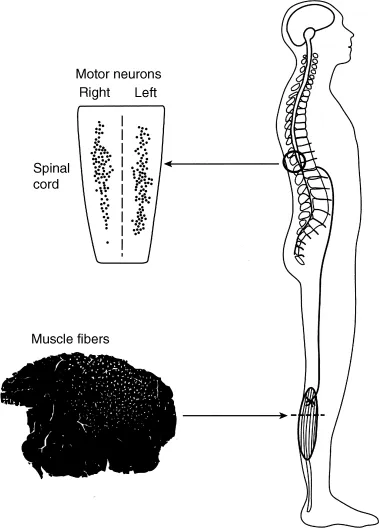
Figure 1.1 Muscle force is controlled by a population of motor units (motor unit pool) located in the spinal cord with each motor unit innervating a number of muscle fibers (muscle unit). The muscle fibers belonging to a single muscle unit are indicated by the white dots in the cross-sectional view of the muscle. A typical motor unit pool spans several spinal segments, and muscle units are usually limited to discrete parts of the muscle. Modified from Enoka [30] with permission.
1.2.2 Muscle Fibers
The muscle fibers innervated by a single axon are known as the muscle unit (Fig. 1.1), the size of which varies across each motor unit pool. The motor units first recruited during a voluntary contraction innervate fewer muscle fibers and hence have smaller muscle units than those that are recruited later in the contraction. Most motor units in a muscle have small muscle units and only a few have large muscle units [76,102,107] (Fig. 1.2A). Based on the association between muscle unit size and maximal motor unit force, Enoka and Fuglevand [32] estimated the innervation numbers (muscle unit size) for the 120 motor units in a human hand muscle (first dorsal interosseus) ranged from 21 to 1770 (Fig. 1.2B). Similar relations likely exist for most muscles [51]. Due to the exponential distribution of innervation number across a motor unit pool, it is necessary to distinguish between the number of motor unit action potentials discharged from the spinal cord and the number of muscle fiber action potentials recorded in the muscle with EMG electrodes. This distinction is indicated with the term “neural drive” to denote the motor unit action potentials and “muscle activation” to indicate the muscle fiber action potentials [26,31,36].
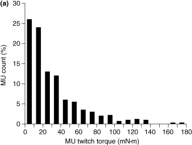
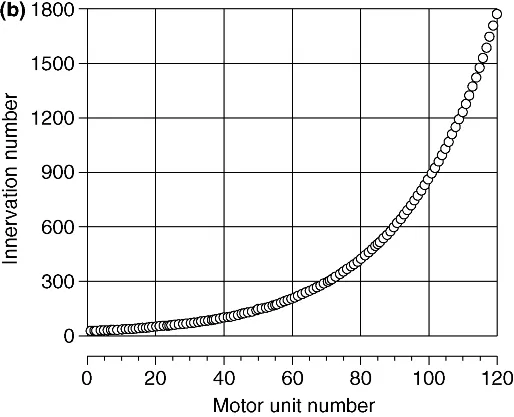
Figure 1.2 Variation in muscle unit size across the motor unit pool. (a) Distribution of motor unit (MU) twitch torques for 528 motor units in the tibialis anterior muscle of 10 subjects [107]. (b) Estimated distribution of innervation numbers across the 120 motor units comprising the first dorsal interosseus muscle [32].
The fibers in each muscle unit are located in a subvolume of the muscle and intermingle with the fibers of other muscle units (Fig. 1.1). The spatial distribution of the fibers belonging to a muscle unit is referred to as the motor unit territory. Counts of muscle unit fibers indicate that motor unit territories can occupy from 10% to 70% of the cross-sectional area of a muscle and that the density of muscle unit fibers ranges from 3 to 20 per 100 muscle fibers [51]. Moreover, the fibers of a single muscle unit often do not extend from one end of the muscle to the other, but instead terminate within a muscle fascicle [48,108]. As a consequence of muscle unit anatomy, the forces generated by individual muscle fibers must be transmitted through various layers of connective tissues before reaching the skeleton and contributing to the movement. Such interactions attenuate the unique contribution of individual fibers to the net muscle force during a movement and thereby reduce the influence of differences in contractile properties among muscle fibers.
1.3 Motor Neuron
The motor unit is classically considered to be the final common pathway in that sensory and descending inputs converge onto a single neuron that discharges an activation signal to the muscle fibers it innervates [28]. The motor neuron has extensive dendritic branches that receive up to 50,000 synaptic contacts with each contact capable of eliciting inward or outward currents across the membrane...