eBook - ePub
Skin Tissue Engineering and Regenerative Medicine
Mohammad Albanna,James H Holmes IV
This is a test
- 466 páginas
- English
- ePUB (apto para móviles)
- Disponible en iOS y Android
eBook - ePub
Skin Tissue Engineering and Regenerative Medicine
Mohammad Albanna,James H Holmes IV
Detalles del libro
Vista previa del libro
Índice
Citas
Información del libro
The skin is the largest human organ system. Loss of skin integrity due to injury or illness results in a substantial physiologic imbalance and ultimately in severe disability or death. From burn victims to surgical scars and plastic surgery, the therapies resulting from skin tissue engineering and regenerative medicine are important to a broad spectrum of patients.
Skin Tissue Engineering and Regenerative Medicine provides a translational link for biomedical researchers across fields to understand the inter-disciplinary approaches which expanded available therapies for patients and additional research collaboration. This work expands on the primary literature on the state of the art of cell therapies and biomaterials to review the most widely used surgical therapies for the specific clinical scenarios.
- Explores cellular and molecular processes of wound healing, scar formation, and dermal repair
- Includes examples of animal models for wound healing and translation to the clinical world
- Presents the current state of, and clinical opportunities for, extracellular matrices, natural biomaterials, synthetic biomaterials, biologic skin substitutes, and adult and fetal stem and skin cells for skin regenerative therapies and wound management
- Discusses new innovative approaches for wound healing including skin bioprinting and directed cellular therapies
Preguntas frecuentes
¿Cómo cancelo mi suscripción?
¿Cómo descargo los libros?
Por el momento, todos nuestros libros ePub adaptables a dispositivos móviles se pueden descargar a través de la aplicación. La mayor parte de nuestros PDF también se puede descargar y ya estamos trabajando para que el resto también sea descargable. Obtén más información aquí.
¿En qué se diferencian los planes de precios?
Ambos planes te permiten acceder por completo a la biblioteca y a todas las funciones de Perlego. Las únicas diferencias son el precio y el período de suscripción: con el plan anual ahorrarás en torno a un 30 % en comparación con 12 meses de un plan mensual.
¿Qué es Perlego?
Somos un servicio de suscripción de libros de texto en línea que te permite acceder a toda una biblioteca en línea por menos de lo que cuesta un libro al mes. Con más de un millón de libros sobre más de 1000 categorías, ¡tenemos todo lo que necesitas! Obtén más información aquí.
¿Perlego ofrece la función de texto a voz?
Busca el símbolo de lectura en voz alta en tu próximo libro para ver si puedes escucharlo. La herramienta de lectura en voz alta lee el texto en voz alta por ti, resaltando el texto a medida que se lee. Puedes pausarla, acelerarla y ralentizarla. Obtén más información aquí.
¿Es Skin Tissue Engineering and Regenerative Medicine un PDF/ePUB en línea?
Sí, puedes acceder a Skin Tissue Engineering and Regenerative Medicine de Mohammad Albanna,James H Holmes IV en formato PDF o ePUB, así como a otros libros populares de Technik & Maschinenbau y Biomedizinwissenschaft. Tenemos más de un millón de libros disponibles en nuestro catálogo para que explores.
Información
Categoría
Technik & MaschinenbauCategoría
BiomedizinwissenschaftChapter 1
Anatomy, Physiology, Histology, and Immunohistochemistry of Human Skin
Justine Fenner, and Richard A.F. Clark Departments of Dermatology and Biomedical Engineering, Stony Brook University, Stony Brook, NY, USA
Abstract
The purpose of this introductory chapter is to discuss the anatomy, physiology, histology, and immunohistochemistry of the skin. The sections on the epidermis cover the structure of its stratified layers, its cellular contents, and the architecture of dermoepidermal junction. The sections on the dermis contain information on its extracellular matrix structure and dermal fibroblasts that are required for its homeostasis and sections on the microvasculature, muscles, and nerves found in the dermis as well as various appendages including hairs, erector pili, adenexal glands, and nails. The adenexal glands include eccrine, apocrine, and sebaceous glands. The hypodermis deep to the dermis is also briefly examined. In addition, the chapter delves into the causes and effects of skin loss in the United States, thereby stressing the importance of finding a cost-effective, transformative therapy. It discusses the importance of planimetry and morphometrics of reepithelialization and granulation tissue ingrowth as tools to measure the progression of wound healing. Finally, the chapter addresses how immunohistochemistry can be used as a tool to monitor wound healing. It examines the various stains and biomarkers used to quantify processes such as angiogenesis, reestablishment of epidermal maturation, basement membrane integrity, and the establishment of nonepidermal cells in the epidermis. The chapter aggregates information across multiple sources in order to provide the most current portrayal of research in the field of the skin. The intent is to inform the reader on the basics of the integument for the purpose of understanding the mechanisms underlying skin response to tissue-engineered constructs and regenerative medicine.
Keywords
Basement membrane; Dermis; Epidermis; Hypodermis; Keratinocyte; Melanocyte; Vasculature; Wound healingIntroduction
The largest organ in the human body is the skin. It composes 16% of a person’s body weight with an average weight of 4 kg, while encompassing a surface area of 1.8 m2. The skin is a metabolically active organ with a variety of vital functions essential to maintenance of homeostasis and protection of the body. It acts as a barrier to chemical and physical agents, prevents the loss of body fluids, and helps to regulate body temperature [1]. The skin also serves as a sensory organ and provides a surface for one to grip. It plays a vital role in vitamin D production and makes antimicrobial peptides [2]. Skin is continuous with themucous membranes of our respiratory system, digestive tract, and urogenital tract and gives rise to nails, hair, and sweat glands.
Skin Anatomy, Histology, and Physiology
Skin is composed of three layers: the epidermis, dermis, and hypodermis (Figure 1). Skin is also often classified as being either thick or thin depending on the width of the epidermis. Thick skin has an epidermis thickness of 0.8–1.5 mm, whereas thin skin has an epidermis thickness of 0.07–0.15 mm [3]. Thick, non-hair-bearing (glabrous) skin is found on palmar and plantar surfaces and has no hair, arrector pili muscles, or sebaceous glands [4]. Thinner skin is found over the rest of the body, but is especially thin over the eyelids, and is composed of less cellular layers [3].
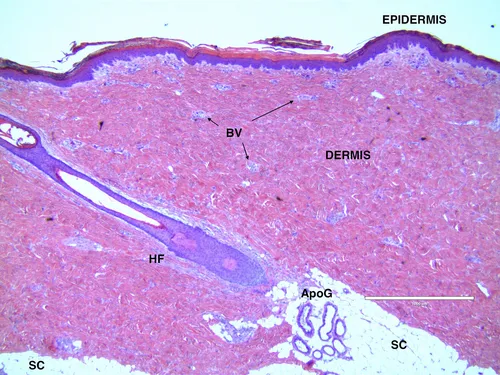
Figure 1 Normal porcine back skin fixed and stained with H&E.
Pig skin, of all animals, is most like human in terms of architecture, thickness, lack of a panniculus carnosis (muscle layer under the subcutaneous adipose tissue), and sparseness of hair; and therefore, a superb model for evaluating tissue-engineered constructs. This specimen of porcine skin comes from the back and is stained with hematoxylin and eosin that renders nuclei blue and keratin and collagen pink. The keratinized EPIDERMIS (pink) is composed of corneocytes that along with the underlying granular cell layer (not seen at this low magnification) provide the permeability barrier to the skin. The underlying stratum spinosum and basal cell layer (blue) are the differentiating and proliferating layers of the skin. Normal skin in humans regenerates every 28 days. The DERMIS is mostly composed of collagen (pink), which provides tensile strength, but contains many blood vessels (BV), nerves (not well-visualized with this stain), and appendageal structures like hair follicles (HF), apocrine glands (ApoG), sebaceous glands that secrete oil, and eccrine glands that generate sweat. The latter are on most skin surfaces in humans but only reside in specialized areas of pig skin, for example, the snout. The hypodermis or subcutaneous adipose tissue (SC) provides a cushioning effect from blunt trauma, as well as insulation.
Epidermis
The epidermis is the outermost layer of the skin and ranges in thickness from 0.05 mm on the eyelids to 1.55 mm on palms and soles [5]. It is composed mostly of stratified squamous epithelium, with the innermost layer consisting of a single row of columnar cells called basal cells that are attached to the basement membrane. The epidermis is constantly regenerating, making itself a durable keratinized boundary [3].
The epidermis is further divided into four layers: the stratum basale, the stratum spinosum, the stratum granulosum, and the stratum corneum. A fifth layer, the stratum lucidum, is found between the stratum corneum and stratum granulosum, only in the thick skin of the palms and soles [6].
The stratum basale is the innermost layer of the epidermis and the location of cell division. Some basal cells are stem cells that slowly generate other basal cells and suprabasal cells that rapidly divide (transient-amplifying cells) to generate more keratinocytes. Human skin regenerates about every 28 days [7]. Basal cells also produce antimicrobial proteins that are imperative in the skin’s defensive role [8].
Melanocytes comprise about 5–10% of the basal cell population [3]. Their principle role is in the production of melanosomes that are transferred to keratinocytes. The type and abundance of melanosomes determine the pigment intensity of the skin [4]. Merkel cells are found, albeit infrequently, in the basal cell layer as well. They are closely associated with terminal filaments of cutaneous nerves and are believed to play a role in sensation [3]. They are found in especially high concentration in areas associated with cutaneous nerves and touch sensation, such as the fingers and lips [9].
Superficial to the stratum basale is the stratum spinosum. The stratum spinosum contains a high concentration of keratin filaments and desmosomes that tightly adhere adjacent cells to one another [5]. During H&E staining the filaments between desmosomes shrink resulting in a “spiny” appearance, thus the name stratum spinosum. Keratinization begins in the basal cells, but the keratin type switches as keratinocytes differentiate and transit into the stratum spinosum [10]. Keratinization, or cornification, is the process of cell differentiation in which keratinocytes transition from their postgerminative state in the stratum basale and suprabasal cell layer to terminally differentiated, hardened cells filled with protein in the stratum corneum [11]. In the stratum basale, keratinocytes also begin to produce lamellar bodies in the golgi [12]. Lamellar bodies are tubulovesicular secretory organelles related to lysosomes. They secrete their contents including lipids, protease inhibitors, hydrolases, and antimicrobial peptides into the upper layers of the epidermis [10]. Consequently, they are important in forming a boundary that prevents the loss of fluids while providing antimicrobial protection [3]. Langerhans cells reside for the most part in the stratum spinosum. These cells are antigen-presenting cells that serve an immunologic role in the skin [4].
Above the stratum spinosum is the stratum granulosum. At the interface between the stratum granulosum and the stratum corneum, keratinocytes become flattened and lose their nuclei. It is here that lamellar bodies secrete their contents forming a lipid barrier. Keratohyalin granules are also formed in the stratum granulosum where they bind to keratin filaments [3]. This binding creates large aggregations that form the electron dense masses within the cytoplasm of keratinocytes resulting in a “granular” appearance [4].
The most superficial layer, the stratum corneum, contains cells completely devoid of nuclei and organelles [13]. The keratinocytes become elongated and flattened to form a lamellar array of corneocytes [4]. Dense bodies, remnants of desmosomes, along with a lipid glue partially derived from lamellar granules, hold the corneocytes together [10]. Corneocytes are enveloped in a l...
Índice
- Cover image
- Title page
- Table of Contents
- Copyright
- Dedication
- List of Contributors
- Foreword
- Chapter 1. Anatomy, Physiology, Histology, and Immunohistochemistry of Human Skin
- Chapter 2. Molecular and Cellular Biology of Wound Healing and Skin Regeneration
- Chapter 3. Tissue Processing and Staining for Histological Analyses
- Chapter 4. Clinical Management of Wound Healing and Hypertrophic Scarring
- Chapter 5. Process Development and Manufacturing of Human and Animal Acellular Dermal Matrices
- Chapter 6. Clinical Applications of Acellular Dermal Matrices in Reconstructive Surgery
- Chapter 7. Advances in Acellular Extracellular Matrices (ECM) for Wound Healing
- Chapter 8. Natural Biomaterials for Skin Tissue Engineering
- Chapter 9. Synthetic Biomaterials for Skin Tissue Engineering
- Chapter 10. Hybrid Biomaterials for Skin Tissue Engineering
- Chapter 11. Biologic Skin Substitutes
- Chapter 12. Wound Healing: A Comprehensive Wound Assessment and Treatment Approach
- Chapter 13. Current Innovations for the Treatment of Chronic Wounds
- Chapter 14. The Surgical Management of Burn Wounds
- Chapter 15. Advances in Isolation and Expansion of Human Cells for Clinical Applications
- Chapter 16. Cutaneous Applications of Stem Cells for Skin Tissue Engineering
- Chapter 17. Advances in Biopharmaceutical Agents and Growth Factors for Wound Healing and Scarring
- Chapter 18. Skin Models for Drug Development and Biopharmaceutical Industry
- Chapter 19. Animal Models for Wound Healing
- Chapter 20. Human Skin Bioprinting: Trajectory and Advances
- Chapter 21. Translational Research of Skin Substitutes and Wound Healing Products
- Index
Estilos de citas para Skin Tissue Engineering and Regenerative Medicine
APA 6 Citation
Albanna, M., & Holmes, J. (2016). Skin Tissue Engineering and Regenerative Medicine ([edition unavailable]). Elsevier Science. Retrieved from https://www.perlego.com/book/1830059/skin-tissue-engineering-and-regenerative-medicine-pdf (Original work published 2016)
Chicago Citation
Albanna, Mohammad, and James Holmes. (2016) 2016. Skin Tissue Engineering and Regenerative Medicine. [Edition unavailable]. Elsevier Science. https://www.perlego.com/book/1830059/skin-tissue-engineering-and-regenerative-medicine-pdf.
Harvard Citation
Albanna, M. and Holmes, J. (2016) Skin Tissue Engineering and Regenerative Medicine. [edition unavailable]. Elsevier Science. Available at: https://www.perlego.com/book/1830059/skin-tissue-engineering-and-regenerative-medicine-pdf (Accessed: 15 October 2022).
MLA 7 Citation
Albanna, Mohammad, and James Holmes. Skin Tissue Engineering and Regenerative Medicine. [edition unavailable]. Elsevier Science, 2016. Web. 15 Oct. 2022.