
eBook - ePub
Diffusion MRI
From Quantitative Measurement to In vivo Neuroanatomy
Heidi Johansen-Berg,Timothy E.J. Behrens
This is a test
- 632 páginas
- English
- ePUB (apto para móviles)
- Disponible en iOS y Android
eBook - ePub
Diffusion MRI
From Quantitative Measurement to In vivo Neuroanatomy
Heidi Johansen-Berg,Timothy E.J. Behrens
Detalles del libro
Vista previa del libro
Índice
Citas
Información del libro
Diffusion MRI remains the most comprehensive reference for understanding this rapidly evolving and powerful technology and is an essential handbook for designing, analyzing, and interpreting diffusion MR experiments.
Diffusion imaging provides a unique window on human brain anatomy. This non-invasive technique continues to grow in popularity as a way to study brain pathways that could never before be investigated in vivo.
This book covers the fundamental theory of diffusion imaging, discusses its most promising applications to basic and clinical neuroscience, and introduces cutting-edge methodological developments that will shape the field in coming years. Written by leading experts in the field, it places the exciting new results emerging from diffusion imaging in the context of classical anatomical techniques to show where diffusion studies might offer unique insights and where potential limitations lie.
- Fully revised and updated edition of the first comprehensive reference on a powerful technique in brain imaging
- Covers all aspects of a diffusion MRI study from acquisition through analysis to interpretation, and from fundamental theory to cutting-edge developments
- New chapters covering connectomics, advanced diffusion acquisition, artifact removal, and applications to the neonatal brain
- Provides practical advice on running an experiment
- Includes discussion of applications in psychiatry, neurology, neurosurgery, and basic neuroscience
- Full color throughout
Preguntas frecuentes
¿Cómo cancelo mi suscripción?
¿Cómo descargo los libros?
Por el momento, todos nuestros libros ePub adaptables a dispositivos móviles se pueden descargar a través de la aplicación. La mayor parte de nuestros PDF también se puede descargar y ya estamos trabajando para que el resto también sea descargable. Obtén más información aquí.
¿En qué se diferencian los planes de precios?
Ambos planes te permiten acceder por completo a la biblioteca y a todas las funciones de Perlego. Las únicas diferencias son el precio y el período de suscripción: con el plan anual ahorrarás en torno a un 30 % en comparación con 12 meses de un plan mensual.
¿Qué es Perlego?
Somos un servicio de suscripción de libros de texto en línea que te permite acceder a toda una biblioteca en línea por menos de lo que cuesta un libro al mes. Con más de un millón de libros sobre más de 1000 categorías, ¡tenemos todo lo que necesitas! Obtén más información aquí.
¿Perlego ofrece la función de texto a voz?
Busca el símbolo de lectura en voz alta en tu próximo libro para ver si puedes escucharlo. La herramienta de lectura en voz alta lee el texto en voz alta por ti, resaltando el texto a medida que se lee. Puedes pausarla, acelerarla y ralentizarla. Obtén más información aquí.
¿Es Diffusion MRI un PDF/ePUB en línea?
Sí, puedes acceder a Diffusion MRI de Heidi Johansen-Berg,Timothy E.J. Behrens en formato PDF o ePUB, así como a otros libros populares de Medicine y Neurology. Tenemos más de un millón de libros disponibles en nuestro catálogo para que explores.
Información
Section III
Diffusion MRI for In vivo Neuroanatomy
Outline
Chapter 16 Mapping Connections in Humans and Non-Human Primates
Chapter 17 Classic and Contemporary Neural Tract-Tracing Techniques
Chapter 18 The Human Connectome
Chapter 19 MR Diffusion Tractography
Chapter 20 Validation of Tractography
Chapter 21 Connectivity Fingerprinting of Gray Matter
Chapter 22 Contribution of Diffusion Tractography to the Anatomy of Language
Chapter 23 Presurgical Tractography Applications
Chapter 24 Comparing Connections in the Brains of Humans and Other Primates Using Diffusion-Weighted Imaging
Chapter 25 Imaging Structure and Function
Chapter 16
Mapping Connections in Humans and Non-Human Primates
Aspirations and Challenges for Diffusion Imaging
David C. Van Essen∗, Saad Jbabdi†, Stamatios N. Sotiropoulos†, Charles Chen∗, Krikor Dikranian∗, Tim Coalson∗, John Harwell∗, Timothy E.J. Behrens† and Matthew F. Glasser∗, ∗Department of Anatomy and Neurobiology, Washington University School of Medicine, St. Louis, MO, USA, †Centre for Functional Magnetic Resonance Imaging of the Brain (FMRIB), Nuffield Department of Clinical Neurosciences, University of Oxford, John Radcliffe Hospital, Oxford, UK
Abstract
Systematic mapping of long-distance pathways in the human brain (the “macro-connectome”) represents a grand challenge for the coming century. Diffusion imaging and resting-state functional MRI represent the two main modalities for examining the macro-connectome in vivo. However, the fidelity with which these two complementary techniques reflect true connectional anatomy is yet to be thoroughly assessed. We review a set of neuroanatomical observations in the macaque monkey that frame our understanding of brain connectivity for primates, including humans. We then describe approaches used by the Human Connectome Project (HCP) to improve data acquisition, analysis, and visualization for diffusion and functional imaging. We also analyze methodological biases and limitations in diffusion imaging and tractography, and discuss options for reducing their impact.
Keywords
Tractography; connectivity; macaque; human; myelination
Outline
16.1 Introduction
16.2 Neuroanatomical Fundamentals
16.2.1 Predominance of Cortico-Cortical Pathways
16.2.2 Highly Distributed Reciprocal Connectivity
16.2.3 Wide Range of Connection Strengths and an Exponential Distance Relationship
16.2.4 Patchy Inter-Areal Connections and a Sparse Connectivity Graph
16.2.5 No Major Gyral/Sulcal Connectivity Bias
16.2.6 Diverse Patterns of Axonal Divergence, Branching, and Dispersion
16.3 Approaches to Imaging Human Brain Connectivity
16.3.1 In Vivo Connectomics
16.3.2 Technical Limitations of Diffusion MRI
16.3.3 Resting-State Functional Connectivity
16.4 Imaging Structural Connectivity: The HCP Strategy
16.4.1 MRI Sequences
16.4.2 Analysis
16.4.3 Visualization
16.5 The Fiber Architecture of Gyral Blades and Deep White Matter
16.5.1 Folding-Related Biases in Apparent Connectivity
16.5.2 Anatomical Underpinnings of the Gyral Bias
16.5.2.1 White Matter Orientation Distribution Near Cortex
16.5.2.2 Interaction of White Matter Orientation Distribution in Gyral Blades with Assumptions of Current Tractography Algorithms
16.5.2.3 Folding-Related Wedges of Gray Matter
16.5.3 Axonal Trajectories in the Vicinity of Convoluted Cortex
16.5.3.1 Structure Tensor Analysis of Myelinated Fiber Orientation
16.5.4 Comparing Diffusion MRI with Axonal Trajectories Near the Gray/White Border
16.5.5 Deep White Matter: Crossing Fibers and Other Complexities
16.6 Discussion
16.6.1 Compensating for Gyral Biases
16.6.2 How Complex is White Matter Architecture?
Acknowledgments
References
Acknowledgments
We thank J. Olney for making postnatal macaque histological sections available; J. L. Price for sharing myelin-stained sections; and P. Bridgman for technical advice. Funded by NIH R01-MH-60974 (DVE); Human Connectome Project (1U54MH091657-01) from the 16 NIH Institutes and Centers that Support the NIH Blueprint for Neuroscience Research; NIH F30 MH097312 (MFG); HD052664 (to J.O.) and grant HD 062171 (J.O.).
16.1 Introduction
Systems neuroscience has long relied on piecemeal efforts to characterize brain circuits by analyzing the connections of one area, region, or system at a time. Recently, an alternative approach has emerged of generating a macroscopic connectome, that is, a “comprehensive” map of long-distance connectivity throughout the brain, down to the spatial resolution afforded by the imaging methods available (Sporns et al., 2005). The nascent field of connectomics received a major boost in 2010 with the launching of the Human Connectome Project (HCP), a large-scale NIH-funded effort to map the macroscopic connectome in a large number of healthy adults (cf. Van Essen and Ugurbil, 2012; Van Essen et al., 2012a). A major strength of the HCP is a focus on state-of-the-art methods that have been optimized for the analysis of brain connectivity. Two complementary methods are being used by the HCP to estimate connectivity in individual subjects. Maps of structural connectivity are generated using diffusion imaging combined with probabilistic tractography. Maps of functional connectivity are generated on the basis of spatiotemporal correlations derived from resting-state functional MRI scans. Each of these approaches is powerful, yet each faces serious limitations in terms of sensitivity and susceptibility to methodological bias.
In this chapter, we focus mainly on tractography and structural connectivity, in keeping with the theme of this book. Section 16.2 describes a set of fundamental neuroanatomical observations in the macaque monkey that provide elements of “ground truth” for analyses of brain connectivity for primates in general, including humans. Section 16.3 briefly summarizes some of the technical challenges associated with inferring structural connectivity from diffusion and from inferring functional connectivity using resting-state functional MRI (rfMRI). Section 16.4 reviews the approaches used by the HCP to improve data acquisition, analysis, and visualization for diffusion imaging. Section 16.5 focuses on the fiber architecture of gyral blades and deep white matter, looking at important issues that have received scant attention in the literature. In the Discussion (Section 16.6) we consider several options that may help compensate for experimental biases.
16.2 Neuroanatomical Fundamentals
Thousands of anatomical studies in non-human primates have used retrograde and/or anterograde tracers to report connections between gray matter subdivisions of the brain. Several of these studies are especially useful in framing key issues pertaining to structural connectivity. This includes two studies (Markov et al., 2011, 2012) that analyzed retrograde tracer injections made into 29 cortical areas and provide the first extensive quantification of the strength of area-to-area connections in the macaque. Another useful approach involves anterograde tracer injections to analyze axonal trajectories within white matter as well as terminations in cortical and subcortical gray matter (Schmahmann and Pandya, 2006; Lehman et al., 2011). By reconstructing the entire trajectories, not just the gray matter origins and terminations, such studies provide a critical substrate for tractography validation. These studies, plus others cited below, support a number of general observations.
16.2.1 Predominance of Cortico-Cortical Pathways
Cortico-cortical circuits comprise the vast majority of long-distance pathways in the macaque brain. Strong evidence for this assertion comes from inspecting the distribution of retrogradely labeled neurons in section contours for the 39 injection sites illustrated by Markov et al. (2012, their figure 6 and supplemental figure S2). For example, Figure 16.1 shows four representative section contours from a tracer injection in area TEO (Markov et al., 2012). Some retrogradely labeled neurons were present in subcortical structures (right side of third and fourth panels), but the vast majority of labeling in this and all other illustrated cases was in the cerebral cortex. We conclude that connections with various subcortical nuclei, while obviously critical for proper brain function, constitute only a small minority of long-distance connections. The predominance of cortico-cortical connections is likely to be even greater in humans, given that cerebral cortex constitutes an even larger proportion of brain mass (Azevedo et al., 2009; Herculano-Houzel, 2009).
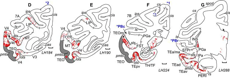
FIGURE 16.1 Representative coronal section drawings showing retrogradely labeled neurons (red) resulting from a tracer injection in area TEO (second panel). Retrograde labeling within the injected area (gray) is not displayed. Adapted, with permission, from Markov et al. (2012), using four of their 16 illustrated section contours from their Case 11 (M119LH).
16.2.2 Highly Distributed Reciprocal Connectivity
Each locus in cortical gray matter has a highly distributed pattern of inputs and outputs. Markov et al. (2012) analyzed their 39 injections into 29 cortical areas in relation to a 91-area cortical parcellation. They reported that each injection revealed inputs from at least 25 areas and up to 84 other cortical areas (average = 55 areas). A large majority of these pathways are bi-directional (reciprocal). The unweighted (binary) cortical graph, or area-to-area connectivity matrix, is much denser (66%) than previously appreciated (e.g. Felleman and Van Essen, 1991). Altogether, Markov et al. (2012) reported 1615 (of a possible 2639) inter-areal pathways in their 29 × 91 graph. Simple extrapolation suggests that a full 91 × 91 area graph might contain about 5500 (of a possible 8281) inter-areal pathways just for one hemisphere. The total number of area-to-area pathways would be even larger if the analysis were based on a finer grained parcellation, such as the 130-area parcellation reported by Van Essen et al. (2012b). It would be larger still if connections with subcortical structures and interhemispheric cortical area-to-area connections were included as well. Hence, the number of pathways with neuroanatomically distinct origins and terminations in the macaque “parcellated connectome” likely exceeds 10 000. The number of pathways in the human parcellated connectome is presumably even larger, as humans are estimated to have more cortical areas than macaques (Van Essen et al., 2012c).
16.2.3 Wide Range of Connection Strengths and an Exponential Distance Relationship
Long-distance area-to-area connections vary in strength over five orders of magnitude and are distributed nearly continuously over this range. As is evident in Figure 16.1, connections are generally strongest between neighboring cortical areas, and the strength of connections declines progressively with increasing inter-areal separation (Markov et al., 2012) with a slope that is approximately exponential (Markov et al., 2013).
16.2.4 Patchy Inter-Areal Connections and a Sparse Connectivity Graph
The connections between any pair of cortical areas are not diffuse but instead are localized in a complex, patchy pattern that reflects the internal organization of each ar...
Índice
- Cover image
- Title page
- Table of Contents
- Copyright
- Foreword
- Contributors
- Section I: Introduction to Diffusion MRI
- Section II: Diffusion MRI for Quantitative Measurement
- Section III: Diffusion MRI for In vivo Neuroanatomy
- Index