Chapter 1 Cellular physiology and histology
Chapter aims
After reading this chapter, you will be able to:
- describe the structure of a typical human cell;
- describe the movement of materials into and out of cells;
- explain how cells can be used to screen for disease;
- provide a description of the major types of human tissue;
- explain the difference between eukaryotic and prokaryotic cells.
Introduction
Case study: Josie – breast cancer
Josie was showering when she noticed a small but hard lump in her right breast. Josie was quickly referred to the local breast screening clinic for further investigation where ultrasound sonography revealed a dense mass around the size of a large garden pea. The consultant immediately recommended a needle biopsy which was carried out the same day under local anaesthetic. The tissue collected was sent for histological examination and a week later Josie was diagnosed with breast cancer and her treatment options were discussed.
The collection of a tissue sample from a patient is termed a biopsy. There are many types of biopsy ranging from the collection of a peripheral blood sample from a finger prick to a more invasive needle or surgical biopsy. As we have seen in Josie’s case, study biopsies may be carried out to look for characteristic tissue changes that may be indicative of diseases such as cancer. Biopsies can also be used to check for infection or to monitor a variety of biochemical parameters in patients.
An average adult body is thought to be constructed from around 50 trillion (50 million million) cells, the majority of which have a finite lifespan, and are continually being replaced as they die. This means that most of the tissues and organs of the human body are not static but in a continual state of flux as senescent, aged cells are replaced.
This chapter will begin by examining the internal structure of a human cell. We will explore how deoxyribonucleic acid (DNA) is organised in the nucleus and the nature of human chromosomes and their use in screening for genetic disease. The individual components of the cytoplasm and structure of the plasma (cell) membrane will be described and mechanisms of transporting materials into and out of cells explored. Once you have a good grasp of cell structure, we will examine how cells are organised into the tissues which are used to construct the human body. Since microbes greatly outnumber human cells, we will examine the nature of bacterial cells which are found colonising the body as part of the microbial biome. To reinforce the key points we will explore the use of cells in detecting disease and examine how certain drugs can target specific cell types to treat disease.
Regions of a cell
Human cells are traditionally split into three distinct regions: the nucleus, the cytoplasm and the plasma (cell) membrane (Figure 1.1).
Figure 1.1 Cell structure
The nucleus
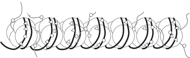
In most cells the nucleus is a centrally located structure that is separated from the cytoplasm by the nuclear membrane (nuclear envelope). The region inside the nuclear membrane is called the nucleoplasm and usually has a granular appearance with a denser inner region called the nucleolus. This granular appearance is due to the presence of condensed chromatin which consists of DNA molecules and histone proteins. The histones function as physical spools around which the DNA is wound and stored in a very compact form (Figure 1.2). This spooling of DNA is essential since each cell, which on average is only around 12 μm in diameter (12 1/000th mm), has to store around 3 metres (10 feet) of DNA.
Figure 1.2 Chromatin and the spooling and storage of DNA
Appearance of chromosomes during cell division
When a cell is preparing to divide, the DNA is progressively wound up tighter and tighter so that it begins to fold up upon itself into tight coils; this tightly wound-up DNA is much denser and thicker and condenses in the nucleus in the form of thread-like structures which are referred to as chromosomes. To help you visualise how chromosomes appear, attempt Activity 1.1.
Activity 1.1 Reflection
Find a rubber elastic band and two pens. Loop the elastic band around both pens and progressively wind up the elastic tighter and tighter.
What do you notice is happening to the elastic?
There are some possible answers to all activities at the end of the chapter, unless otherwise indicated.
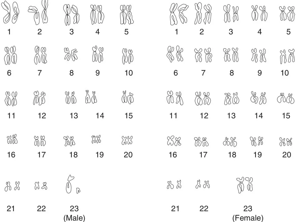
Now that you understand how chromosomes become visible during cell division, we can examine how they can be used to screen for diseases. Nucleated human cells usually have 23 pairs of chromosomes, giving a total of 46, which is referred to as the diploid number (the normal expected number). The only major exceptions to this rule are the sperm and egg cells (ova) which by necessity must have half the diploid number of chromosomes. Half the diploid number in humans is 23 and this is referred to as the haploid number. Having haploid sperm and ova ensures that during fertilisation the diploid number of 46 is restored and the number of chromosomes remains constant down the generations (Chapter 13). Photographs of human chromosomes can be taken during cell division and placed into their ordered pairs according to size; these photographs are called karyographs and reveal the individual’s chromosomal make-up, which is referred to as their karyotype.
Figure 1.3 Chromosomes and karyotypes
The first 22 pairs of human chromosomes are referred to as autosomes and these appear structurally the same in both males and females (Figure 1.3). The final 23rd pair determines the physical gender of the individual and for this reason these are referred to as the sex chromosomes. Females usually have two XX chromosomes (XX) and males usually have an X and a Y chromosome (XY). However, as we will see in Chapter 13, there are frequently variations in the patterns of sex chromosomes and for this reason not all females are XX and not all males are XY. The sex chromosomes only determine the physical gender of the individual and it is recognised that gender identity can be very fluid; frequently the gender that someone feels aligned to may not necessarily reflect their inherited sex chromosomes.
Examining an individual’s chromosomes is most frequently carried out before they are born. During pregnancy foetal cells may be collected by procedures such as amniocentesis or chorionic villus sampling. During amniocentesis the amniotic fluid that surrounds the developing foetus is collected; this will contain cells that have become detached from the foetus as it moves. Since the foetus is continually growing, a large number of the cells harvested will be dividing and therefore have chromosomes visible. The process of karyotyping that follows commonly reveals chromosomal abnormalities such as Down’s syndrome and Turner’s syndrome (Chapter 14).
The cytoplasm and cytoplasmic organelles
The cytoplasm is the region between the plasma membrane and the nuclear membrane. It is predominantly composed of the endoplasmic reticulum (ER) which consists of a system of interconnected flattened membranes. In diagrams of the cell (see Figure 1.1) only small portions of the ER are usually shown, but in reality this complex labyrinth-like system occupies a large proportion of the cell. The ER is split into two distinct types: the rough ER has a multitude of tiny specialised organelles termed ribosomes embedded within its membranes, which are responsible for its characteristic rough, uneven appearance. Ribosomes are the organelles where amino acids are linked together to form proteins according to the instructions encoded in DNA (Chapter 14); for this reason the rough ER is referred to as a region of protein synthesis within cells. The second type of ER is termed smooth ER since it lacks ribosomes; smooth ER is primarily involved in lipid (fat) synthesis. Fats have a multitude of functions within the body including: synthesis of cell membranes, storage of energy, insulation and cushioning and protecting fragile organs such as the kidneys.
The cytoplasm is an aqueous environment and is filled with a watery fluid called the cytosol. The cytosol functions as a transport medium within cells containing dissolved sugars for energy, amino acids for protein synthesis and a variety of intracellular chemical signals and growth factors which are involved in coordinating the internal biochemistry and physical activities of the cell.
The Golgi apparatus
The Golgi apparatus is a specialised region of smooth ER resembling a series of crescent-shaped stacked membranes (Figure 1.1). The Golgi is frequently referred to as the cell’s ‘packaging and export’ region since it is involved in preparing material for release from cells. Its key role is refining proteins from the rough ER; this usually involves adding sugar residues to the crude amino acid sequences via a process termed glycosylation. The refined proteins may be used within the cell or may leave the Golgi in membranous sacs called secretory vesicles which travel to the cell membrane before their contents are discharged out of the cell. Cells that have a secretory role such as those within endocrine glands may each have several well-developed regions of Golgi apparatus; a good example would be the insulin-producing beta cells of the pancreas. The Golgi is also responsible for packaging digestive enzymes required for intracellular digestion into small membrane-bound sacs called lysosomes (see below).
Mitochondria
Mitochondria are small bean-/boat-shaped cellular organelles (Figure 1.1) responsible for releasing energy within cells. Each mitochondrion consists of an outer smooth membrane and a highly folded inner membrane. The prominent folds of the inner membrane are termed cristae and associated with these folds are the enzymes responsible for cellular respiration. Within the mitochondria, glucose, which is derived from carbohydrate-rich foods, is reacted with oxygen acquired by our respiratory system to release energy. This energy is then used to synthesise the energy storage molecule adenosine triphosphate (ATP) from adenosine diphosphate (ADP) and free phosphate. This process results in the production of water and carbon dioxide as waste products. Since these biochemical reactions occur in the presence of oxygen, the process is referred to as aerobic respiration.
Glucose (C6H12O6) + Oxygen (O2) → Carbon dioxide (CO2) + Water (H2O) + Energy (38ATP)
In theory each molecule of glucose can yield 38 molecules of ATP but in reality this is never achieved, and a yield of around 30 ATPs per glucose molecule is typical. From a nursing point of view the simple equation above tells us something essential about human physiology: to generate the energy necessary to keep us alive we must eat (glucose) and breathe (oxygen). Indeed, a key role that nurses play is in ensuri...