Comprehensive Atlas of High-Resolution Endoscopy and Narrowband Imaging
Jonathan Cohen, Jonathan Cohen
This is a test
This is a test
English
ePUB (apto para móviles)
Disponible en iOS y Android
eBook - ePub
Comprehensive Atlas of High-Resolution Endoscopy and Narrowband Imaging
Jonathan Cohen, Jonathan Cohen
Detalles del libro
Vista previa del libro
Índice
Citas
Información del libro
Previous edition won First Prize in the Gastroenterology category of the 2008 BMA Medical Book Competition
High-resolution endoscopy and narrowband imaging have revolutionized the field. Edited by a gastroenterologist with a reputation for delivering outstanding material, this new edition of an award-winning atlas provides you with an outstanding collection of images, videos, and expert diagnostic guidance to enhance your decision making.
To accelerate your learning, Dr. Cohen offers more than 2000 endoscopic images, emphasizing conditions for which NBI is particularly useful – such as finding dysplasia in Barrett's mucosa, and diagnosing adenomatous colon polyps – and providing exceptional preparation for the future of endoscopy practice, with a broad new look at normal and abnormal findings throughout the GI tract.
The book is divided into three main parts:
The basics of NBI
Clinical applications of NBI
Atlas of 1600 color images, broken into sections on the pharynx and esophagus, stomach, small intestine, and colon, including correlating histopathology and multiple examples of key pathologies
The accompanying website features more than 85 video clips containing over 3 hours of annotated video, to give you a complete sense of how HRE and NBI work and look in real time, including during therapeutic procedures.
All of the over 1000 new images appear in much brighter color, reflecting the advance in scope technology since the first edition. New chapters have been added to present the data supporting increased use of NBI in optical diagnosis and in the context of therapeutic procedures. For the first time, brilliant images of the bile duct and pancreas are included as the imaging revolution has expanded to reach these new locations. This spectacular new imaging modality promises to enhance endoscopic decision making in real time, facilitate therapeutic maneuvers, make tissue sampling more precise, and make resection of mucosal neoplasia more complete.
Expertly guiding you through the latest advances, this book facilitates your mastery of the field, and provides an up-to-date reference for gastroenterologists and endoscopists to improve their practice.
Preguntas frecuentes
¿Cómo cancelo mi suscripción?
Simplemente, dirígete a la sección ajustes de la cuenta y haz clic en «Cancelar suscripción». Así de sencillo. Después de cancelar tu suscripción, esta permanecerá activa el tiempo restante que hayas pagado. Obtén más información aquí.
¿Cómo descargo los libros?
Por el momento, todos nuestros libros ePub adaptables a dispositivos móviles se pueden descargar a través de la aplicación. La mayor parte de nuestros PDF también se puede descargar y ya estamos trabajando para que el resto también sea descargable. Obtén más información aquí.
¿En qué se diferencian los planes de precios?
Ambos planes te permiten acceder por completo a la biblioteca y a todas las funciones de Perlego. Las únicas diferencias son el precio y el período de suscripción: con el plan anual ahorrarás en torno a un 30 % en comparación con 12 meses de un plan mensual.
¿Qué es Perlego?
Somos un servicio de suscripción de libros de texto en línea que te permite acceder a toda una biblioteca en línea por menos de lo que cuesta un libro al mes. Con más de un millón de libros sobre más de 1000 categorías, ¡tenemos todo lo que necesitas! Obtén más información aquí.
¿Perlego ofrece la función de texto a voz?
Busca el símbolo de lectura en voz alta en tu próximo libro para ver si puedes escucharlo. La herramienta de lectura en voz alta lee el texto en voz alta por ti, resaltando el texto a medida que se lee. Puedes pausarla, acelerarla y ralentizarla. Obtén más información aquí.
¿Es Comprehensive Atlas of High-Resolution Endoscopy and Narrowband Imaging un PDF/ePUB en línea?
Sí, puedes acceder a Comprehensive Atlas of High-Resolution Endoscopy and Narrowband Imaging de Jonathan Cohen, Jonathan Cohen en formato PDF o ePUB, así como a otros libros populares de Medicina y Gastroenterologia ed epatologia. Tenemos más de un millón de libros disponibles en nuestro catálogo para que explores.
1 Narrowband imaging: historical background and basis for its development
Shigeaki Yoshida
In Japan, where the incidence of gastric cancer is very much higher than in the rest of the world, greater attention has been paid to early diagnosis since the beginning of the 1950s when the “gastrocamera” was first introduced. In those days, the finding of early gastric cancer (EGC) was not frequent and most of these lesions were identified from the differential diagnosis of deeply ulcerated (type III) or polypoid (type I) lesions, which can be easily detected. In the 1970s, early diagnosis progressed and it became possible to detect those cancers showing the appearance of ulcer scar (type IIc) and plateau-like elevation (type IIa). Furthermore, at the beginning of the 1980s, early diagnosis of gastritis-like malignancy (type IIb-like) became more readily possible following the results of retrospective studies of rapidly growing advanced cancer [1]. With this increased appreciation of the appearance of early superficial lesions, the widespread use of biopsy and with careful scrutiny of the mucosa using dye-spraying techniques, EGCs appearing as just faint mucosal irregularities or discoloration came to be the most frequent EGC being diagnosed by the late 1980s [2].
Such results were also applied to esophageal and colorectal malignancies, and there has been a general acceptance in Japan that early malignancies in the alimentary tract may not appear polypoid or ulcerative. The desire to better recognize such malignancies, which may be difficult to distinguish from nonspecific inflammation or trauma, had prompted us to envision new endoscopic technology capable of revealing cancer-specific images of the surface structure of the mucosa. It is within this context that the field of narrowband imaging (NBI) was developed as a promising way to facilitate the endoscopic diagnosis of early neoplastic and pre-cancerous lesions in the alimentary tract.
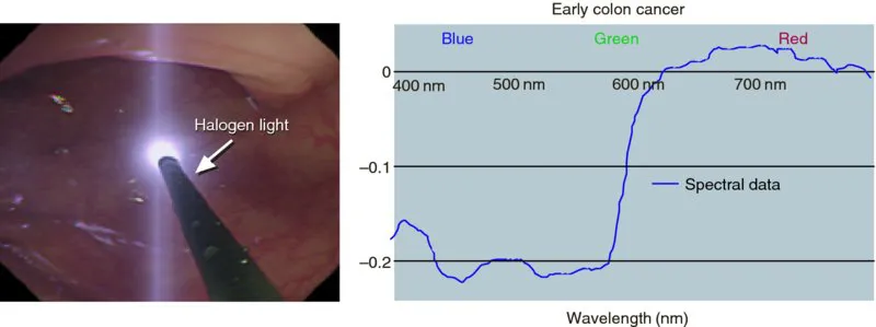
NBI is an optical image enhancement technology that visualizes vessels on the surface of the mucosa and patterns on the surface of mucosa by employing the characteristics of the visible light spectrum. The development of NBI goes back to the study of spectroscopy more than 20 years ago. The Japanese government implemented the Second Term Comprehensive 10-Year Strategy for Cancer Control in 1994. Together with Professor N. Oyama of the Tokyo Institute of Technology and Olympus Medical Systems Corp., we received funding from the project and started the study in which we intended to digitalize the color and structure of mucosa in order to establish a more objective/quantitative pathologic diagnosis and hence better diagnostic yield. At that time, multiple facilities and industries had conducted studies to achieve optical biopsy using the characteristics of the visible light spectrum. We aimed to achieve differentiation of normal and abnormal mucosa using a custom-made spectrophotometer developed by Olympus Medical Systems Corp.
Using the method described in Figure 1.1, we obtained and analyzed more than 2000 samples from esophagus, stomach, and colon. However, we faced multiple challenges to establish a stable diagnostic standard. The spectrum showed different patterns in normal and abnormal tissues but the spectral pattern differed from patient to patient, so that it was quite difficult to achieve stable classification between normal and abnormal. Furthermore, spectral data were not stable under the measuring conditions employed.
Figure 1.1 Spectral reflectance analysis. Spectral data were sampled at intervals of 2 nm ranging from 400 to 800 nm. In each examination, we measured spectral reflectance in both normal and neoplastic areas. (Copyright S. Yoshida.)
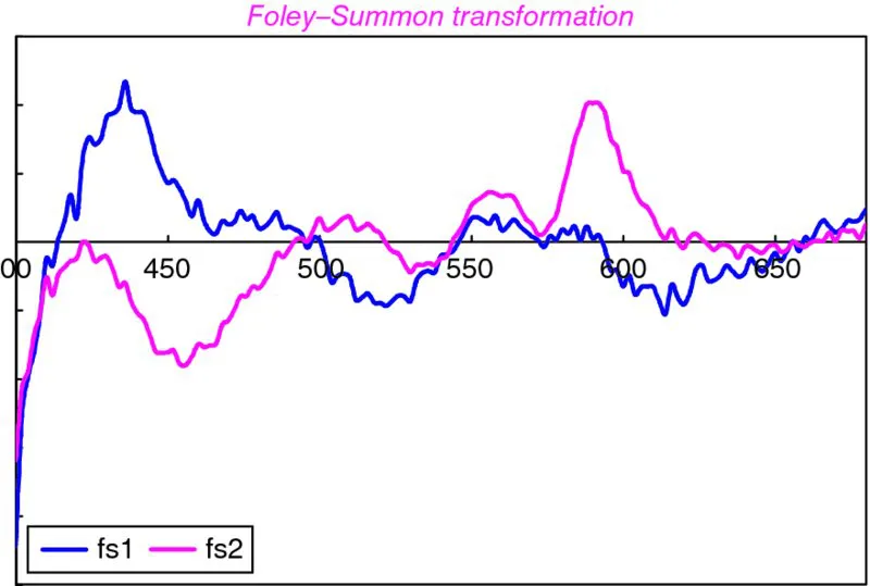
However, throughout the study we noticed a specific spectral pattern when selecting certain narrowband wavelengths (Figure 1.2). To highlight the specific pattern, we shifted our study from qualitative analysis using spectroscopy to qualitative imaging that enhanced details of the mucosal surface. As a result, when employing a narrowband filter, we found excellent light enhancement deep in the mucosa at red light wavelengths, shallow mucosal surface features at blue light wavelengths, and levels in between at green light wavelengths [3]. Based on the findings, we continued the study with the research and development group at Olympus and finally found that narrowband blue light wavelengths matched the light absorption characteristics of blood hemoglobin and enhanced details of the mucosal surface.
Figure 1.2 Spectral sensitivity functions for discrimination of cancerous regions. (Copyright S. Yoshida.)
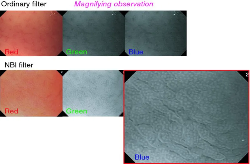
In December 1999, we obtained the world’s first clinical images using NBI in our facility (Figures 1.3–1.6). The original technology only generated black and white monochrome images with limited information for diagnosis, making it impractical for clinical applications. The challenge was shortly solved by the introduction of newer improved filters and the development of a prototype incorporating a circuit board exclusively for NBI color display.
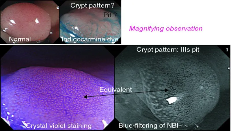
Figure 1.3 Normal gastric mucosa: mucosal crypt pattern of the stomach can be observed without dye spraying by blue-filtering of NBI. (Copyright S. Yoshida.)
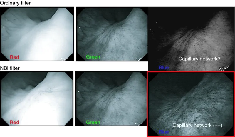
Figure 1.4 Gastric ulcer scar: capillary network can be observed without dye spraying by blue-filtering of NBI. (Copyright S. Yoshida.)
Figure 1.5 Flat adenoma of sigmoid colon: crypt pattern of the depressed area can be observed without dye spraying by blue-filtering of NBI. (Copyright S. Yoshida.)
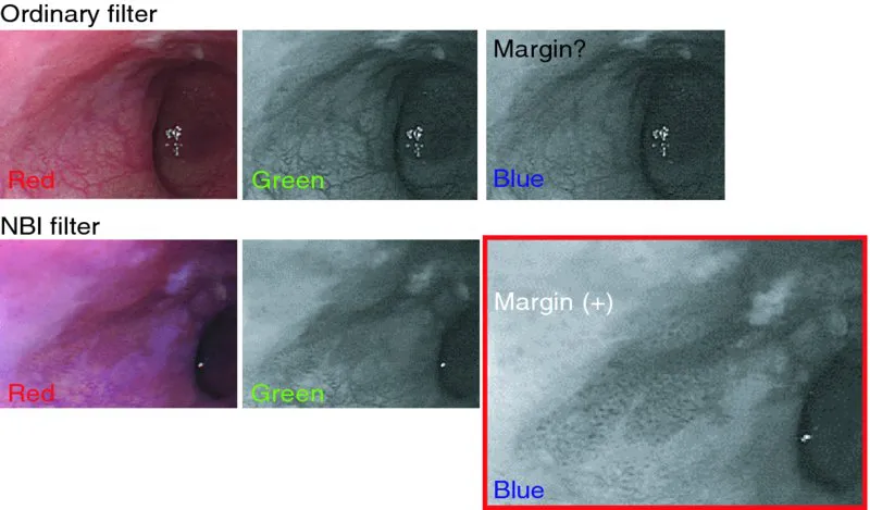
Figure 1.6 Esophageal cancer (type 0–IIc): the margin of the lesion is clearly detected by blue-filtering of NBI. (Copyright S. Yoshida.)
Since these first clinical NBI pictures were achieved, we have actively expanded the study in cooperation with multiple research facilities. As a result of this collaborative investigation, the application of NBI diagnosis has expanded rapidly [4, 5]. Starting with the diagnosis of colonic tumor and squamous cell carcinoma of esophagus, the applications of NBI were established in other fields such as superficial carcinoma in pharynx, Barrett’s esophagus and adenocarcinoma, stomach cancer, and inflammatory bowel disease. Multiple studies have been published in these areas; the results have been published in academic society proceedings, research committee reports and clinical papers in peer-reviewed journals. Much of this data is discussed in detail in subsequent chapters of this book.
In December 2005, the NBI system became commercially available from Olympus, and the technology and diagnosis expanded further, not only in Japan but also worldwide.
In summary, endoscopic diagnosis has been rapidly progressing. Beyond technical advances such as chromoendoscopy and improvements in image quality, endoscopic diagnosis has now advanced to the area of pathology. This is possible because the imaging technology now allows assessment of the three-dimensional architecture of tissue by fine examination of the mucosal surface with magnif...