Pocket Guide to Echocardiography
eBook - ePub

Pocket Guide to Echocardiography

Andro G. Kacharava, Alexander T. Gedevanishvili, Guram G. Imnadze, Dimitri M. Tsverava, Craig M. Brodsky, Andro G. Kacharava, Alexander T. Gedevanishvili, Guram G. Imnadze, Dimitri M. Tsverava, Craig M. Brodsky

Partager le livre
  1. English
  2. ePUB (adapté aux mobiles)
  3. Disponible sur iOS et Android
eBook - ePub

Pocket Guide to Echocardiography

Andro G. Kacharava, Alexander T. Gedevanishvili, Guram G. Imnadze, Dimitri M. Tsverava, Craig M. Brodsky, Andro G. Kacharava, Alexander T. Gedevanishvili, Guram G. Imnadze, Dimitri M. Tsverava, Craig M. Brodsky

DĂ©tails du livre
Aperçu du livre
Table des matiĂšres
Citations

À propos de ce livre

With its easy accessibility, low cost, and ability to deliver, essential bedside information about the cardiac structure and function, echocardiography has become one of the most relied-upon diagnostic tools in clinical medicine. As a result, more clinicians than ever before must be able to accurately interpret echocardiographic information in order to administer appropriate treatment.

Based on the authors' experience teaching echocardiography in busy clinical settings, this new pocketbook provides reliable guidance on everyday clinical cardiac ultrasound and the interpretation of echocardiographic images. It has been designed to help readers develop a stepwise approach to the interpretation of a standard transthoracic echocardiographic study and teach how to methodically gather and assemble the most important information from each of the standard echocardiographic views in order to generate a complete final report of the study performed.

What's included:

‱A summary of TTE examination protocol and a comprehensive listing of useful formulas and normal values

‱Atrial and ventricular dimensions, LV and RV systolic function, LV diastolic patterns

‱Echocardiographic findings in the most commonly encountered cardiac diseases and disorders, including various cardiomyopathies, cardiac tamponade, constrictive pericarditis, valvular heart disease, pulmonary hypertension, infective endocarditis, and congenital heart disease

‱Companion website with video clips and over 70 self-assessment questions


Packed with essential information and designed for quick look-up, this pocketbook will be of great assistance for anyone who works in busy clinical settings and who needs a ready and reliable guide to interpreting echocardiographic information to help deliver optimal patient care.

Foire aux questions

Comment puis-je résilier mon abonnement ?
Il vous suffit de vous rendre dans la section compte dans paramĂštres et de cliquer sur « RĂ©silier l’abonnement ». C’est aussi simple que cela ! Une fois que vous aurez rĂ©siliĂ© votre abonnement, il restera actif pour le reste de la pĂ©riode pour laquelle vous avez payĂ©. DĂ©couvrez-en plus ici.
Puis-je / comment puis-je télécharger des livres ?
Pour le moment, tous nos livres en format ePub adaptĂ©s aux mobiles peuvent ĂȘtre tĂ©lĂ©chargĂ©s via l’application. La plupart de nos PDF sont Ă©galement disponibles en tĂ©lĂ©chargement et les autres seront tĂ©lĂ©chargeables trĂšs prochainement. DĂ©couvrez-en plus ici.
Quelle est la différence entre les formules tarifaires ?
Les deux abonnements vous donnent un accĂšs complet Ă  la bibliothĂšque et Ă  toutes les fonctionnalitĂ©s de Perlego. Les seules diffĂ©rences sont les tarifs ainsi que la pĂ©riode d’abonnement : avec l’abonnement annuel, vous Ă©conomiserez environ 30 % par rapport Ă  12 mois d’abonnement mensuel.
Qu’est-ce que Perlego ?
Nous sommes un service d’abonnement Ă  des ouvrages universitaires en ligne, oĂč vous pouvez accĂ©der Ă  toute une bibliothĂšque pour un prix infĂ©rieur Ă  celui d’un seul livre par mois. Avec plus d’un million de livres sur plus de 1 000 sujets, nous avons ce qu’il vous faut ! DĂ©couvrez-en plus ici.
Prenez-vous en charge la synthÚse vocale ?
Recherchez le symbole Écouter sur votre prochain livre pour voir si vous pouvez l’écouter. L’outil Écouter lit le texte Ă  haute voix pour vous, en surlignant le passage qui est en cours de lecture. Vous pouvez le mettre sur pause, l’accĂ©lĂ©rer ou le ralentir. DĂ©couvrez-en plus ici.
Est-ce que Pocket Guide to Echocardiography est un PDF/ePUB en ligne ?
Oui, vous pouvez accĂ©der Ă  Pocket Guide to Echocardiography par Andro G. Kacharava, Alexander T. Gedevanishvili, Guram G. Imnadze, Dimitri M. Tsverava, Craig M. Brodsky, Andro G. Kacharava, Alexander T. Gedevanishvili, Guram G. Imnadze, Dimitri M. Tsverava, Craig M. Brodsky en format PDF et/ou ePUB ainsi qu’à d’autres livres populaires dans Medicine et Cardiology. Nous disposons de plus d’un million d’ouvrages Ă  dĂ©couvrir dans notre catalogue.

Informations

Éditeur
Wiley-Blackwell
Année
2012
ISBN
9781118480038
Édition
1
Sous-sujet
Cardiology
Chapter 1
Comprehensive Transthoracic Echocardiographic Examination Protocol

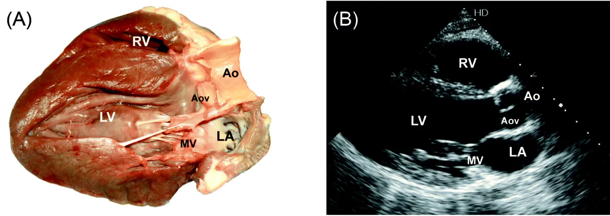
Parasternal Long-axis view (Fig. 1)

1. 2D image (4 beats) (measure LVOT diameter).
2. Color Doppler through MV and AoV (4 beats).
3. M-mode through Aortic root (measure root and LA diameter, and aortic cusp separation).
4. Color Doppler M-mode through the aortic root (4 beats).
5. M-mode through MV (± Valsalva test to check for MV prolapse and SAM; measure “E” to “S” separation distance).
6. Color Doppler M-mode through MV (4 beats).
7. M-mode through mid LV (4 beats) (measure septal and inferolateral wall thickness LVEDD and LVESD).
Figure 1 (A and B) Parasternal long-axis view.
img

RA/RV view

From parasternal LAX view tilt transducer to point it to right hip:
1. 2D image (4 beats);
2. Color Doppler through TV (4 beats);
3. CW Doppler through TV to measure max TR velocity if TR jet present.

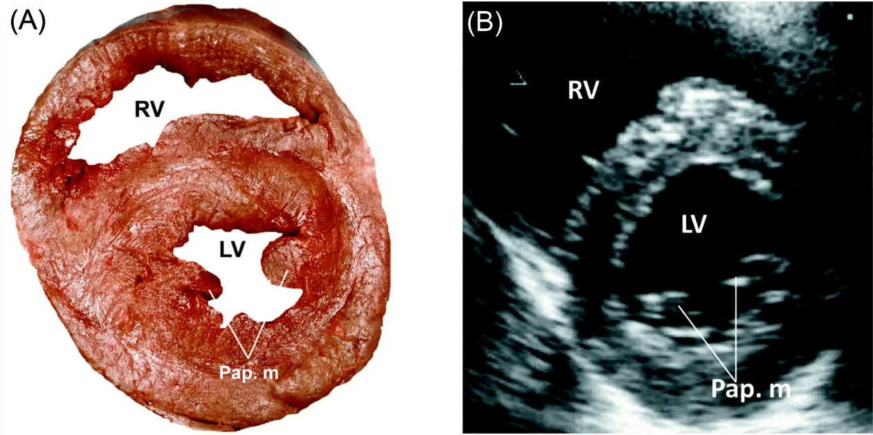
Parasternal Short-axis view (Fig. 2 and Fig. 3)
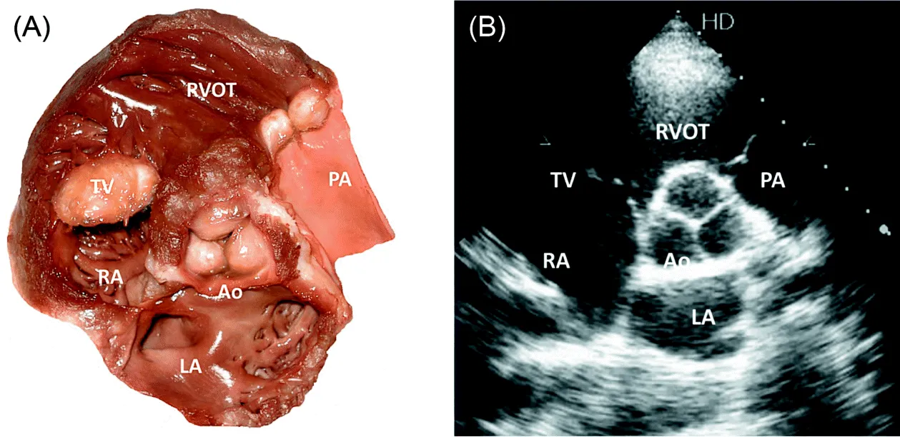
1. 2D through AoV (4 beats) (to assess structure and mobility; use zoom).
2. Color Doppler through AoV (4 beats).
3. 2D through PV (4beats).
4. Color Doppler through PV (4 beats).
5. PW Doppler at the tips of PV to measure PAT (4 beats).
6. CW Doppler through PV to measure PR velocity if present, and maximum outflow velocity through PV.
7. 2D through TV (4 beats).
8. Color Doppler through TV (4 beats).
9. PW Doppler at the tips of TV leaflets to assess inflow pattern (4 beats).
10. If inflow jet max velocity is >1.5 m/sec, trace the diastolic flow to measure mean transvalvular gradient.
11. CW Doppler through TV to measure max TR velocity if TR jet present.
12. Serial 2D short axis images through the LV from base toward apex.
Figure 2 (A and B) Parasternal short-axis view.
img
Figure 3 (A and B) Parasternal short-axis view.
img

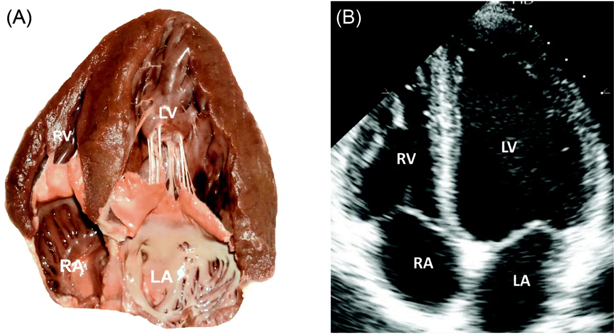
Apical 4-chambers view (Fig. 4)

1. 2D image (4 beats).
2. Color Doppler through the MV (4 beats).
3. Color Doppler M-mode through the MV at the end expiration (4 beats).
4. PW Doppler at the tips of MV leaflets to assess inflow pattern and velocity (4 beats); if pseudonormal or restrictive inflow pattern observed, decrease the preload and reassess the inflow pattern; in impaired relaxation act opposite.
5. PW Doppler at the right/left upper PV to assess inflow pattern (4 beats).
6. PW tissue Doppler at the basal and mid septal and lateral walls (4 beats).
7. If inflow jet max velocity is > 1.9 m/sec, trace the diastolic flow to measure mean transvalvular gradient, then measure PHT of the jet in CW mode.
8. CW Doppler through MV to measure max MR velocity if MR jet present. (Obtain simultaneous SBP measurement to calculate mean LAP.)
9. PW tissue Doppler at basal lateral walls of the RV to assess TEI index and systolic velocity.
10. M-mode through the RV basal lateral wall to measure TAPSE.
Figure 4 (A and B) Apical 4-chambers view.
img

Apical 5-chambers view

From apical 4-chambers view tilt transducer slightly anteriorly; examiner's hand moves downward toward the patient's bed:
1. 2D image (4 beats).
2. Color Doppler through AoV and MV (4 beats).
3. PW Doppler through LVOT and trace the flow (4 beats).
4. CW Doppler through AoV and if outflow max velocity is >1.9 m/sec, trace...

Table des matiĂšres