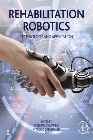
Rehabilitation Robotics
eBook - ePub

Rehabilitation Robotics

Technology and Application

Roberto Colombo,Vittorio Sanguineti

  1. 382 pages
  2. English
  3. ePUB (adapté aux mobiles)
  4. Disponible sur iOS et Android
eBook - ePub

Rehabilitation Robotics

Technology and Application

Roberto Colombo,Vittorio Sanguineti

DĂ©tails du livre
Aperçu du livre
Table des matiĂšres
Citations

À propos de ce livre

Rehabilitation Robotics gives an introduction and overview of all areas of rehabilitation robotics, perfect for anyone new to the field. It also summarizes available robot technologies and their application to different pathologies for skilled researchers and clinicians. The editors have been involved in the development and application of robotic devices for neurorehabilitation for more than 15 years. This experience using several commercial devices for robotic rehabilitation has enabled them to develop the know-how and expertise necessary to guide those seeking comprehensive understanding of this topic.

Each chapter is written by an expert in the respective field, pulling in perspectives from both engineers and clinicians to present a multi-disciplinary view. The book targets the implementation of efficient robot strategies to facilitate the re-acquisition of motor skills. This technology incorporates the outcomes of behavioral studies on motor learning and its neural correlates into the design, implementation and validation of robot agents that behave as 'optimal' trainers, efficiently exploiting the structure and plasticity of the human sensorimotor systems. In this context, human-robot interaction plays a paramount role, at both the physical and cognitive level, toward achieving a symbiotic interaction where the human body and the robot can benefit from each other's dynamics.

  • Provides a comprehensive review of recent developments in the area of rehabilitation robotics
  • Includes information on both therapeutic and assistive robots
  • Focuses on the state-of-the-art and representative advancements in the design, control, analysis, implementation and validation of rehabilitation robotic systems

Foire aux questions

Comment puis-je résilier mon abonnement ?
Il vous suffit de vous rendre dans la section compte dans paramĂštres et de cliquer sur « RĂ©silier l’abonnement ». C’est aussi simple que cela ! Une fois que vous aurez rĂ©siliĂ© votre abonnement, il restera actif pour le reste de la pĂ©riode pour laquelle vous avez payĂ©. DĂ©couvrez-en plus ici.
Puis-je / comment puis-je télécharger des livres ?
Pour le moment, tous nos livres en format ePub adaptĂ©s aux mobiles peuvent ĂȘtre tĂ©lĂ©chargĂ©s via l’application. La plupart de nos PDF sont Ă©galement disponibles en tĂ©lĂ©chargement et les autres seront tĂ©lĂ©chargeables trĂšs prochainement. DĂ©couvrez-en plus ici.
Quelle est la différence entre les formules tarifaires ?
Les deux abonnements vous donnent un accĂšs complet Ă  la bibliothĂšque et Ă  toutes les fonctionnalitĂ©s de Perlego. Les seules diffĂ©rences sont les tarifs ainsi que la pĂ©riode d’abonnement : avec l’abonnement annuel, vous Ă©conomiserez environ 30 % par rapport Ă  12 mois d’abonnement mensuel.
Qu’est-ce que Perlego ?
Nous sommes un service d’abonnement Ă  des ouvrages universitaires en ligne, oĂč vous pouvez accĂ©der Ă  toute une bibliothĂšque pour un prix infĂ©rieur Ă  celui d’un seul livre par mois. Avec plus d’un million de livres sur plus de 1 000 sujets, nous avons ce qu’il vous faut ! DĂ©couvrez-en plus ici.
Prenez-vous en charge la synthÚse vocale ?
Recherchez le symbole Écouter sur votre prochain livre pour voir si vous pouvez l’écouter. L’outil Écouter lit le texte Ă  haute voix pour vous, en surlignant le passage qui est en cours de lecture. Vous pouvez le mettre sur pause, l’accĂ©lĂ©rer ou le ralentir. DĂ©couvrez-en plus ici.
Est-ce que Rehabilitation Robotics est un PDF/ePUB en ligne ?
Oui, vous pouvez accĂ©der Ă  Rehabilitation Robotics par Roberto Colombo,Vittorio Sanguineti en format PDF et/ou ePUB ainsi qu’à d’autres livres populaires dans Technology & Engineering et Biomedical Science. Nous disposons de plus d’un million d’ouvrages Ă  dĂ©couvrir dans notre catalogue.

Informations

Éditeur
Academic Press
Année
2018
ISBN
9780128119969
Chapter 1

Physiological basis of neuromotor recovery

Kevin C. Elliott; David T. Bundy; David J. Guggenmos; Randolph J. Nudo University of Kansas Medical Center, Kansas City, KS, United States

Abstract

Planning and execution of movement require coordinated activity from several interconnected cortical motor areas. When an area in this specialized motor network is damaged (e.g., through traumatic brain injury or ischemic event), motor network activity can be disrupted, leading to functional deficits. How the surviving motor network reorganizes to compensate for the injury and functional deficits can vary as a pathological consequence of the location and extent of the brain injury. The current chapter summarizes how neuroplasticity modifies motor networks in response to injury by focusing on the changes after an ischemic event in the primary motor cortex. Neuroanatomical and neurophysiological evidence in animal models and human stroke survivors is reviewed to demonstrate how injuries functionally impair motor networks, how motor networks compensate for injury to improve motor function, and how select therapies help facilitate recovery. Further research into these neuroplasticity mechanisms may one day help to develop more effective rehabilitation strategies.

Keywords

Stroke; Motor cortex; Neuronal plasticity; Animal models; Infarction; Synapses; Primates; Hand; Movement

Introduction

Each year in the United States, an estimated 795,000 people experience an acute, localized deprivation of oxygen and nutrient-rich blood that impairs the long-term structure and function of the brain: a phenomenon known as a stroke [1]. While a subset of strokes result from a blood vessel hemorrhage, the overwhelming majority (~ 87%) of stroke incidences are ischemic, resulting from a partial or complete blockage of a main arterial branch in the brain. Ischemic strokes typically involve the middle cerebral artery, a major arterial branch that supplies blood to the frontal, temporal, and parietal cortices and striatal and capsular subcortical areas. Approximately 51% of strokes occur in the cortex as a result of middle cerebral artery occlusion [2], and overall, there is a large variability in stroke type, injury location, severity, and subsequent cell death or infarction that induces a wide range of sensory, motor, and/or cognitive impairments among stroke survivors. As such, this chapter will focus on the effects of ischemic strokes on motor activity in normal and impaired cortices and further elaborate on potential therapeutic options to restore normal motor function after a stroke.
The emphasis of this chapter is on motor function, both in normal and impaired cortex, and the neurophysiological basis of neuromotor recovery following stroke. This chapter will (1) review the functional connectivity and activity within the motor network that underlies movement generation, (2) describe the phenomenon of infarction and explain how it alters motor network function, and (3) discuss the influence of rehabilitation on motor recovery following stroke.

The Functional Organization of the Motor Network

Neural processes, such as those that underlie the generation of arm movement, are thought to rely on distributed networks throughout cortical and subcortical brain areas. Though many of the pathways involved in the generation of simple and complex movements require extensive activity from these subcortical areas, the focus here is on the cortex. Studies in humans largely confirm the broad connections between primary motor cortex, M1 (see Table 1 for all anatomical abbreviations), and other cortical motor areas by the extensive white matter connections measured with diffusion tensor imaging and the strong functional interactions observed as part of resting state functional connectivity MRI networks [3]. For a more comprehensive description of connections between motor areas, see Fig. 1.
Fig. 1

Fig. 1 Neuroanatomical studies in the motor areas of nonhuman primates reveal extensive connectivity between M1, premotor, supplementary, and other association motor areas within each hemisphere. As motor areas are bilaterally symmetrical, the medial view of one hemisphere and lateral view of the other hemisphere are used to show all relevant areas. The smaller, whole brain diagram shows the motor areas with reference to the brain in gray. Medial (MCA), anterior (ACA), and posterior (PCA) arteries are shown in black. The larger diagram shows the connectivity between motor areas. Arrows represent bidirectional connectivity between corresponding motor areas. M1 interconnects with PMd and PMv, SMA, S1, posterior parietal cortex, cingulate cortex, and contralateral M1. PMd and PMv—regions that input extensively to M1—interconnect with SMA, prefrontal cortex, orbitoprefrontal cortex, S1, and CMA. Like premotor areas, SMA and anterior “pre-SMA” similarly interconnect with M1, PMd/PMv, and CMA.
Table 1
Motor cortex nomenclature
Modern functional nomenclatureModern abbreviationBrodmann area nomenclatureMatteli et al. nomenclature
Primary motor cortex M1 Area 4 F1
Dorsal premotor cortex PMd Area 6 F2,F7
Ventral premotor cortex PMv Area 6 F4,F5
Supplementary motor area SMA Area 6m F3
Presupplementary motor area Pre-SMA Area 6 F6
Cingulate motor area CMA Area 23c, 24c -
Somatosensory cortex S1 Area 1,2,3 -
Prefrontal cortex PFC Area 8, 9,10 -
Posterior parietal cortex PPC Area 5 -
The M1, premotor, and supplementary motor areas contain a subset of neurons that target spinal motoneurons, and though these corticospinal (CS) neurons are distributed throughout the cortex, about half reside in M1 [4]. Compared with other mammals, primate CS neurons monosynaptically target a greater number of motoneurons that drive distal forelimb musculature, and an overwhelming 80% of these CS neurons originate directly from the caudal half of M1 [4,5]. A subset of CS neurons that target single motoneurons, referred to as corticomotoneuronal cells, functionally encode specific criteria, such that a single corticomotoneuronal cell may fire to activate, rigidly lock, or brake a specific muscle movement, but are unlikely to be involved in all three actions [6]. The functional significance of this organization is that M1 is uniquely specialized to drive fine motor control such as digit manipulation or complex forelimb movements and many other parameters associated with movement. It follows that M1 injury severely impairs these capabilities relative to other motor regions.

Motor Network Activity and Movement

To better understand the movement parameters encoded by M1, the activity of single CS neurons has been studied for several decades. Early work decoding the activity of individual M1 CS neurons showed that CS activity precedes muscle contraction of small groups of muscles, and the rate of firing of these neurons is related to the force exerted by distal forelimb movements. This suggests that individual CS neurons can encode kinetic-based parameters, where specific neurons correlate to the contraction of muscle groups involved in functional movement execution, such as arm flexion or extension [7]. While CS activity initiates gross muscle movements, the subset of CS neurons that project directly to spinal motoneurons (corticomotoneuronal cells) are recruited during precision movements that require finer muscle control [8]. Subsequent studies analyzed the population codes of M1 neuronal ensembles to demonstrate that most M1 neurons are tuned broadly to movement direction, with increases or decreases in firing rate during limb movements aimed toward or away from the neuron's preferred direction, respectively. These results suggest that in addition to simple muscle-/kinetic-based parameters, M1 encodes extrinsic movement kinematics that includes related features of direction, velocity, position, orientation, and specific muscle activations related to the completion of movement [9]. Thus, M1 contains neuronal populations that encode both “simple” kinetic activity and more “complex” kinematics [9].
In addition to recording neuronal activity during movement, stimulation of the motor areas noninvasively either via transcranial magnetic stimulation at the cortical surface (electrocortical stimulation mapping) or via intracortical microstimulation (ICMS) at the motor output layers can elicit movements that denote the area's functional representation. ICMS techniques have revealed that M1 contains prominent proximal (e.g., elbow/shoulder) and distal (e.g., digit) forelimb and hind limb representations for the contralateral side of the body [10]. These generated movement representations are not static, and even short-term behavioral experience is sufficient to reorganize the ICMS-defined borders of each representation [11], suggesting that functional organization—and neurophysiological activity generally—may be sensitive to the perturbations by a stroke within the motor network or subsequent recovery and rehabilitation.
While the preceding understanding of motor activity has been largely based on animal models, several human studies have examined motor physiology using noninvasive electrophysiological recordings a...

Table des matiĂšres

  1. Cover image
  2. Title page
  3. Table of Contents
  4. Copyright
  5. Contributors
  6. Rehabilitation Robotics: Technology and Applications
  7. Chapter 1: Physiological basis of neuromotor recovery
  8. Chapter 2: An overall framework for neurorehabilitation robotics: Implications for recovery
  9. Chapter 3: Biomechatronic design criteria of systems for robot-mediated rehabilitation therapy
  10. Chapter 4: Actuation for robot-aided rehabilitation: Design and control strategies
  11. Chapter 5: Assistive controllers and modalities for robot-aided neurorehabilitation
  12. Chapter 6: Exoskeletons for upper limb rehabilitation
  13. Chapter 7: Exoskeletons for lower-limb rehabilitation
  14. Chapter 8: Performance measures in robot assisted assessment of sensorimotor functions
  15. Chapter 9: Computational models of the recovery process in robot-assisted training
  16. Chapter 10: Interactive robot assistance for upper-limb training
  17. Chapter 11: Promoting motivation during robot-assisted rehabilitation
  18. Chapter 12: Software platforms for integrating robots and virtual environments
  19. Chapter 13: Twenty + years of robotics for upper-extremity rehabilitation following a stroke
  20. Chapter 14: Three-dimensional, task-oriented robot therapy
  21. Chapter 15: Robot-assisted rehabilitation of hand function
  22. Chapter 16: Robot-assisted gait training
  23. Chapter 17: Wearable robotic systems and their applications for neurorehabilitation
  24. Chapter 18: Robot-assisted rehabilitation in multiple sclerosis: Overview of approaches, clinical outcomes, and perspectives
  25. Chapter 19: Robots for cognitive rehabilitation and symptom management
  26. Chapter 20: Hybrid FES-robot devices for training of activities of daily living
  27. Chapter 21: Robotic techniques for the assessment of proprioceptive deficits and for proprioceptive training
  28. Chapter 22: Psychophysiological responses during robot-assisted rehabilitation
  29. Chapter 23: Muscle synergies approach and perspective on application to robot-assisted rehabilitation
  30. Chapter 24: Telerehabilitation Robotics: Overview of approaches and clinical outcomes
  31. Index
Normes de citation pour Rehabilitation Robotics

APA 6 Citation

[author missing]. (2018). Rehabilitation Robotics ([edition unavailable]). Elsevier Science. Retrieved from https://www.perlego.com/book/1828503/rehabilitation-robotics-technology-and-application-pdf (Original work published 2018)

Chicago Citation

[author missing]. (2018) 2018. Rehabilitation Robotics. [Edition unavailable]. Elsevier Science. https://www.perlego.com/book/1828503/rehabilitation-robotics-technology-and-application-pdf.

Harvard Citation

[author missing] (2018) Rehabilitation Robotics. [edition unavailable]. Elsevier Science. Available at: https://www.perlego.com/book/1828503/rehabilitation-robotics-technology-and-application-pdf (Accessed: 15 October 2022).

MLA 7 Citation

[author missing]. Rehabilitation Robotics. [edition unavailable]. Elsevier Science, 2018. Web. 15 Oct. 2022.