The Paracellular Channel
eBook - ePub

The Paracellular Channel

Biology, Physiology, and Disease

Jianghui Hou

  1. 248 pages
  2. English
  3. ePUB (adapté aux mobiles)
  4. Disponible sur iOS et Android
eBook - ePub

The Paracellular Channel

Biology, Physiology, and Disease

Jianghui Hou

Détails du livre
Aperçu du livre
Table des matières
Citations

À propos de ce livre

The Paracellular Channel: Biology, Physiology and Disease serves as the first volume to offer a cohesive and unifying picture of the critical functions of paracellular channels (tight junctions) in different tissues. This new class of ion channel utilizes a completely different mechanism to create ion passage pathways across the cell junction. This volume outlines common principles that govern the organization and regulation of these diverse cellular structures, describes the methodology of study, and highlights the pathophysiologic consequence of abnormal structure and functions of the paracellular channels in human diseases.

Coverage includes biochemical, biophysical, structural, physiologic analyses of the paracellular channel, and new technologies for recording and characterization.

  • Offers integrated coverage of all key aspects of the paracellular channel, an understudied field that may hold key insights into some of the most mysterious aspects of physiology
  • Targets different levels of expertise, spanning from graduate students, interns and clinical fellows, to seasoned researchers that study functions, regulation and dysfunctions of different tissue barriers
  • Provides a cohesive and unifying picture that describes the critical functions of paracellular channels (tight junctions) in different tissues

Foire aux questions

Comment puis-je résilier mon abonnement ?
Il vous suffit de vous rendre dans la section compte dans paramètres et de cliquer sur « Résilier l’abonnement ». C’est aussi simple que cela ! Une fois que vous aurez résilié votre abonnement, il restera actif pour le reste de la période pour laquelle vous avez payé. Découvrez-en plus ici.
Puis-je / comment puis-je télécharger des livres ?
Pour le moment, tous nos livres en format ePub adaptés aux mobiles peuvent être téléchargés via l’application. La plupart de nos PDF sont également disponibles en téléchargement et les autres seront téléchargeables très prochainement. Découvrez-en plus ici.
Quelle est la différence entre les formules tarifaires ?
Les deux abonnements vous donnent un accès complet à la bibliothèque et à toutes les fonctionnalités de Perlego. Les seules différences sont les tarifs ainsi que la période d’abonnement : avec l’abonnement annuel, vous économiserez environ 30 % par rapport à 12 mois d’abonnement mensuel.
Qu’est-ce que Perlego ?
Nous sommes un service d’abonnement à des ouvrages universitaires en ligne, où vous pouvez accéder à toute une bibliothèque pour un prix inférieur à celui d’un seul livre par mois. Avec plus d’un million de livres sur plus de 1 000 sujets, nous avons ce qu’il vous faut ! Découvrez-en plus ici.
Prenez-vous en charge la synthèse vocale ?
Recherchez le symbole Écouter sur votre prochain livre pour voir si vous pouvez l’écouter. L’outil Écouter lit le texte à haute voix pour vous, en surlignant le passage qui est en cours de lecture. Vous pouvez le mettre sur pause, l’accélérer ou le ralentir. Découvrez-en plus ici.
Est-ce que The Paracellular Channel est un PDF/ePUB en ligne ?
Oui, vous pouvez accéder à The Paracellular Channel par Jianghui Hou en format PDF et/ou ePUB ainsi qu’à d’autres livres populaires dans Biological Sciences et Biophysics. Nous disposons de plus d’un million d’ouvrages à découvrir dans notre catalogue.

Informations

Année
2018
ISBN
9780128146361
Chapter 1

Introduction

Abstract

The paracellular channel is a new concept in ion channel biology. Its structure and function are vastly different from the membrane channel. The paracellular channel is located within the tight junction. Several key milestones in paracellular channel biology are recounted, including the discovery of its molecular component, the establishment of its connection to human disease, the elucidation of its assembling mechanism, the resolution of its crystal structure, the separation of its water from ion permeability, and the development of its recording approach. The paracellular channel is being recognized as an essential component of the cellular transport machinery that evolves to cope with various transport needs.

Keywords

crystal structure
electrophysiology
genetic disease
ion channel
mutation
protein interaction
tight junction
water channel

1.1 A new class of ion channel

The cell membrane is a biologic lipid bilayer that separates the interior of the cell from the exterior environment. Ion channels are pore forming membrane proteins that allow passage of ions through the cell membrane (Fig. 1.1). When life evolves from unicellular to multicellular organisms, coordination of cell growth by a process known as morphogenesis creates complex three-dimensional structures, such as the blood vessel, renal tubule, pulmonary alveolus, and so on. These tissue structures, made of continuous layers of cells, separate the exterior environment into two independent compartments. The paracellular channel is a new class of ion channel oriented perpendicular to the plasma membrane plane and serving to join the two exterior compartments (Fig. 1.2). The paracellular channel, now believed to be made of the class of claudin proteins, conducts ions in a similar way to the conventional membrane channel by displaying size and charge selectivity (Tang & Goodenough, 2003; Tsukita & Furuse, 2000). However, the paracellular channel is fundamentally different from the membrane channel in many aspects.
image
Figure 1.1 Membrane ion channel.
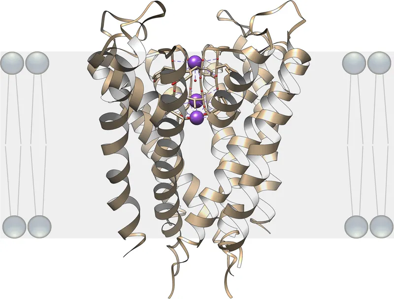
The ion channel in the lipid bilayer permits passage of potassium ions (labeled in violet) but not sodium ions. The oxygen atoms (labeled in red) of the amino acid residues forming the channel pore interact with and stabilize the potassium ions by creating an environment very similar to the aqueous environment outside the lipid bilayer. Cells may open or close the channel by employing additional gating mechanisms. The depicted ion channel structure is based upon the X-ray analysis of the KcsA K+ channel from Streptomyces lividans (MacKinnon, 2004).
image
Figure 1.2 Paracellular ion channel.
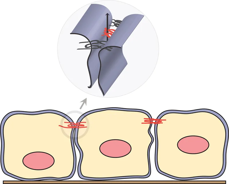
The paracellular channel is found within the TJ structure and is part of the paracellular diffusion barrier between the two plasma membranes. The paracellular channel conducts ions on the basis of size and charge. The paracellular channel is thought ...

Table des matières

  1. Cover
  2. Title page
  3. Table of Contents
  4. Copyright
  5. Dedication
  6. Author Biography
  7. Preface
  8. Acknowledgment
  9. Chapter 1: Introduction
  10. Chapter 2: Paracellular Channel Formation
  11. Chapter 3: Paracellular Channel Recording
  12. Chapter 4: Paracellular Cation Channel
  13. Chapter 5: Paracellular Anion Channel
  14. Chapter 6: Paracellular Water Channel
  15. Chapter 7: Paracellular Channel in Organ System
  16. Chapter 8: Paracellular Channel in Human Disease
  17. Chapter 9: Paracellular Channel as Drug Target
  18. Chapter 10: Paracellular Channel Evolution
  19. Chapter 11: Perspective
  20. Index
Normes de citation pour The Paracellular Channel

APA 6 Citation

Hou, J. (2018). The Paracellular Channel ([edition unavailable]). Elsevier Science. Retrieved from https://www.perlego.com/book/1829172/the-paracellular-channel-biology-physiology-and-disease-pdf (Original work published 2018)

Chicago Citation

Hou, Jianghui. (2018) 2018. The Paracellular Channel. [Edition unavailable]. Elsevier Science. https://www.perlego.com/book/1829172/the-paracellular-channel-biology-physiology-and-disease-pdf.

Harvard Citation

Hou, J. (2018) The Paracellular Channel. [edition unavailable]. Elsevier Science. Available at: https://www.perlego.com/book/1829172/the-paracellular-channel-biology-physiology-and-disease-pdf (Accessed: 15 October 2022).

MLA 7 Citation

Hou, Jianghui. The Paracellular Channel. [edition unavailable]. Elsevier Science, 2018. Web. 15 Oct. 2022.