eBook - ePub
Hematology
Henry
This is a test
Partager le livre
- 44 pages
- English
- ePUB (adapté aux mobiles)
- Disponible sur iOS et Android
eBook - ePub
Hematology
Henry
Détails du livre
Aperçu du livre
Table des matières
Citations
À propos de ce livre
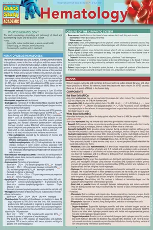
We love providing medical professionals, nurses, and nursing students products that make their jobs or studies easier. They live to help others, so we want to help them. By popular demand, this reference guide covers an immense amount of information in 6 laminated pages that will help anyone in the medical field that works with or around the study of blood (morphology, physiology and pathology). For more, get our Pathology 1 and 2 guides, Nursing Lab Values and Lymphatic System anatomy for a set.
6-page laminated reference guide includes:
- Hematopoiesis/Hemopoiesis
- Organs of the Lymphatic System
- Blood
- Components
- Hemostasis
- Blood Groups
- ABO System
- RH System
- Specimen Collection
- Laboratory Assessment of Blood Formation & Disorders
- Abnormal Blood Cell Morphology
- Blood Disorders (over 2 full pages)
Suggested uses:
- NCLEX – great for reviewing hematologic disorders leading up to the exam
- Students – a very lightweight, inexpensive tool for boosting grades that can be slipped between your notebook pages for quick and easy answers
- Medical Professionals – great tool to have handy for a memory jog for you or as a nursing station reference for the team
Foire aux questions
Comment puis-je résilier mon abonnement ?
Il vous suffit de vous rendre dans la section compte dans paramètres et de cliquer sur « Résilier l’abonnement ». C’est aussi simple que cela ! Une fois que vous aurez résilié votre abonnement, il restera actif pour le reste de la période pour laquelle vous avez payé. Découvrez-en plus ici.
Puis-je / comment puis-je télécharger des livres ?
Pour le moment, tous nos livres en format ePub adaptés aux mobiles peuvent être téléchargés via l’application. La plupart de nos PDF sont également disponibles en téléchargement et les autres seront téléchargeables très prochainement. Découvrez-en plus ici.
Quelle est la différence entre les formules tarifaires ?
Les deux abonnements vous donnent un accès complet à la bibliothèque et à toutes les fonctionnalités de Perlego. Les seules différences sont les tarifs ainsi que la période d’abonnement : avec l’abonnement annuel, vous économiserez environ 30 % par rapport à 12 mois d’abonnement mensuel.
Qu’est-ce que Perlego ?
Nous sommes un service d’abonnement à des ouvrages universitaires en ligne, où vous pouvez accéder à toute une bibliothèque pour un prix inférieur à celui d’un seul livre par mois. Avec plus d’un million de livres sur plus de 1 000 sujets, nous avons ce qu’il vous faut ! Découvrez-en plus ici.
Prenez-vous en charge la synthèse vocale ?
Recherchez le symbole Écouter sur votre prochain livre pour voir si vous pouvez l’écouter. L’outil Écouter lit le texte à haute voix pour vous, en surlignant le passage qui est en cours de lecture. Vous pouvez le mettre sur pause, l’accélérer ou le ralentir. Découvrez-en plus ici.
Est-ce que Hematology est un PDF/ePUB en ligne ?
Oui, vous pouvez accéder à Hematology par Henry en format PDF et/ou ePUB ainsi qu’à d’autres livres populaires dans Medicina et Teoria, pratica e riferimenti medici. Nous disposons de plus d’un million d’ouvrages à découvrir dans notre catalogue.
Informations
Sujet
MedicinaSous-sujet
Teoria, pratica e riferimenti medici
BLOOD DISORDERS
- ABO hemolytic disease of newborn (ABO HDN): ABO incompatibility between mother and fetus (~20% of births). The majority of the cases of ABO HDN are caused by “immune” IgG antibodies in mothers with group O blood. It may potentially only be found in the first pregnancy. In red cell alloimmunization, IgG Abs pass from mother to fetus, where they bind to red cells, leading to their destruction. While more frequent than Rh HDN, ABO HDN is usually mild and does not require treatment
- Agranulocytosis: Also called bone marrow failure, it is the deficiency of granulocytes; patient is more susceptible to infection
- Amyloidosis: Extracellular deposition (focal, localized, or systemic) of abnormal fibrillary protein. Can be hereditary or acquired. All deposits, except for intracerebral plaques, contain non-fibrillar glycoprotein amyloid P; otherwise, composition differs with disease
- Systemic amyloid light chain (AL): Monoclonal light chains produced from the clonal proliferation of plasma cells are deposited
- Antibody (Ab)-mediated coagulation factor deficiency: Caused by autoantibody binding. The most commonly targeted coagulation factor is factor VIII (e.g., acquired hemophilia A)
- Anti-phospholipid syndrome (APS): Occurrence of venous and arterial thrombosis and/or recurrent miscarriage coupled with persistent antiphospholipid Ab
- Anemia: Reduction in Hb concentration; can be caused by the loss, destruction, and/or reduced production of RBCs
- Anemia of chronic disorders (ACD): Anemia occurring in patients with chronic inflammatory/ malignant disease
- Aplastic (bone marrow failure); can be idiopathic acquired
- Bacterial infection associated
- Chronic renal failure associated
- Congenital; includes dyserythropoietic and fanconi anemia (FA)
- Congestive heart failure associated
- Folate-deficiency (folic acid deficiency)
- Hemolytic
- Alloimmune (fetus/newborn)
- Autoimmune: Either warm or cold, such as primary cold agglutinin disease
- Chemical agent, such as a drug (e.g., dapsone and sulfasalazine), copper, lead, chlorate, and arsine
- G6PD deficiency: Red cell is susceptible to oxidative stress
- Hereditary elliptocytosis
- Hereditary spherocytosis (HS)
- Immune injury
- Infection
- March hemoglobinuria: Damage to red cells between the small bones of the feet caused by marching or running for a long time
- Paroxysmal nocturnal hemoglobinuri
- Physical agent, such as a burn causing acanthocytosis or spherocytosis
- Red cell fragmentation syndrome
- Hypothyroidism associated
- Iron deficiency in chronic hemodialysis
- Lead poisoning associated
- Liver disease associated
- Macrocytic: Circulating RBCs have a higher average volume (i.e., mean corpuscular volume (MCV))
- Megaloblastic
- Non-megaloblastic
- Marrow infiltration
- Microcytic: Often hypochromic. Circulating RBCs have a lower average mass of Hb (i.e., mean corpuscular hemoglobin (MCH)). It can be caused by an inherited mutation of matriptase-2 (no inhibition of hepcidin secretion) or DMT1 genes
- Neonatal
- Decreased production
- Hemorrhage
- Increased destruction
- Normocytic (normochromic)
- Nutritional deficit
- Paroxysmal nocturnal hemoglobinuria (PNH): Acquired disorder in which synthesis of a glycosylphosphatidylinositol (GPI) anchor is deficient
- Pernicious anemia (PA): Stomach atrophies due to an autoimmune attack on gastric mucosa; leads to malabsorption of vitamin B12
- Sideroblastic: Ring sideroblasts in bone marrow Hereditary:
- Hypochromic, microcytic blood
- Refractory with ring sideroblasts
- Pregnancy (during), premature birth (infants)
- Rheumatoid arthritis associated
- Sickle cell
- Viral infection associated, such as HIV
- Bacterial infection: Most common cause of neutrophil leukocytosis is acute infection. Prolonged infection may lead to mild anemia; gram- bacteria can lead to severe hemolytic anemia in bacterial septicemia. Coagulation factors increase and natural anticoagulants decrease in an acute infection. If the acute infection is severe, there may be thrombocytopenia
- BernarD-Soulier syndrome: Defective glycoprotein 1b on the surface of a platelet; rare autosomal recessive coagulopathy. It is characterized by thrombocytopenia, large platelets, defective binding to vWF, defective adherence to subendothelial connective tissue, and no platelet aggregation with ristocetin
- Congenital factor XIII (FXIII) A-subunit deficiency: Rare autosomal recessive disorder; catalytic A subunit of FXIII is defective; causes hemorrhagic diathesis
- Disseminated intravascular coagulation (DIC): Also called disseminated intravascular coagulopathy and consumptive coagulopathy; widespread activation of clotting cascade. Clots form in small blood vessels, compromising tissue blood flow, which can cause multiple organ damage. While clotting factors and platelets are involved in abnormal coagulation, normal clotting is disrupted and can lead to bleeding. Large amounts of the smallest fragments of fibrin/ fibrinogen are detected in plasma
- Drug effect on platelet function: Aggregation/ Blood clot formation
- Aspirin
- Dipyridamole
- Clopidogrel
- Prasugrel
- Dyshemoglobinemia: Derivative of Hb that cannot reversibly associate with oxygen; primarily heme prosthetic group is stereochemically altered
- Carboxyhemoglobinemia: CO, Hb covalent bond
- Methemoglobinemia: Ferrous iron is oxidized
- Elevated plasmic uric acid level in an adult with malignancy
- Essential thrombocythemia: Sustained increase in platelets due to megakaryocyte proliferation and an increased production of platelets. The majority of patients have JAK2 (V617F) mutation; the majority of patients without JAK2 mutation have mutations in the CALR gene
- Glanzmann’s disease: Also called thrombasthenia, it involves a defective glycoprotein IIb/IIIa (αIIb and β3) on the surface of a platelet. It is an autosomal recessive disorder leading to the failure of primary platelet aggregation.
- Glycogen storage disease: Defect in the glycogen synthesis or breakdown; can be genetic or acquired
- Granulocytosis: Increased level of granulocytes in peripheral blood
- Heavy-chain disease (HCD): Plasma cell disorder/ malignancy in which incomplete monoclonal Ig heavy chains are overproduced. Typically involves deletion in N-terminal region, leaving the heavy chain unable to form a disulfide bond with a light chain
- α chain disease (IgA; Seligmann’s disease or αHCD)
- γ chain disease (IgG; Franklin’s disease or γHCD)
- μ chain disease (IgM; μHCD)
- Hemochromatosis: Can be genetic, primary, or hereditary. Iron is excessively absorbed from the gastrointestinal tract leading to iron overload
- Most common cause is a homozygous missense mutation in the high iron Fe (HFE) gene (C282Y)
- Hemoglobin disease
- Hemoglobin C (HbC): E6K mutation in β-globin chain; mild hemolytic anemia occur...