Biological Sciences
Cell Membrane Structure
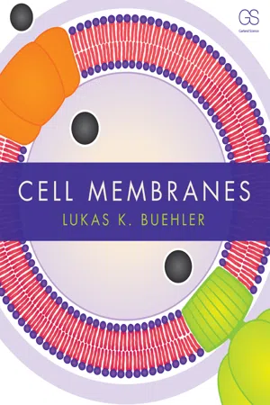
The cell membrane is composed of a phospholipid bilayer with embedded proteins, cholesterol, and carbohydrates. This structure provides a selectively permeable barrier that regulates the movement of substances in and out of the cell. The fluid mosaic model describes the dynamic nature of the cell membrane, with proteins and other molecules moving within the lipid bilayer.
Written by Perlego with AI-assistance
Related key terms
1 of 5
12 Key excerpts on "Cell Membrane Structure"
 eBook - PDF
- Lukas Buehler(Author)
- 2015(Publication Date)
- Garland Science(Publisher)
The structure of cell membranes is a composite of lipids and proteins, with lipids forming the basic bilayer scaf-fold that holds proteins, which carry out most of a membrane’s functions. Membranes are self-renewing structures that form self-enclosed vesicu-lar compartments. They have a unique combination of physical properties that makes them fluid and elastic, electrically charged, and insulating. With thousands of different building blocks of lipids and proteins, membranes are linked to diseases and serve as therapeutic targets. To appreciate how mem-branes contribute to the cellular organization of life in health and disease, we will next explore what cell membranes are made of and what they do, and analyze their structure and physical properties. Further Reading Bangham AD, Standish MM & Weissmann G (1965) The action of steroids and streptolysin S on the permeability of phospholipid structures to cations. J Mol Biol 13:253–259. Borst P & Elferink RO (2002) Mammalian ABC transporters in health and disease. Annu Rev Biochem 71:537–592. Cavalier-Smith T (2001) Obcells as proto-organisms: membrane heredity, lithophosphorylation, and the origins of the genetic code, the first cells, and photosynthesis. J Mol Evol 53:555–595. De Weer P (2000) A century of thinking about cell membranes. Annu Rev Physiol 62:919–926. Fagerberg L, Jonasson K, von Heijne G et al. (2010) Prediction of the human membrane proteome. Proteomics 10:1141–1149 (doi: 10.1002/pmic.200900258). Gennis RB (2005) Biomembranes : Molecular Structure and Function. Springer. Lehnert U, Xia Y, Royce TE et al. (2004) Computational analysis of membrane proteins: genomic occurrence, structure prediction and helix interactions. Q Rev Biophys 37:121–146. Luckey M (2008) Membrane Structural Biology. Cambridge University Press. Mizuno N, Niwa T, Yotsumoto Y & Sugiyama Y (2003) Impact of drug transporter studies on drug discovery and development. eBook - ePub
- Subrata Pal(Author)
- 2019(Publication Date)
- Academic Press(Publisher)
Chapter 14Membrane structure and function
Abstract
Biomembranes consist of a hydrophobic matrix formed by a lipid bilayer with surface-bound and embedded proteins. The bilayer is both diverse and asymmetric in respect to its lipid composition. However, the amphipathic nature of all kinds of lipid molecules present in the membrane thermodynamically favors the formation of the common bilayer structure. Both lipids and proteins in the bilayer are in constant motion. As the boundary of a cell, the membrane displays a property of selective permeability, allowing only certain substances to be passively or actively transported across it. It also plays a crucial role in the cell’s response to environmental signals.Keywords
Biomembrane; Lipid bilayer; Membrane protein; Fluid mosaic model; Transport across membrane; Signal transductionIn order to sustain life, cells need to carry out a wide variety of chemical reactions. Earlier, we have discussed the synthesis of some of the most important biomolecules—DNA, RNA, and protein. However, we have to understand that the cell is able to synthesize its molecular constituents as well as carry out a number of other metabolic processes, efficiently and in an organized manner, since it is partially secluded from its more random environment by a biomembrane. The hydrophobic core of the membrane is formed by lipid assemblies.14.1 Composition of the membrane
In a living cell, lipids perform three general functions: (a) storage of energy as triglycerol ester and steryl esters, (b) as first and second messengers in signal transduction and molecular recognition processes, and (c) formation of the matrix of cellular membranes. It is the third function that we are concerned with in this chapter.Some bacterial cells (Gram-positive) contain just one membrane, whereas others (Gram-negative) are surrounded by two membranes—inner and outer. Eukaryotic cells are enclosed in a single plasma membrane. Some of the internal organelles of eukaryotic cells such as the nucleus, mitochondria, and chloroplasts are surrounded by double lipid bilayers, while endoplasmic reticula, Golgi apparatus, and lysosomes each contains a single lipid bilayer. Nevertheless, in all cases, lipids form the common core. Besides, the cell membranes also contain proteins and carbohydrates of different types and in varying amounts. eBook - ePub
Drug-Biomembrane Interaction Studies
The Application of Calorimetric Techniques
- Rosario Pignatello(Author)
- 2013(Publication Date)
- Woodhead Publishing(Publisher)
All living cells, prokaryotic and eukaryotic, possess a thin cell or plasma membrane (also known as plasmalemma), which encloses their contents and acts as a semi-porous barrier to the outside environment. It also serves as the communications interface between the cell and its environment. Biological membranes also compartmentalize cellular organelles and their functions. Inside a cell, the endoplasmic reticulum, Golgi apparatus, lysosomes, vesicles and vacuoles are surrounded by a single biomembrane sheet. Mitochondria and the nucleus are surrounded by two membrane layers. Finally, the membrane regulates the flow of materials into and out of the cell, mediates intercellular communication, contacts and adhesion, and performs a multitude of other tasks.Various scientific hypotheses have been used to describe the structure of plasma membranes and Eichman wrote a good historical description.9 Of the various models, the most accepted theory is the fluid mosaic model, which was developed by Singer and Nicolson in 1972. According to this theory, a cell membrane consists of a continuous, fluid, double layer of phospholipid (PL), containing or attaching to in different ways other components like proteins, carbohydrates and cholesterol (CHOL) (see below). In a very simplified visualization, the PLs form a thin, flexible sheet, while the proteins float in this sheet like icebergs, and the carbohydrates protrude out from the surface (Figure 1.1 ).Figure 1.1 The fluid mosaic model of cell membranesThe name of this model arises from the assumption that the plasma membrane is not rigid, but exists in a fluid state where the different molecules are arranged like a mosaic pattern. The arrangement of these molecules is, however, not random, but regulated (in the physiological state) or dysregulated (in a disease state), which affects the coexistence, movement, trafficking and function of each component and of the entire membrane. Alterations to cell membrane dynamics and strength are often associated with disease. eBook - ePub
- Britannica Educational Publishing, Kara Rogers(Authors)
- 2010(Publication Date)
- Britannica Educational Publishing(Publisher)
CHAPTER 2Cell Membranes and Cell WallsT he contents of all cells are enveloped by a highly specialized cell membrane. This membrane is a chemically complex structure and consists of multiple components, each of which contributes to the unique functions performed by the membrane. One of the most important of these functions is to serve as a protective layer, defending the cell interior against physical insult. In plants, a rigid cell wall reinforces this function, endowing stems, leaves, and roots with exceptional strength in the face of physical challenges ranging from wind to rain to sunlight.Contrasted against this physical role is the intricate regulatory nature of the membrane, which influences a variety of cellular activities, including the uptake of nutrients, cell-to-cell adhesion, and cell communication. Membrane-based regulatory capacity is made possible by the presence of diverse arrays of receptors and channels that recognize specific types of molecules, allowing them to enter and leave cells only under certain conditions, which are dictated by both intracellular and extracellular factors. This regulatory ability is one of the most complex and amazing features of cell membranes.THE CELL MEMBRANE
A thin membrane surrounds every living cell, delimiting the cell from the environment around it. Enclosed by this cell membrane (also known as the plasma membrane) are the cell’s constituents, often large, water-soluble, highly charged molecules such as proteins, nucleic acids, carbohydrates, and substances involved in cellular metabolism. Outside the cell, in the surrounding water-based environment, are ions, acids, and alkalis that are toxic to the cell, as well as nutrients that the cell must absorb in order to live and grow. The cell membrane, therefore, has two functions: first, to be a barrier keeping the constituents of the cell in and unwanted substances out and, second, to be a gate allowing transport into the cell of essential nutrients and movement from the cell of waste products. eBook - PDF
Cells and Tissues
An Introduction to Histology and Cell Biology
- Rogers(Author)
- 2012(Publication Date)
- Academic Press(Publisher)
It appears to function in cell recognition, permitting or preventing contact between the membranes of adjacent cells. The extracellular position of this glycocalyx reminds us that the cell membrane has recognizable polarity; this can also be deduced from biochemical observations, such as those on ion transport by N a -K ATPase referred to above. The cell membrane, then, defines the boundaries of the cell, creating and maintaining surprising differences in composition between the cell and the surrounding fluid. These include the creation of a difference in electrical potential between the inner and outer surfaces of the membrane, which is the basis of excitability in, for instance, nerve and muscle (to be discussed more fully in Chapter 12). 3. The anatomy of the cell 25 Fig. 3.3 Cytoplasm of two cells in the luminal epithelium of the rat uterus: cell membranes arrowed. Many membrane-bound structures are visible, including part of a nucleus (bottom left). (X 18000.) (Courtesy, DrJ.B. Furness.) Phospholipid membranes exist within the cell: wherever you see them, they define volumes of fluid which differ in composition from the fluid surrounding them (Fig. 3.3). The nucleus The nucleus is the most obvious membrane-bound structure within the cell. The nucleus contains the genetic material — in man, this is grouped into 46 chromosomes, each a complex of DNA with proteins. The activation of particular genes may result in considerable changes in cell behaviour — those for cell division are one example. We do not know the precise events that activate genes, but it makes good sense for the cell to control strictly the micro-environment inside the nucleus, so that activation becomes a deliberate act rather than the result of chance fluctuations in the composition of the fluid around the genetic material. The nuclear membrane, then, separates the genetic material from the hurly-burly of the cytoplasm.
- A. Kleinzeller(Author)
- 2012(Publication Date)
- Elsevier Science(Publisher)
The described basic concepts of the (mammalian) cell mem-brane were subsequently found to apply to cell membranes in general as well as to subcellular membranous structures. The views (and questions) held on the structure of the cell membrane in the early 60ties prompted further development. Several fundamental properties of biological membranes had to be addressed in structural terms, e.g. (a) the capability of large molecules (e.g. proteins) and particles passing through cell membranes (in the course of the phagocytic and pinocy-18 At the Frascati meeting in 1965, prominent electron microscopists argued for a whole day, accusing each other of producing artifacts. 19 The membrane models of 1960 visualized an essentially static membrane. Phys-ico-chemical studies on lipid membranes demonstrated {cf. Luzzati, 1968) that the lamellar phase of the lipid bilayer may be labile, permitting changes in its properties, including permeability (Seddon, 1990, see below). THE POSTULATE OF THE CELL MEMBRANE 45 totic processes), or protein entry into cells (e.g. antigens) or secretion; and (b) Nageli's observation in 1855 that the break-ing-up of cells produces a rapid 'de novo' formation of plasma membranes. 1.1. Asymmetric structure of the cell membrane The models put forward by Dewey and Barr (1970) and Singer and Nicolson (1972) (the fluid-mosaic membrane) visualized membrane proteins coating both lipid faces (ecto-and endo-proteins), with some molecules spanning through the lipid bi-layer (integral proteins); in addition to providing mechanical stability of the fluid structure, the integral proteins might be involved in the passage of solutes and solvents, as suggested by the Stein-Danielli model (1956). eBook - PDF
Nerve Membranes
A Study of the Biological and Chemical Aspects of Neuron–Glia Relationships
- Patricia V. Johnston, Betty I. Roots, G. A. Kerkut(Authors)
- 2013(Publication Date)
- Pergamon(Publisher)
C H A P T E R 6 STRUCTURE AND FUNCTION IN BIOLOGICAL M E M B R A N E S : RECENT THEORIES THE purpose of this chapter is to review recent theories of biological membrane structure and to consider how these contribute towards understanding the mechan-isms by which membranes perform their role. In a few cases we shall consider the particular features of a specific membrane that may allow it to perform its specific function. In the next chapter this aspect will be considered in more detail with respect to structure and function of neural membranes. I. Membrane Composition We cannot say with any certainty that biological membranes in situ are distinct entities amenable to separation. They could be continuous with non-membranous parts of the cytoplasm. As noted earlier (Chaper 1, p. 4), the appearance of some structure at cell surfaces, and at the surface of cellular organelles, does not preclude a continuous relationship and/or exchange of material between these structures and the apparently unstructured part of the cytoplasm. Moreover, when membranes are isolated it is extremely difficult to determine whether or not material has been lost or gained during the isolation procedure. Can, then, any useful analytical information be obtained from isolated membranes? This depends upon the source and type of membrane being isolated. Plasma membranes, with one excep-tion, are possibly the most difficult to isolate in any conceivably pure form. The exception is the plasma membrane of the erythrocyte. Erythrocytes of many species are relatively simple cells and are lacking in intracellular membranous systems likely to contaminate a surface membrane preparation. Loss or gain of material during isolation still remains a problem, however. Neuronal and glial plasma membranes lie at the other extreme. As discussed later, the complex relationship of most neurons with their supporting glia make it difficult to separate the cells, let alone their individual plasma membranes. eBook - PDF
Infrastructure and Activities of Cells
Biotechnology by Open Learning
- M.C.E. van Dam-Mieras, B C Currell, R C E Dam-Mieras(Authors)
- 2016(Publication Date)
- Butterworth-Heinemann(Publisher)
60 Chapter 3 The structure and function of membranes 3.1 Introduction Cells in multicellular organisms are maintained as distinct entities by the plasma membrane. This acts both as a barrier, separating the cell's internal solutions from those around it, and as a transport system, allowing the passage of certain compounds into or out of the cell. The functioning of this membrane is of paramount importance to the organism as a whole because the specialised functioning of cells depends upon the proper regulation of what goes in and what goes out. If cells are treated with chemicals which alter the permeability properties of the plasma membrane they can no longer maintain the correct internal environment and cease to function properly. In this chapter we will explore the structure of the plasma membrane, and indirectly, that of other cellular membranes, to see if we can understand how it carries out its various functions. 3.2 Early studies of the membrane needed a readily available source of cells If a scientist is planning to study plasma membranes a suitable source of material is necessary. One particular source has been used much more than any other. n Can you suggest what source of plasma membranes have been used more than any other? Would it be of plant or animal origin? Ideally this source will be readily available and it must be reasonably easy to obtain plasma membrane material from it. If the plasma membrane can be obtained in relatively pure form so much the better. I I Do you know of a cell type which satisfies these criteria? The answer is the human red blood cell. This is a quite unusual cell type because when it is fully mature it no longer contains a nucleus and has virtually no other internal membranes. It is also available from blood banks in considerable quantity. Thus red blood cells are a potentially useful source of membranes. Having got the cells, we now break them open by lysis, using the process of osmosis. eBook - PDF
- Lon J. Van Winkle(Author)
- 1999(Publication Date)
- Academic Press(Publisher)
C H A P T E R 2 I Biomembrane Composition, Structure, and Turnover 1. INTRODUCTION For many years it was unclear how phospholipid bilayers only about 5 nm thick could nevertheless be strong enough to withstand the stresses on the plasma membranes of most cells. True, the sizes of most animal cells are small enough (i.e., --~20/zm in diameter) for adhesive forces between water molecules to maintain a more or less spherical cell shape inside a lipid bilayer surface. For this reason, plasma membranes might, as first approximations, be able to lie relatively passively as unreinforced thin lipid barriers at the surface's of cells. A few moments reflection on the requirements of the membranes of most cells in their natural environ- ments leads, however, immediately away from any such notion of a placid existence for most cell membranes. For example, monocytes surrounded by thin membranes are greatly deformed as they migrate between vascular endothelial cells in response to injury or infection (Fig. 2.1). The endothelial cells, on the other hand, must withstand powerful hemodynamic sheer stresses on their lumenal surfaces that would occur at arterial branch points. In fact, when such stresses are excessive, as in hypertension, they seem to initiate or contribute to development of atherosclerosis (Fig. 2.1). While each cell type may have its own specific requirements for movement, reinforcement, and signaling, all eukarocytic cells benefit from the normally inconspicuous cytoskele- tal components that support their membrane structures and functions (Fig. 2.2). Cell membranes also face continuous challenge to their integrity from within. An area of membrane about equal to the area of the entire cell surface turns over about every 30 min in many cells due to endocytosis and exocytosis. eBook - PDF
Channels, Carriers, and Pumps
An Introduction to Membrane Transport
- Wilfred D. Stein(Author)
- 2012(Publication Date)
- Academic Press(Publisher)
CHAPTER 1 Structural Basis of Movement across Cell Membranes How do molecules and ions move across cell membranes? How does the cell membrane act as a barrier to such movements? How do special components of the membrane enable specific substrates to overcome this barrier and even allow metabolites to be concentrated within the cell or actively extruded from it? We shall try to answer these questions in this book. But first we need to look at the structure of cell membranes, since this structural information is essential in order to understand how all membrane transport takes place. 1.1. MEMBRANE STRUCTURE: ELECTRON MICROSCOPY OF BIOLOGICAL MEMBRANES All living cells are enclosed by one or more membranes, which define the cell as a living unit—cutting it off from its environment. Figure 1.1(a) shows a cross section of the membrane that encloses the human red blood cell. (The picture was made using an electron microscope, the cell having been fixed prior to microscopy by treatment with a solution of potas-sium permanganate, which stabilizes the components of the membrane.) The cell membrane is seen as a double line of material lying diagonally across the photograph. The cytoplasm of the cell is the darker material lying to the right and above this line, while the extracellular environment lies below and to the left of the double line. The thickness of the mem-brane is some 7.5 nanometers (nm) [75 angstroms (A)]. 1 2 1. Structural Basis of Movement across Cell Membranes Fig. 1.1. Structure of the cell membrane, (a) An electron micrograph of portion of a human red blood cell, fixed with permanganate and sectioned. Magnification: x280,000. Taken, with kind permission, from J. D. Robertson, in Cellular Membranes in Develop-m e n t (M. Locke, ed.), pp. 1-81. Academic Press, N e w York, 1964. (b) Etched, freeze-fracture image of a ferritin-conjugated red cell membrane. Magnification: x57,000. Taken, with kind permission, from D. eBook - PDF
Dynamic Aspects of Cell Surface Organization
Cell Surface Reviews
- George Poste, Garth L. Nicolson(Authors)
- 2013(Publication Date)
- North Holland(Publisher)
1.1. Some basic principles Within the past few years a general consensus has been reached that the structure of different cellular membranes conforms to a number of basic principles (reviews, Korn, 1969a,b; Goldup et al., 1970; Singer, 1971, 1974a; Singer and Nicolson, 1972; Wallach, 1972; Bretscher, 1973; Overath and Träu-ble, 1973; Fox, 1975a). The first of these is that the major membrane lipids, such as the phospholipids, are arranged in a planar bilayer configuration which is predominantly in a ''fluid'' state under physiological conditions (reviews, Singer, 1971; Bangham, 1972; Singer and Nicolson, 1972; Träuble, 1972; Edidin, 1974; Fox, 1975a; Nicolson, 1976a; and Kimelberg, this volume, p. 205). G. Poste 8c G.L. Nicolson (eds.) Dynamic Aspects of Cell Surface Organization © Elsevierj North-Holland Biomedicai Press, 1977 2 TABLE I SOME RECENT REVIEWS ON CELL SURFACE STRUCTURE AND DYNAMICS (1) Physical methods for studying cell membranes Weiss (1969) McConnell and McFarland (1970) Gitler (1972) Horwitz (1972) Wallach (1972) (2) Lipids of cell membranes Bangham (1972) Law and Snyder (1972) Träuble (1972) Bretscher (1973) (3) Proteins of cell membranes Branton and Deamer (1972) Guidotti (1972) Razin (1972) Singer and Nicolson (1972) Steck and Fox (1972) (4) Oligosaccharides of cell membranes Roseman (1970) Winzler (1970) Kraemer (1971) Nigam and Cantero (1972) (5) Dynamics of cell surface receptors Unanue and Karnovsky (1973) Cuatrecasas (1974) Edidin (1974) (6) Specialized structures of cell surfaces Branton (1971) McNutt and Weinstein (1973) Turner and Burger (1973) Chapman (1973) Oseroff et al. (1973) Edidin (Ì974) Wallach and Winzler (1974) Papahadjopoulos and Kimelberg (1973) Zwaal et al. (1973) Fox (1975a) Juliano (1973) Singer (1974a,b) Steck (1974) Vanderkooi (1974) Parsons and Subjeck (1972) Kefalides (1973) Nicolson (1974) Hughes (1975) Nicolson (1974, 1976a,b) Singer (1974b) Gilula (1974) Staehelin (1974) Weinstein et al. eBook - PDF
- Gerald Karp, Janet Iwasa, Wallace Marshall(Authors)
- 2018(Publication Date)
- Wiley(Publisher)
(b) The same experiment shown in a is carried out for 33 ms in an artificial bilayer, which lacks the “picket fences” present in a cellular membrane. The much more open, extended trajectory of the phospholipid can now be explained by simple, unconfined Brownian movement. For the sake of comparison, fake compartments were assigned in b and indicated by different colors. SOURCE: From Takahiro Fujiwara et al., Courtesy of Akihiro Kusumi, Nagoya, J. Cell Biol. 157:1073, 2002; Reproduced with permission of The Rockefeller University Press. 8.7 • The Dynamic Nature of the Plasma Membrane 333 334 CHAPTER 8 • Cellular Membrane 8.8 The Red Blood Cell: An Example of Plasma Membrane Structure Of all the diverse types of membranes, the plasma membrane of the human erythrocyte (red blood cell) is the most studied and best understood (FIGURE 8.32a). There are several reasons for the popularity of this membrane. The cells are inexpensive to obtain and readily available in huge numbers from whole blood. They are already present as single cells and need not be dissoci- ated from a complex tissue. The cells are simple by comparison with other cell types, lacking nuclear and cytoplasmic mem- branes that inevitably contaminate plasma membrane prepara- tions from other cells. In addition, purified, intact erythrocyte plasma membranes can be obtained simply by placing the cells in a dilute (hypotonic) salt solution. The cells respond to this osmotic shock by taking up water and swelling, a phenomenon termed hemolysis. As the surface area of each cell increases, the cell becomes leaky, and the contents, composed almost totally of dissolved hemoglobin, flow out of the cell leaving behind a plasma membrane “ghost” (Figure 8.32b). Once erythrocyte plasma membranes are isolated, the pro- teins can be solubilized and separated from one another (frac- tionated), providing a better idea of the diversity of proteins within the membrane.
Index pages curate the most relevant extracts from our library of academic textbooks. They’ve been created using an in-house natural language model (NLM), each adding context and meaning to key research topics.