Languages & Linguistics
Oral Cavity
The oral cavity, also known as the mouth, is the initial part of the digestive system and the primary site for speech production. It includes the lips, teeth, tongue, and palate, and plays a crucial role in articulating sounds and forming words in human language. The oral cavity is essential for phonetics and phonology studies in linguistics.
Written by Perlego with AI-assistance
Related key terms
1 of 5
5 Key excerpts on "Oral Cavity"
 No longer available |Learn more
Language, Literacy and Communication in the Early Years:
A critical foundation
- Carol Hayes(Author)
- 2016(Publication Date)
- Critical Publishing(Publisher)
the hard palate, which is the roof of the mouth; 4. the alveolar ridge, situated behind the teeth; 5. the tongue; The oral tradition • 49 6. the teeth; 7. lips, which can readily change the resonance of the speech sounds. Lips Pharynx Larynx Nasal Cavity Hard Palate Soft Palate Vocal Cords Alveolar Ridge Jaw Tongue Figure 3.1 Vocal articulators. However, there are also three other areas which can contribute to the changing sounds which are not usually described as articulators: 1. the jaw; 2. the nose and nasal cavity; 3. the larynx, the primary function of which is to stop food from getting into the lungs and causing choking. Pinker ( 1994 ) suggests that the tongue is the most important organ of speech. The tongue is one large muscular structure, capable of making movements both vertical and horizontal at the rate of up to nine per second. Pinker ( 1994 ) describes the tongue as three organs in one: 1. the hump or body, with a blade front and back; 2. the tip; 3. the root. The tongue allows the exit of the air from the lungs to be channelled for longer, or down a narrower outlet, by changing the shape of the throat and mouth. The flow of air can also be interrupted by the teeth or the lips. 50 • Language, Literacy and Communication in the Early Years To articulate a phoneme, the commands must be executed with precise timing, the most complicated gymnastics are called upon to perform. (Pinker, 1994 , p 171) It should be remembered that the articulation equipment in a baby is very different from that of an adult or an older child. In the first instance there are likely to be no teeth, and the cav -ities are smaller and not always shaped in quite the same way. Vitally, the production of speech and its understanding requires the brain to extract the meaning from a highly variable acoustic signal, taking into account the variability of speech sounds, including the difference between male and female pitch, tempo, accent and dia-lect.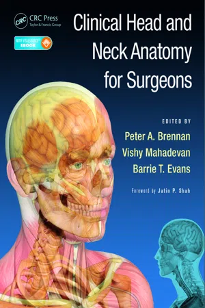 eBook - PDF
- Peter A. Brennan, Vishy Mahadevan, Barrie T. Evans, Peter A. Brennan, Vishy Mahadevan, Barrie T. Evans(Authors)
- 2015(Publication Date)
- CRC Press(Publisher)
77 9 Oral Cavity MADAN G. ETHUNANDAN The Oral Cavity is the entry point to the alimen-tary tract and has an important role to play in the preparation of food, swallowing, speech, breathing and structural support to the face and aesthetics. The shape and size changes with age and is influenced by the eruption and loss of teeth and age-related changes of the surrounding skin, muscles, ligaments and bone. Lined by mucous membrane of differing characters, it is lubricated by major and minor salivary glands. A detailed appreciation of its anatomy is mandatory for safe surgical practice. The Oral Cavity can be divided into two parts: the Oral Cavity proper and the oral vestibule. The vesti-bule is a narrow slit-like space between the lips and cheek on one side and the teeth and gingivae on the other. It communicates with the exterior through the oral fissure and the Oral Cavity proper through the retro-molar region and interdental spaces. The Oral Cavity proper is the area limited by the dental arches and the oropharyngeal isthmus, i.e. the junc-tion of the Oral Cavity with the oropharynx. It is principally occupied by the tongue and is a ‘potential space’ that only exists when the mouth is open. The oropharyngeal isthmus is defined by the junction of the hard and soft palate superiorly, palatoglossal folds laterally and sulcus terminalis delineating the oral from the pharyngeal tongue, inferiorly. This transition from the Oral Cavity to the oropharynx is a watershed in terms of function and innervation. Functionally, the transition is from the voluntary to the reflex (involuntary) phase of swallowing. The motor supply to the musculature of the Oral Cavity, i.e. orbicularis oris and buccinator (buccal and marginal mandibular branches of the facial), tongue (hypoglossal) and mylohyoid (motor root of trigeminal) is under voluntary control. eBook - PDF
- Marjorie Short, Deborah Levin-Goldstein, Marjorie Short(Authors)
- 2021(Publication Date)
- Cengage Learning EMEA(Publisher)
Due to electronic rights, some third party content may be suppressed from the eBook and/or eChapter(s). Editorial review has deemed that any suppressed content does not materially affect the overall learning experience. Cengage Learning reserves the right to remove additional content at any time if subsequent rights restrictions require it. 14 Section I ● Introduction to the Oral Cavity Key Terms Anterior tonsillar pillar Buccal Circumvallate Filiform Foliate Foramen caecum Fordyce granules Fovea palatinus Frenum Fungiform Gingiva Hard palate Incisive papilla Labial Labial commissure Labial tubercle Labiomental groove Linea alba Lingual frenum Mandibular tori Maxillary tuberosity Median sulcus Nasolabial groove Oral Cavity Oral mucosa Oral vestibule Palatine raphe Palatine rugae Palatine torus Philtrum Posterior tonsillar pillar Retromolar area Soft palate Stensen’s papilla Sublingual fold Sublingual caruncle Taste buds Tonsils Uvula Vermilion zone RELATED TERMINOLOGY The names of many Oral Cavity structures, as well as associated and descrip- tive terms, are derived from Latin words. As these words appear repeatedly throughout the readings, it is helpful to become familiar with them. (See Table 2–1.) Copyright 2022 Cengage Learning. All Rights Reserved. May not be copied, scanned, or duplicated, in whole or in part. Due to electronic rights, some third party content may be suppressed from the eBook and/or eChapter(s). Editorial review has deemed that any suppressed content does not materially affect the overall learning experience. Cengage Learning reserves the right to remove additional content at any time if subsequent rights restrictions require it. Chapter 2 ● Structures of the Oral Cavity 15 THE Oral Cavity The term Oral Cavity is used when referring to the inner portion of the mouth. The Oral Cavity extends from the anterior opening at the lips to the oropharynx, or throat posteriorly.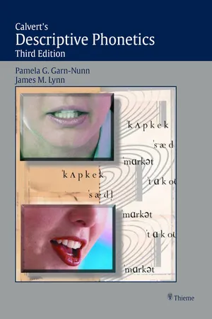 eBook - PDF
- Pamela G. Garn-Nunn, James M. Lynn(Authors)
- 2011(Publication Date)
- Thieme(Publisher)
In producing consonants and vowels, the tongue shape can vary from broad to narrow, flat to curled, and whole tongue positioning to differential positioning of tongue segments. All the vowels and most of the consonants require tongue action. Only /m/, /p/, /b/, /f/, and /v/ do not. M ANDIBLE The mandible, or lower jaw, plays both an active and a passive role in articulation of speech sounds. It forms the base for the tongue and houses the man-dibular teeth. Biologically, its rotary action is necessary for chewing. For speech, the mandible can be raised or lowered by varying degrees, contributing to changes in vowel articulation. N ASAL CAVITY The nasal cavity lies directly superior to the Oral Cavity. Horizon-tally, it extends from the external nares (nostrils) to the posterior pharyngeal wall. Vertically, it is bounded by the base of the skull and the palate and velum. Its pri-mary purpose is to receive inhaled air, filter it, warm it, and direct it toward the trachea (windpipe).With its soft, moist lining, it contributes to the distinctive res-onance characteristics of the cavity. The nasal cavity participates in speech reso-nance with either closure or opening of the velopharyngeal port. It is always open Lingual frenum Middle Back Front/ Blade tip Figure 2–2 Tongue surface landmarks. THE SPEECH PRODUCTION MECHANISM AND PROCESSES 15 anteriorly, at the nostrils, unless you have a cold or other infection. Even if the velopharyngeal port is closed, the nasal cavity resonates the vibrating airstream from the larynx. In this case, the combined resonation of oral and nasal air pro-duces an individual speaker’s distinctive voice quality. Production of /m/, /n/, and / √ / requires closure somewhere in the Oral Cavity combined with opening of the velopharyngeal port (lowering the velum). This allows the nasal cavity to serve as the primary resonator. eBook - PDF
Introducing Linguistics
Theoretical and Applied Approaches
- Joyce Bruhn de Garavito, John W. Schwieter(Authors)
- 2021(Publication Date)
- Cambridge University Press(Publisher)
Speech sounds are generated when air is pushed through the vocal tract. The vocal tract is the area from the nose and the nasal cavity down to the vocal 2 Phonetics Christine Shea and Sarah Ollivia O’Neill 26 Phonetics cords deep in the throat. As the air passes through the vocal tract, it moves through the larynx, the pharynx, the Oral Cavity, and the nasal cavity. 2.2.1 Articulators In spite of the diverse number of sounds that exist in the world’s languages, humans use a small set of articulators, or speech organs (e.g., jaw, tongue, teeth, lips, hard palate) to produce all human speech sounds. The articulators are all located in the upper por- tion of the speech production system, above the larynx. Figure 2.1 shows the parts of the speech production system. Larynx Nasal cavity Palate Pharynx Velum Oral Cavity Tongue Epiglottis Trachea Lungs Lungs Supralaryngeal vocal tract Subglottal Figure 2.1 Speech production system 27 2.2 Speech Production There are two types of articulators: • Active articulators: move during the production of a speech sound to form a closure of some type in the vocal tract. For example, when we articulate the first sound in the word tea, our tongue moves to the roof of our mouth, just behind our teeth. The tongue, in this case, is the active articulator because it moves towards the roof of our mouth, which does not move and is the passive articulator. • Passive articulators: do not (typically) move and are often the point of contact for an active articulator. For example, when English speakers produce the sound ‘g’, the back of the tongue is the active articulator and the velum (see Figure 2.1) is the passive articulator. When we name sounds, we typically refer to the passive articulator. In Table 2.1 you can see some examples of speech sounds produced by different com- binations of passive and active articulators.
Index pages curate the most relevant extracts from our library of academic textbooks. They’ve been created using an in-house natural language model (NLM), each adding context and meaning to key research topics.