![]()
PART 1
FUNDAMENTALS
1 History of MV imaging
Marcel van Herk
2 Detector construction
Daniel Morf
3 Monte Carlo simulation of EPIDs
Jeffrey V. Siebers and I. Antoniu Popescu
![]()
1
History of MV imaging
MARCEL VAN HERK
1.1 Motivation
1.2 Early devices
1.3 Amorphous silicon devices
1.4 Early clinical experience
1.5 Summary
References
The purpose of radiotherapy is to eradicate a tumor with high-energy X-rays while sparing the surrounding organ-at-risks (OAR) as much as possible. Typically, there will always be some discrepancy between the planning situation and treatment. Treatment margins are employed to make the treatment robust against these discrepancies. A misplacement of the tumor with respect to the beams that exceeds the treatment margins can cause a geometric miss. The importance of this effect has been recognized for some time. Marks (1974) compared field placement errors (FPE) with and without immobilization. Byhardt in 1978 reported FPE per site, whereas Brahme in 1984 demonstrated the effect of field shifts on estimated tumor control probability (TCP), and Kinzie et al. (1983) showed that recurrences occurred more frequently when inadequate margins are applied (Marks 1974, Byhardt et al. 1978, Brahme 1984, Kinzie et al. 1983).
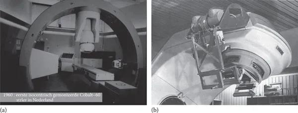
Rabinowitz et al. (1985) related FPE to clinical outcome where major protocol variations were observed frequently and were shown to affect the survival. The only available method to measure FPE at that time was portal film, although early attempts had been made to integrate X-ray on linear accelerators (LINAC) or cobalt sources (Figure 1.1) (Lokkerbol and Smit 1961, Biggs et al. 1985). In addition, a rare report exists of remote visual evaluation of setup based on a fluorescent screen mirror system without a camera (https://www.historad.com/en/#!/en/100-years-radiotherapy-netherlands-cancer-institute-rebuilding/image-guided-rotational-therapy/). These approaches were not widely disseminated, mainly due to a lack of means to digitize the images and insufficient computing power to quickly process and analyze the films. A complicating factor was the difference in perspective between the imaging and treatment beamlines. This made it very difficult to interpret the images, as computed tomography (CT) and digitally reconstructed radiographs (DRRs) were not yet available.
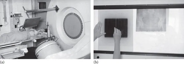
At this time (mid-1980s), X-ray film was used for megavoltage (MV) imaging; however, the film was expensive, cumbersome, and error prone due to the low contrast. Investigators such as Galbraith (1989) pursued optimization of the film-screen cassettes. In 1985, Meertens showed the feasibility of digital image processing to enhance contrast and sharpness of MV films (Meertens 1985b). However, the home-built laser scanner employed in that study took several hours to digitize one film and the limited computing power available (e.g., Digital PDP-11) further increased the time needed to process images, limiting these approaches to research only. Grayscale display devices were not common, so processed data needed to be printed on film for review. The necessary film printers for digital images appeared with CT scanners in the early 1980s. In those days, most common computer equipment (terminals and printers) supported text only. Analysis of portal films was done by manual comparison with 2D kilovoltage (kV) radiographs or digital reconstructed radiographs using, for example, rulers or templates (Figure 1.2). Around this time, it became clear that some digital solution would be highly preferable. The introduction and availability of microprocessors facilitated this technological shift. The Intel 8086 processor, one of the first 16 bits microprocessors with sufficient computing power for image processing, had appeared in 1978 enabling digital image processing on small and low cost computer systems. The time was ripe for electronic portal imaging devices (EPIDs).
Figure 1.1 (a) Early reported integration of imaging on a radiotherapy machine often utilized diagnostic X-rays. An X-ray tube and image intensifier-based X-ray detector are placed perpendicular to the beamline of this early isocentric Co60 machine developed at the Netherlands Cancer Institute. The device can be seen in action here: https://www.youtube.com/v/9_B8DfvBvKY&hl=en_US&feature=player_embedded&version=3. (b) Biggs et al. reported in 1985 the integration of an X-ray imaging chain (film-based) on a linear accelerator. For both systems, image analysis was hampered by the use of a different imaging and treatment beamline.
Figure 1.2 (a) Portal film-screen cassette mounted on a linear accelerator. (b) Portal films were visually compared with reference images (e.g., simulator images) often with the aid of templates or rulers. The hands shown are of Dr. J. Lebesque. (Courtesy of the Harm Meertens, the Netherlands Cancer Institute.)
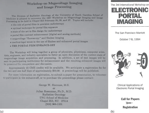
Coinciding with the initiation of the EPID development, a number of workshops were held that eventually evolved into the recurring Electronic Portal Imaging (EPI) conferences. This meeting series is still continuing to date—every other year, mostly in Europe, United States, or Australia. The memorable first workshop (not counted in the series though) was held in Chapel Hill, North Carolina in 1987 (Figure 1.3a), organized by George Sherouse. Norman Bailey and Arthur Boyer organized the subsequent meeting in Las Vegas in 1989. At this meeting, physicist Shlomo Shalev organized an image quality competition and enhance-off. The Las Vegas phantom was introduced there, which is still commonly used, and was later combined with commercial analysis software, called PIPS (Rajapakshe 1996). Subsequent meetings were held in Newport Beach, San Francisco (Figure 1.3b), Amsterdam, Houston, Brussels, Vancouver, Brighton, Melbourne, Leuven, Sydney, Aarhus, and St. Louis. In its current form, the workshop is now called Electronic Patient Imaging and covers all forms of in-room patient imaging.
Over the years, the meetings changed focus from detector development to image analysis, clinical application, setup error correction protocols, treatment margins, use of implanted markers, portal dosimetry, tracking, and magnetic resonance (MR) guidance. Most important, the meetings always have had a multidisciplinary character, with physicists, physicians, and radiographers in the organizing committee, presenting and in attendance. Noteworthy is the 2006 meeting chaired by Kay Hatherley, the radiographer greatly responsible for the early introduction of EPID in Australia. She opened the meeting giving a welcome speech from inside the shark tank of the Melbourne aquarium.
Figure 1.3 (a) Invitation for the first workshop on Electronic Portal Imaging in Chapel Hill that brought together about 25 pioneers in the field. (b) The 3rd workshop was visited by around 200 people. The logo, designed by Shlomo Shalev, very aptly visualized the goal of electronic portal imaging: hitting the target. (Courtesy of Marcel van Herk.)
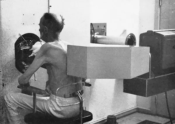
The most obvious design for an EPID is to combine a fluorescent screen with a video camera similar to the devices used for low-energy X-rays. For compactness and to keep the camera out of the high-energy radiation beam, a tilted mirror is typically placed below the fluorescent screen, while the camera is looking in a direction parallel to the screen. One of the earliest reported systems using this technology is by Benner et al. (1962) (Figure 1.4).
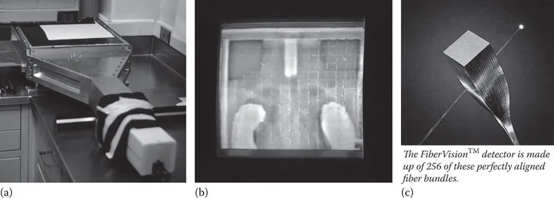
However, video systems have several drawbacks. The relatively low light output of the fluorescent screen, in combination with the poor light collection due to the lens aperture leads to a low detective quantum efficiency (DQE) of well below 1%, while the devices are very bulky. Larger lens apertures would lead to poor resolution due to spherical aberrations. Yet, because of their simple design, it was these kinds of systems that were first adopted as commercial solutions for companies such as Elekta and Siemens (Benner et al. 1962, Baily 1980, Leong 1986, Visser 1990). Add-on systems using this technology were developed by Eliav and Cablon (de Boer 2000, Odero 2009). The problem of low light collection was addressed by using amplified cameras and later cooled CCD cameras (Franken 2004). Cooled cameras also alleviated issues with radiation damage to the CCD chip. Cablon produces such a system, with software developed in collaboration with the Erasmus Medical Center in Rotterdam, the Netherlands, although Elekta also produces a CCD camera device intended for lower income markets. To address the bulkiness of the device, Wong et al. (then in St. Louis) proposed the use of optical fiber coupling instead of a mirror and started a company called FiberVision (Figure 1.4) (Wong et al. 1990). However, interestingly the fiber technology did not overcome low DQE because the light-acceptance angle of the tapered fibers was similar to a lens system (Boyer et al. 1992) and the difficulty of manufacturing and large weight made this approach impractical (Figure 1.5).
Figure 1.4 Possibly the first reported EPID. The basic design and mechanical construction is very similar to later screen/mirror devices, although the video camera (gray box on the right) is very bulky. Here the device is used in the fixed beamline of a 30 MV Betatron. (Courtesy of Radium Hospital, Oslo, Norway, 16 December 1961; From Benner S. et al., Phys Med Biol., 7, 29–34, 1962.)
Figure 1.5 The FiberVision EPID is a screen/video camera system, where the mirror has been replaced by a tapered bundle of optical fibers. Images (a) and (b) were made by Marcel van Herk during a visit to St. Louis in 1988, image (c) was provided by John Wong.
The pursuit of compact solutions led to several subsequent innovations. The first truly compact system was based on a scanning line array of diodes and was introduced by Lam et al. (1986). However, due to the scanning approach and the low DQE of the applied diodes, this device had poorer quantum efficiency than video-based systems (~0.01%). Morton and Swindell combined photocells with heavy tungsten alloy scintillators bringing the DQE of such a scanning device to practical levels of about 0.5% (Morton et al. 1991), and first clinical use of the device was reported, in particular in breast (Gildersleve et al. 1994).
However, low sampling efficiency still limited the DQE, as the detectors are located only for a very short time at any array location. Ioniz...