Ultrasound is, per definition, the sound of a frequency higher than the hearing limit of the human ear, i.e., above 16 to 20 kHz. Bat’s definition of ultrasound would be different. In medical diagnostics, one normally uses ultrasound waves of frequencies between 2 and 10 MHz. Basic physical principles are equally valid for audible sound as for ultrasound, only at different scales. Ultrasound is a mechanical wave, i.e., it consists of mechanical vibrations of medium particles through which it propagates. In soft tissues, the medium particles vibrate along the direction of wave propagation creating their densifications and rarefactions in space. Such a wave is called a longitudinal wave. The particles (molecules) oscillate around their (stochastic) balance positions with no net flow of matter, however, the energy flows. At very high energy densities some net flow can be induced, but this does not apply to energies of ultrasound used in diagnostics. Other types of waves like transversal and Raileigh cannot propagate to any appreciable distance in soft tissues. Ultrasound waves are characterized by parameters like frequency, wavelength, propagation speed, intensity, and pressure. Frequency is expressed in hertz (Hz), i.e., cycles per second. The physical dimension is 1/s; 1 Hz = 1 c/s, 1 kHz = 1000 c/s, and 1 MHz = 1 million c/s. The frequency used in diagnostics largely influences their properties. Wavelength is the distance between the same phases of compression of the medium in two consecutive cycles in space and is measured in meters or its subunits like millimeters. The propagation speed depends mainly on the media (tissue) properties through which the wave propagates and is related with frequency and wavelength as follows:
where c = propagation velocity in meters per second (m/s), f = frequency in hertz (Hz), and λ = wavelength in meters (m).
Strictly speaking, sounds of different frequencies travel at different velocities (frequency velocity dispersion), but these effects are negligible at frequencies, intensities, and circumstances of medical echography. The average propagation speed in soft tissues is 1540 m/s. The speed depends on both density and elastic properties of tissues.
When traveling through tissues, ultrasound causes a variation of the total pressure, i.e., the sonic pressure oscillations are superimposed upon the static pressure (atmospheric pressure). The measure of energy density flowing through a unity area in one unit of time is called intensity and is expressed in watts per square meter or centimeter (W/cm2 or W/m2).
When speaking about ultrasound intensity in an ultrasound beam, it is not sufficient to simply state the intensity, one must as well state “where” and “when”; i.e., at what position in space and within which interval. Simply stating the intensity applies only to a plane wave in a nonattenuating medium. In echography, one uses pulsed ultrasound and focused beams in attenuating media. After we discuss characteristics of ultrasound beams, we shall turn back to definitions of ultrasound intensities in the beam.
Table 1
ORIENTATIONAL VALUES OF PROPAGATION SPEEDS IN SOFT TISSUES1,2 Tissue | Speed (m/s) |
Muscle | 1570 |
Kidney | 1560 |
Liver | 1560 |
Brain | 1510 |
Fat | 1440 |
Note: The speed values are for orientation only. Inspection of the available literature shows fairly divergent results. There is a consensus that the average speed for soft tissues should be taken as 1540 m/s.
Likewise, when speaking about frequency, one can define a single frequency only as a continuous wave. When speaking about pulses of some frequency, we usually think of frequency obtained by counting the number of zero-level crossings during the pulse. However, a pulse by physical laws contains a mixture of frequencies which we call the frequency spectrum of the pulse, and which is broader — the shorter the pulse. This fact must be born in mind in the practical use of ultrasound echography because, as we shall show, one uses different frequencies for different purposes, so it may sometimes be useful to bear in mind that when using, e.g., a probe declared 3.5 MHz central frequency, one actually transmits and receives a spectrum of frequencies, which may range from below 1 MHz to more than 5 MHz.
B. Ultrasound Propagation in Tissues
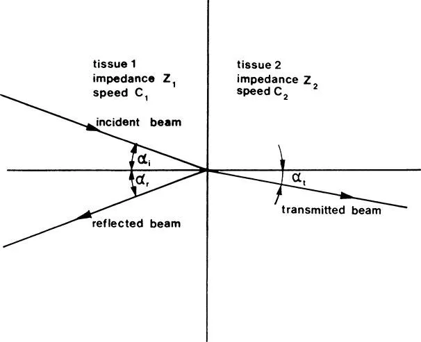
Human tissues are not homogenous in respect to ultrasound wave propagation, so that in transversing the tissues, ultrasound waves are being reflected, refracted, scattered, and absorbed. Angle of refraction depends on the respective velocities in the tissues.
The amount of reflected energy depends on characteristic acoustic impedances and angle of incidence upon the boundary of tissues. The tissues we are speaking about are different tissues in terms of elasticity and other physical properties, but can be parts of the same biological tissue. Characteristic acoustic impedance is a property of a medium and is defined as the ratio of the momentary acoustic pressure and particle velocity induced by this pressure.
In different tissues, ultrasound velocities and impedance are generally different (see Table 1). Differences between different soft tissues are much smaller than between soft tissues and bones, respectively, gas. This fact has important consequences in the use of ultrasound in medicine.
1. Refraction and Reflection of Ultrasound Waves
In discussing reflection and refraction of ultrasound, we shall take the simplest case — the plane wave — which, although an idealization, will give us a good idea of what happens when ultrasound waves transverse a boundary of media which is much larger than the wavelength.
As illustrated in Figure 1, a part of the energy is reflected and some of the energy is transmitted across the boundary. The angle of refraction depends on the ratio of propagation speeds in the two respective media, as described by Equation 1, while the refraction of energy reflected depends on the difference of characteristic acoustic impedances, as well as on the angle of incidence. (Equation 2a). If the speeds of ultrasound in the first and the second medium are c1 and c2, respectively, and the incidence angle is αi and the transmission angle is αt, then the refraction angle can be calculated from Equation 1:
Equation 2 describes the ratio of the amplitudes of the reflected and the incident wave:
| (2a) |
If the incidence is perpendicular to the media boundary, Equation 2a degenerates to
In many practical cases when we consider ultrasound waves, which are reflected back to the transmitting probe, Equation 2b gives a fairly accurate picture of what goes on.
Intensity of ultrasound is proportional to the square of its pressure so that the ratio of the reflected to the incident intensity is at 90°
| (3) |
If the characteristic acoustic impedances are very different like in the case of soft tissue and gas (e.g., in lungs), from the above equation one can see that virtually all ultrasound energy will be reflected. A consequence of this is that organs containing gas, such as the lungs, cannot be examined by ultrasound, and gas in the stomach and intestines will present serious problems. Another consequence is that some sort of contact gel, oil, or other coupling agent should be applied between the ultrasound transmitter and the body to avoid air bubbles being trapped between the probe and the skin and to allow ultrasound to enter the body. It is interesting to see that ultrasound will be reflected irrespective of whether it enters from a lower or a higher impedance. The absolute value of reflected wave is the same for the same difference in characteristic acoustic impedances. On the other hand, the sign changes indicate a difference in phase.
The energy that is not reflected will be transmitted across the impedance boundary. Thus, in many cases it is the characteristic acoustic impedance that is decisive for the intensity of an echo. Sometimes the acoustic impedance is mixed up with the density by a vague analogy with X-ray imaging. This is wrong because the characteristic impedance depends on the density and propagation speed (c):
and not only on the density.
So ...