Visually Memorable Neuroanatomy for Beginners takes a close look at the anatomy of the human brain and teaches readers to identify and examine its structures in a relatable way. Unlike large textbooks that deliver a superficial overview of the subject, this book explores the anatomy and physiology of the brain using mnemonic techniques and informative comic figures that present brain regions at an introductory level, allowing readers to easily identify different parts of the brain. This volume is appropriate for undergraduate and graduate students, postdoctoral fellows, and researchers in the medicine, health sciences, and biological sciences.
Beginning with the morphology of the brain and spinal cord, this book then explores the somatic nerve and autonomic nerve, the cranial nerve and spinal nerve, the function of the brain, and concludes with the development of the nervous system.
Features simplified illustrations for understanding the complicated neuroanatomy structures
Introduces memorizing tips (mnemonics) to help students learn
Describes how best to identify structures in cadaver specimens
Includes comic-style figures to make neuroanatomy approachable for newcomers
Häufig gestellte Fragen
Wie kann ich mein Abo kündigen?
Gehe einfach zum Kontobereich in den Einstellungen und klicke auf „Abo kündigen“ – ganz einfach. Nachdem du gekündigt hast, bleibt deine Mitgliedschaft für den verbleibenden Abozeitraum, den du bereits bezahlt hast, aktiv. Mehr Informationen hier.
(Wie) Kann ich Bücher herunterladen?
Derzeit stehen all unsere auf Mobilgeräte reagierenden ePub-Bücher zum Download über die App zur Verfügung. Die meisten unserer PDFs stehen ebenfalls zum Download bereit; wir arbeiten daran, auch die übrigen PDFs zum Download anzubieten, bei denen dies aktuell noch nicht möglich ist. Weitere Informationen hier.
Welcher Unterschied besteht bei den Preisen zwischen den Aboplänen?
Mit beiden Aboplänen erhältst du vollen Zugang zur Bibliothek und allen Funktionen von Perlego. Die einzigen Unterschiede bestehen im Preis und dem Abozeitraum: Mit dem Jahresabo sparst du auf 12 Monate gerechnet im Vergleich zum Monatsabo rund 30 %.
Was ist Perlego?
Wir sind ein Online-Abodienst für Lehrbücher, bei dem du für weniger als den Preis eines einzelnen Buches pro Monat Zugang zu einer ganzen Online-Bibliothek erhältst. Mit über 1 Million Büchern zu über 1.000 verschiedenen Themen haben wir bestimmt alles, was du brauchst! Weitere Informationen hier.
Unterstützt Perlego Text-zu-Sprache?
Achte auf das Symbol zum Vorlesen in deinem nächsten Buch, um zu sehen, ob du es dir auch anhören kannst. Bei diesem Tool wird dir Text laut vorgelesen, wobei der Text beim Vorlesen auch grafisch hervorgehoben wird. Du kannst das Vorlesen jederzeit anhalten, beschleunigen und verlangsamen. Weitere Informationen hier.
Ist Visually Memorable Neuroanatomy for Beginners als Online-PDF/ePub verfügbar?
Ja, du hast Zugang zu Visually Memorable Neuroanatomy for Beginners von Min Suk Chung,Beom Sun Chung im PDF- und/oder ePub-Format sowie zu anderen beliebten Büchern aus Ciencias biológicas & Neurociencia. Aus unserem Katalog stehen dir über 1 Million Bücher zur Verfügung.
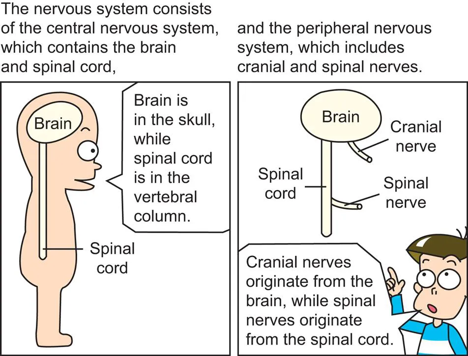
The nervous system consists of the central nervous system (brain, spinal cord) and the peripheral nervous system (cranial nerve, spinal nerve). This chapter explores the gross morphology of the central nervous system, in preparation for further study of the neuronal connections. This chapter details the blood supply and cerebrospinal fluid flow of the central nervous system. Then it sequentially describes the morphology of the cerebral hemisphere, limbic system, basal nuclei, diencephalon, cerebellum, brainstem, and spinal cord. It is necessary to correlate external features of the structures to their sectional planes. It is suggested to review this chapter with other learning materials such as realistic neuroanatomy atlases, plastic specimens, three-dimensional computer models, and cadavers.
The nervous system is a complex network of nerves that carry impulses between the brain, spinal cord, and various parts of the body.
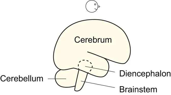
Fig. 1.2 Brain components.
When the brain is viewed laterally, its three components are identifiable: cerebrum, cerebellum, and brainstem. The diencephalon is hidden by the cerebrum (cerebral hemisphere) (Fig. 1.11).
The blood supply, the cerebrospinal fluid flow
Fig. 1.3 Cerebral arteries, cerebellar arteries.
The basilar artery arises from the confluence of the two vertebral arteries at the junction between the pons and medulla oblongata. Branches of the basilar artery, named pontine arteries, feed the pons (Fig. 1.51).
The posterior inferior cerebellar artery branches off from the vertebral artery, while the anterior inferior cerebellar artery and superior cerebellar artery branch off from the basilar artery. This is because the basilar artery is on the pons (Fig. 1.54) which is right in front of the cerebellum (Figs. 1.44, 5.6).
There are three cerebral arteries as well as three cerebellar arteries on each side. The posterior cerebral artery is a terminal division of the basilar artery, while the middle and anterior cerebral arteries are two divisions of the internal carotid artery.
The “posterior” cerebral arteries and internal carotid arteries are connected by the “posterior” communicating arteries, while the bilateral “anterior” cerebral arteries are connected by the “anterior” communicating artery.
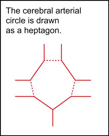
Fig. 1.4
The cerebral arterial circle (circle of Willis) is composed of the posterior cerebral arteries, posterior communicating arteries, anterior cerebral arteries, and anterior communicating artery (Exactly, a short segment of internal carotid artery is included.) (Fig. 1.3). The circle is an anastomosis that guarantees blood supply to the cerebrum.
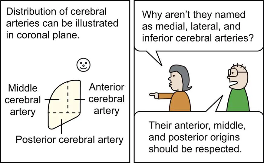
Fig. 1.5 Anterior, middle, and posterior cerebral arteries.
The anterior cerebral artery passes along the medial surface of the cerebral hemisphere anteriorly, superiorly, and then posteriorly. The middle cerebral artery emerges from the lateral sulcus to take charge of most of the lateral surface of the cerebral hemisphere (Fig. 1.23). The posterior cerebral artery passes posteriorly along its inferomedial surface (Figs. 1.6, 1.30).
Fig. 1.6
The anterior and middle cerebral arteries supply blood to the cerebral hemisphere above a certain horizontal plane; the posterior cerebral artery feeds the cerebral hemisphere below the plane (Fig. 1.5). In other words, the horizontal plane is a territorial border between the internal carotid artery and the vertebral artery (Fig. 1.3).
Fig. 1.7
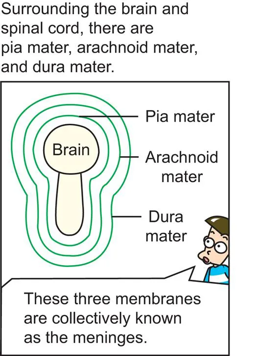
The meninges which cover the brain and spinal cord are like PAD. The meninges are composed of Pia, Arachnoid, and Dura maters.
Fig. 1.8 Meninges of spinal cord.
The pia mater is adhesive to the brain (Figs. 1.14, 1.31) and spinal cord, the arachnoid (spider’s) mater is entangled like a spider’s web, and the dura mater is thick (Fig. 1.17).
Fig. 1.9
The DURA mater reminds us of a DURAble mother.
The subarachnoid space of brain and spinal cord is an actual space containing cerebrospinal fluid ...