Visually Memorable Neuroanatomy for Beginners takes a close look at the anatomy of the human brain and teaches readers to identify and examine its structures in a relatable way. Unlike large textbooks that deliver a superficial overview of the subject, this book explores the anatomy and physiology of the brain using mnemonic techniques and informative comic figures that present brain regions at an introductory level, allowing readers to easily identify different parts of the brain. This volume is appropriate for undergraduate and graduate students, postdoctoral fellows, and researchers in the medicine, health sciences, and biological sciences.
Beginning with the morphology of the brain and spinal cord, this book then explores the somatic nerve and autonomic nerve, the cranial nerve and spinal nerve, the function of the brain, and concludes with the development of the nervous system.
Features simplified illustrations for understanding the complicated neuroanatomy structures
Introduces memorizing tips (mnemonics) to help students learn
Describes how best to identify structures in cadaver specimens
Includes comic-style figures to make neuroanatomy approachable for newcomers
Foire aux questions
Comment puis-je résilier mon abonnement ?
Il vous suffit de vous rendre dans la section compte dans paramètres et de cliquer sur « Résilier l’abonnement ». C’est aussi simple que cela ! Une fois que vous aurez résilié votre abonnement, il restera actif pour le reste de la période pour laquelle vous avez payé. Découvrez-en plus ici.
Puis-je / comment puis-je télécharger des livres ?
Pour le moment, tous nos livres en format ePub adaptés aux mobiles peuvent être téléchargés via l’application. La plupart de nos PDF sont également disponibles en téléchargement et les autres seront téléchargeables très prochainement. Découvrez-en plus ici.
Quelle est la différence entre les formules tarifaires ?
Les deux abonnements vous donnent un accès complet à la bibliothèque et à toutes les fonctionnalités de Perlego. Les seules différences sont les tarifs ainsi que la période d’abonnement : avec l’abonnement annuel, vous économiserez environ 30 % par rapport à 12 mois d’abonnement mensuel.
Qu’est-ce que Perlego ?
Nous sommes un service d’abonnement à des ouvrages universitaires en ligne, où vous pouvez accéder à toute une bibliothèque pour un prix inférieur à celui d’un seul livre par mois. Avec plus d’un million de livres sur plus de 1 000 sujets, nous avons ce qu’il vous faut ! Découvrez-en plus ici.
Prenez-vous en charge la synthèse vocale ?
Recherchez le symbole Écouter sur votre prochain livre pour voir si vous pouvez l’écouter. L’outil Écouter lit le texte à haute voix pour vous, en surlignant le passage qui est en cours de lecture. Vous pouvez le mettre sur pause, l’accélérer ou le ralentir. Découvrez-en plus ici.
Est-ce que Visually Memorable Neuroanatomy for Beginners est un PDF/ePUB en ligne ?
Oui, vous pouvez accéder à Visually Memorable Neuroanatomy for Beginners par Min Suk Chung,Beom Sun Chung en format PDF et/ou ePUB ainsi qu’à d’autres livres populaires dans Ciencias biológicas et Neurociencia. Nous disposons de plus d’un million d’ouvrages à découvrir dans notre catalogue.
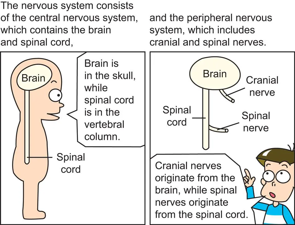
The nervous system consists of the central nervous system (brain, spinal cord) and the peripheral nervous system (cranial nerve, spinal nerve). This chapter explores the gross morphology of the central nervous system, in preparation for further study of the neuronal connections. This chapter details the blood supply and cerebrospinal fluid flow of the central nervous system. Then it sequentially describes the morphology of the cerebral hemisphere, limbic system, basal nuclei, diencephalon, cerebellum, brainstem, and spinal cord. It is necessary to correlate external features of the structures to their sectional planes. It is suggested to review this chapter with other learning materials such as realistic neuroanatomy atlases, plastic specimens, three-dimensional computer models, and cadavers.
The nervous system is a complex network of nerves that carry impulses between the brain, spinal cord, and various parts of the body.
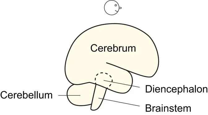
Fig. 1.2 Brain components.
When the brain is viewed laterally, its three components are identifiable: cerebrum, cerebellum, and brainstem. The diencephalon is hidden by the cerebrum (cerebral hemisphere) (Fig. 1.11).
The blood supply, the cerebrospinal fluid flow
Fig. 1.3 Cerebral arteries, cerebellar arteries.
The basilar artery arises from the confluence of the two vertebral arteries at the junction between the pons and medulla oblongata. Branches of the basilar artery, named pontine arteries, feed the pons (Fig. 1.51).
The posterior inferior cerebellar artery branches off from the vertebral artery, while the anterior inferior cerebellar artery and superior cerebellar artery branch off from the basilar artery. This is because the basilar artery is on the pons (Fig. 1.54) which is right in front of the cerebellum (Figs. 1.44, 5.6).
There are three cerebral arteries as well as three cerebellar arteries on each side. The posterior cerebral artery is a terminal division of the basilar artery, while the middle and anterior cerebral arteries are two divisions of the internal carotid artery.
The “posterior” cerebral arteries and internal carotid arteries are connected by the “posterior” communicating arteries, while the bilateral “anterior” cerebral arteries are connected by the “anterior” communicating artery.
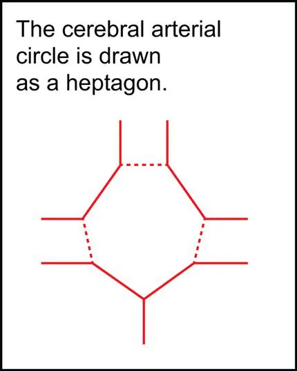
Fig. 1.4
The cerebral arterial circle (circle of Willis) is composed of the posterior cerebral arteries, posterior communicating arteries, anterior cerebral arteries, and anterior communicating artery (Exactly, a short segment of internal carotid artery is included.) (Fig. 1.3). The circle is an anastomosis that guarantees blood supply to the cerebrum.
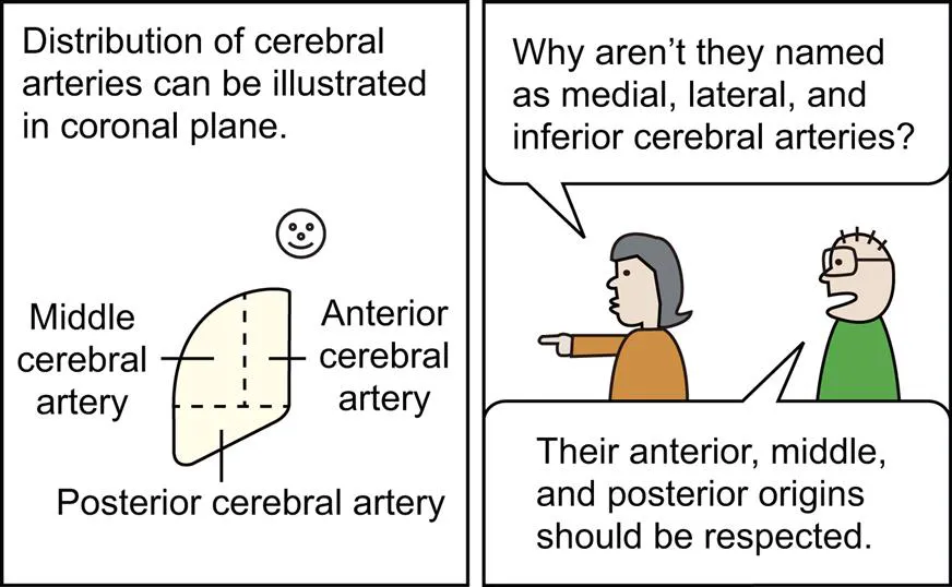
Fig. 1.5 Anterior, middle, and posterior cerebral arteries.
The anterior cerebral artery passes along the medial surface of the cerebral hemisphere anteriorly, superiorly, and then posteriorly. The middle cerebral artery emerges from the lateral sulcus to take charge of most of the lateral surface of the cerebral hemisphere (Fig. 1.23). The posterior cerebral artery passes posteriorly along its inferomedial surface (Figs. 1.6, 1.30).
Fig. 1.6
The anterior and middle cerebral arteries supply blood to the cerebral hemisphere above a certain horizontal plane; the posterior cerebral artery feeds the cerebral hemisphere below the plane (Fig. 1.5). In other words, the horizontal plane is a territorial border between the internal carotid artery and the vertebral artery (Fig. 1.3).
Fig. 1.7
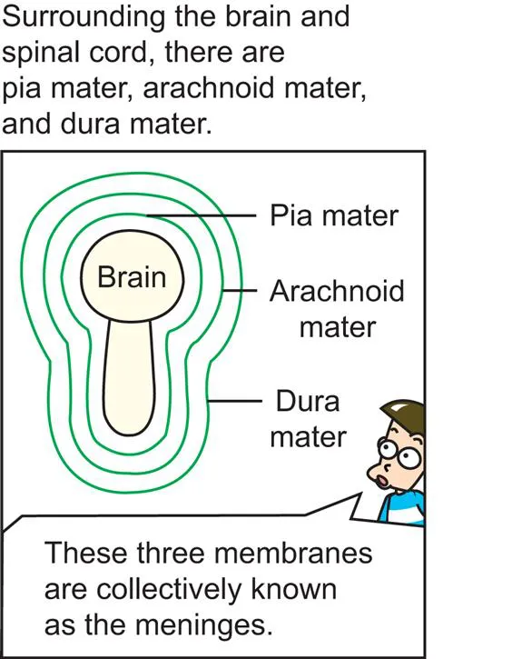
The meninges which cover the brain and spinal cord are like PAD. The meninges are composed of Pia, Arachnoid, and Dura maters.
Fig. 1.8 Meninges of spinal cord.
The pia mater is adhesive to the brain (Figs. 1.14, 1.31) and spinal cord, the arachnoid (spider’s) mater is entangled like a spider’s web, and the dura mater is thick (Fig. 1.17).
Fig. 1.9
The DURA mater reminds us of a DURAble mother.
The subarachnoid space of brain and spinal cord is an actual space containing cerebrospinal fluid ...