Medical Sciences at a Glance
eBook - ePub

Medical Sciences at a Glance

  1. English
  2. ePUB (mobile friendly)
  3. Available on iOS & Android
eBook - ePub

Medical Sciences at a Glance

Book details
Book preview
Table of contents
Citations

About This Book

Medical Sciences at a Glance

The market-leading at a Glance series is used world-wide by medical students, residents, junior doctors and health professionals for its concise and clear approach and superb illustrations.

Each topic is presented in a double-page spread with clear, easy-to-follow diagrams, supported by succinct explanatory text.

Covering the whole medical curriculum, the series now includes workbooks and case books, which allow you to put your knowledge to the test.

Everything you need to know about Medical Sciencesā€¦ at a Glance!

The definitive companion for medical science study and revision

Medical Sciences at a Glance consolidates the scientific knowledge a student needs to provide a solid framework of key facts to build on. Concise, easy to follow, written specifically for medical students, and conveying key concepts through the unique at a Glance style, Medical Sciences at a Glance also demonstrates vital links between different topics and across systems. It is the perfect resource for bridging the gap between A-Level and university, studying a new topic, revising for exams, or refreshing knowledge while on placement.

Key features:

  • Fully cross-referenced to Medicine at a Glance ā€“ together they cover the core concepts of an entire medical degree
  • Highlights key points and their clinical relevance for quick revision and retention of what's most important
  • Brings together all the scientific content on a medical course in one easy-to-read, highly-illustrated title

Medical Sciences at a Glance provides the vital scientific grounding needed to Ā­succeed at medical school.

All content reviewed by students for students

Wiley-Blackwell Medical Education books are designed exactly for their intended audience. All our books are developed in collaboration with students, which means our books are always published with you, the student, in mind.

Frequently asked questions

Simply head over to the account section in settings and click on ā€œCancel Subscriptionā€ - itā€™s as simple as that. After you cancel, your membership will stay active for the remainder of the time youā€™ve paid for. Learn more here.
At the moment all of our mobile-responsive ePub books are available to download via the app. Most of our PDFs are also available to download and we're working on making the final remaining ones downloadable now. Learn more here.
Both plans give you full access to the library and all of Perlegoā€™s features. The only differences are the price and subscription period: With the annual plan youā€™ll save around 30% compared to 12 months on the monthly plan.
We are an online textbook subscription service, where you can get access to an entire online library for less than the price of a single book per month. With over 1 million books across 1000+ topics, weā€™ve got you covered! Learn more here.
Look out for the read-aloud symbol on your next book to see if you can listen to it. The read-aloud tool reads text aloud for you, highlighting the text as it is being read. You can pause it, speed it up and slow it down. Learn more here.
Yes, you can access Medical Sciences at a Glance by Michael D. Randall in PDF and/or ePUB format, as well as other popular books in Medicine & Medical Theory, Practice & Reference. We have over one million books available in our catalogue for you to explore.

Information

Year
2013
ISBN
9781118360965
1
Cells
Sebastiaan Winkler
c1-fig-5001
A cell is the smallest functional and structural unit capable of replicating itself. As such, a cell is considered the basic unit of life. The boundary of a cell is the plasma membrane (see Chapter 2), while the cytoskeleton provides structural support (Figure 1.1). Cells of the human body have a characteristic nuclear compartment, which contains the genetic material, and are thus classified as eukaryotic cells as opposed to prokaryotic bacterial cells, which do not contain a nucleus. Cellular structures with specific functions, organelles, are discussed in Chapter 3.

Tissues

There are more than 200 different types of cells in the human body. These types are highly specialised (differentiated) to carry out specific functions. Groups of cells carrying out a similar function and forming a structure are called tissue. Different tissues combine to form organs. There are four main types of tissue.
  • Epithelial tissue is found at the boundaries of structures in the body. The basal (basolateral) side faces the underlying tissue, which provides nutrients and support, while the apical side is exposed to a different environment. For instance, the apical side of intestinal epithelium faces the inside of the intestine and is exposed to constituents from (digested) food; the apical side of stomach epithelium is exposed to the acidic environment inside the stomach; lung and skin are exposed to air. Secreting glands are also formed by epithelial tissue. There are distinct types of epithelial structures:
    • stratified epithelium: formed by layers of epithelial cells;
    • simple epithelium: formed by a single layer of cells;
    • squamous epithelium: formed by cells which are wider than tall;
    • columnar epithelium: formed by cells which are taller than wide.
  • Stratified squamous epithelium can be keratinised, forming a hard and dry layer as found in the skin, nails and hair, or non-keratinised, which is found in soft tissue such as the inside of the mouth. An example of simple columnar epithelium is the lining of the stomach.
  • Connective tissue provides structure and rigidity. It is characterised by a large space between cells, which is filled with fibrous material that is part of the extracellular matrix. Fibroblasts are the most common cell type in connective tissue. Adipose tissue stores energy in the form of fat, but is also important for the protection and insulation of organs and is now recognised as having an endocrine role. Other types of connective tissue include blood, cartilage and bone.
  • Muscle cells (myocytes) are characterised by their ability to contract when they receive appropriate signals. There are three different types of muscle tissue: skeletal muscle is directly attached to bones; cardiac muscle is the muscle of the heart; smooth muscle lines blood vessels and organs of the body.
  • Nerve cells can sense stimuli and transfer electrical signals to chemical signals, which are sensed by surrounding cells. Nerve cells can have long extensions that are involved in the sensing and transmittance of signals (Chapters 18 and 19).
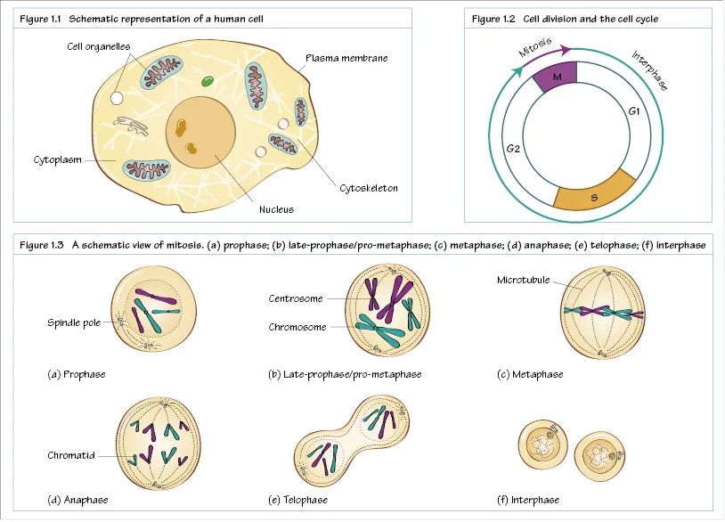
Cell division

The mass and volume of tissues can increase by cell growth, the increase of cell mass and volume by taking up nutrients and synthesising new cell structures, and cell proliferation, the increase of cell numbers by cell division. There are two types of cell division.
  • Mitosis, in which each daughter cell acquires the same amount of genetic material as the parental cell.
  • Meiosis, in which each daughter cell acquires half the amount of genetic material of the parental cell, only occurs in specialised germ tissue.
When cells receive signals to divide they progress through the cell cycle (Figure 1.2). In adult tissue, most cells reside in the interphase. The interphase can be further divided into three phases. In the G1 phase (first gap phase), cells prepare for the duplication of the genetic material. When cells start duplicating their DNA, they progress through the S phase (synthesis phase). Following the G2 phase (second gap), cells undergo mitosis (M phase). Compared with the interphase, the mitotic phase is very short. Mitosis (Figure 1.3) can be separated into the following distinct stages.
  • Prophase: the DNA/chromatin condenses and the characteristic chromosomes become visible. The mitotic spindle starts to form outside the nucleus.
  • In the late prophase/pro-metaphase, the nuclear membrane starts to degrade and the mitotic spindle body moves into the nuclear region. Regions in the chromosomes, centrosomes, are attached to the mitotic spindle.
  • Metaphase: the chromosomes are aligned in the centre plane between the spindle poles.
  • Anaphase: the chromatids of the sister chromosomes start to separate while the spindle poles move further apart.
  • Telophase: the chromatids reach the spindle poles, which starts to disappear. The chromatids decondense and a nuclear membrane is formed around the chromatids.
During mitosis, the division of the cytoplasm between the two daughter cells is called cytokinesis. Cytokinesis starts during prophase, but is not completed until after the end of telophase when the two daughter cells have formed.

Stem cells

Stem cells are undifferentiated cells with two characteristics.
  • Self-renewal: the ability to repeatedly divide while maintaining an undifferentiated state. Stem cells undergo asymmetric cell division: one of the daughter cells retains stem cell characteristics while the other daughter cell undergoes differentiation.
  • The ability to differentiate into specialised cell types.
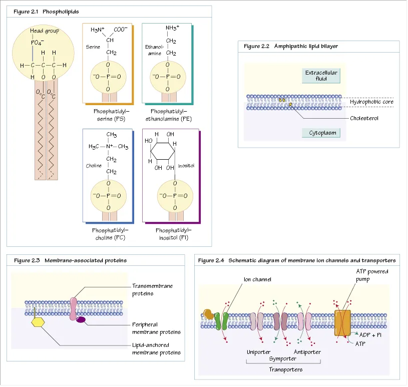
Stem cells are pluripotent if they retain the ability to differentiate into all cell types and tissues. For example, embryonic stem cells are pluripotent stem cells derived from early-stage embryos. Adult stem cells are not pluripotent, but are more specialised and can only form one or several tissues. For example, adult haematological stem cells isolated from the bone marrow can differentiate into the various cell types found in blood. Other adult stem cells are more specialised, e.g. skin stem cells found in the epidermis, which can only differentiate into skin cells. Induced pluripotent stem cells are derived from differentiated tissue that is genetically reprogrammed to return to their undifferentiated state.
2
Organisation of cell membranes
Sebastiaan Winkler
c2-fig-5001

Cell membranes

Cell membranes are large cellular structures that constitute the boundary of a cell or a cell organelle. In contrast to proteins or nucleic acids, membranes are not made up of polymers, but a large number of diverse, relatively small molecules that form non-covalent interactions.

Phospholipids

The principal building blocks of cell membranes are a variety of compounds that are collectively known as phospholipids. Phospholipids are composed of three different parts: the backbone, a polar head group and a fatty acid chain (Figure 2.1). Different combinations of backbone, head groups and fatty acids result in a wide variety of phospholipids. In mammalian cells, glycerol is the backbone of the most abundant class of phospholipids, termed phosphoglycerides, although sphingolipids are also abundant. Backbone moieties have three hydroxyl groups, which are available for the conjugation of the polar head group, and two fatty acid chains. The polar head group is linked to the backbone via a phosphoester bond. In addition, two fatty acids are conjugated to the remaining hydroxyl groups of the backbone. The fatty acids can be broadly divided into two groups. The saturated fatty acids do not contain double bonds and always contains an even number of carbon atoms (usually 16ā€“20). Due to the free rotation of the single carbonā€“carbon bonds, these lipid chains can adopt linear configurations. By contrast, the unsaturated fatty acids contain one or more double bonds. When the groups that lie on either side of the double bond are on opposite sides of the double bond (trans configuration) they are called trans fatty acids (Chapter 5). Like unsaturated fatty acids, trans fatty acids can adopt (near) linear configurations. However, when the groups next to the double bond are on the same side of the double bond (cis configuration) the fatty acids adopt very different shapes. The cis fatty acids have characteristic bends and cannot adopt linear configurations (Chapter 5).
A characteristic feature of the phospholipids is that they are amphipathic: they are both hydrophilic (due to the polar head group) and lipophilic (due to their fatty acid chain).

The lipid bilayer

Due to their amphipathic nature, phospholipids spontaneously organise in such a way that they form two sheets, with the polar head groups facing the aqueous exterior and the fatty acid chains forming a hydrophobic core (Figure 2.2). This structure is termed the lipid bilayer. Phospholipids can diffuse freely in the lipid bilayer, which behaves as a two-dimensional fluid. The lipid bilayer is asymmetrical, because phospholipids on one side of the bilayer do not freely flip to the other side. Thus, on the outside of cell membranes, phospholipids with PC head groups are enriched, while the cytoplasmic side is enriched with PE and PS lipids. The composition of the lipid bilayer is not homogeneous. The properties of cell membranes is influenced by the properties of the locally enriched phospholipids, e.g. the length of fatty acid chains influences the thickness of the lipid bilayer, and the presence of phospholipids containing cis fatty acids reduces the density of phospholipids in the lipid bilayer.
Sterols are another class of lipid components. Cholesterol is the most abundant sterol found in cell membranes (Chapter 5). Cholesterol impacts on the fluidity of th...

Table of contents

  1. Cover
  2. Website ad
  3. Title page
  4. Copyright page
  5. Preface
  6. Contributors
  7. Abbreviations
  8. How to use your textbook
  9. About the companion website
  10. Part 1: Cellular structure and function
  11. Part 2: Cellular metabolism
  12. Part 3: Molecular and medical genetics
  13. Part 4: Nerve and muscle
  14. Part 5: Respiratory system
  15. Part 6: Cardiovascular system
  16. Part 7: Renal system
  17. Part 8: Digestive system
  18. Part 9: Endocrine system
  19. Part 10: Reproductive function
  20. Part 11: Central nervous system
  21. Part 12: Infections and immunity
  22. Part 13: Cancer
  23. Appendix 1: Cross references to Medicine at a Glance (Davey)
  24. Index