Periodontology at a Glance
eBook - ePub

Periodontology at a Glance

  1. English
  2. ePUB (mobile friendly)
  3. Available on iOS & Android
eBook - ePub
Book details
Book preview
Table of contents
Citations

About This Book

Periodontology at a Glance
Valerie Clerehugh, Aradhna Tugnait and Robert J. Genco

Periodontology at a Glance adds another dentistry title to this popular series. It provides readers with an easy-to-use tool to assimilate the key information about periodontics by illustrating each topic with a double-page spread that encapsulates essential knowledge.??Clear diagrams and clinical pictures are accompanied by succinct text, providing a highly visual format to facilitate ease of learning. Suggestions for further reading are provided selectively where appropriate, and the authors present a useful distillation of the salient research literature.

Structured over 43 topics, Periodontology at a Glance covers:

  • Anatomy of the periodontium
  • Aetiology, diagnosis and classification of periodontal diseases
  • Surgical and non-surgical therapies
  • Patient management
  • Risk factors, including systemic disease

Periodontology at a Glance provides a brief but comprehensive overview of periodontology. It will be a popular and useful resource for dentistry students, qualified dentists seeking a refresher, and other dental professionals.

Frequently asked questions

Simply head over to the account section in settings and click on ā€œCancel Subscriptionā€ - itā€™s as simple as that. After you cancel, your membership will stay active for the remainder of the time youā€™ve paid for. Learn more here.
At the moment all of our mobile-responsive ePub books are available to download via the app. Most of our PDFs are also available to download and we're working on making the final remaining ones downloadable now. Learn more here.
Both plans give you full access to the library and all of Perlegoā€™s features. The only differences are the price and subscription period: With the annual plan youā€™ll save around 30% compared to 12 months on the monthly plan.
We are an online textbook subscription service, where you can get access to an entire online library for less than the price of a single book per month. With over 1 million books across 1000+ topics, weā€™ve got you covered! Learn more here.
Look out for the read-aloud symbol on your next book to see if you can listen to it. The read-aloud tool reads text aloud for you, highlighting the text as it is being read. You can pause it, speed it up and slow it down. Learn more here.
Yes, you can access Periodontology at a Glance by Valerie Clerehugh, Aradhna Tugnait, Robert J. Genco in PDF and/or ePUB format, as well as other popular books in Medicine & Dentistry. We have over one million books available in our catalogue for you to explore.

Information

Year
2013
ISBN
9781118691335
Edition
1
Subtopic
Dentistry

1

Anatomy of the periodontium

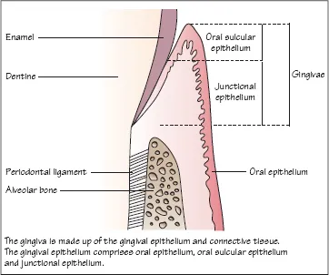
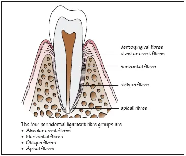
Figure 1.1 Longitudinal section through part of a tooth showing healthy periodontal tissues.
ch01_002_img0011.gif
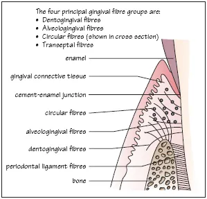
Figure 1.2 Dentogingival fibres, alveolar crest fibres and circular fibres in the gingival connective tissue.
ch01_002_img0012.gif
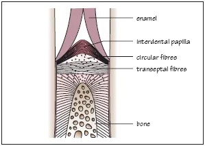
Figure 1.3 Interdental area showing transeptal and circular fibre groups in the gingival connective tissue.
ch01_002_img0013.gif
Figure 1.4 The periodontal ligament.
ch01_002_img0014.gif
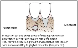
Figure 1.5 Bony fenestration and dehiscence.
ch01_002_img0015.gif
The periodontal tissues form the supporting structures of the teeth. The principal components of the periodontium are shown in Fig. 1.1:
ā€¢ Gingivae (including epithelium and connective tissue).
ā€¢ Periodontal ligament.
ā€¢ Cementum.
ā€¢ Alveolar bone.
Gingivae
The gingivae in health are pink and firm with a knife-edge appearance, scalloped around the teeth. In certain ethnic groups the gingivae may be pigmented. In health, the gingival margin is a few millimetres coronal to the cementā€“enamel junction. The gingival sulcus (or crevice) is a shallow groove which may be between 0.5 and 3 mm in depth around a fully erupted tooth. The gingival tissues are keratinised and appear paler pink than sites of non-keratinised oral epithelium.
Gingival epithelium
The gingival epithelium comprises (Fig. 1.1):
ā€¢ Oral epithelium (OE).
ā€¢ Oral sulcular epithelium (SE).
ā€¢ Junctional epithelium (JE).
The gingival sulcus is lined by SE and JE.
Oral epithelium
ā€¢ The OE is an orthokeratinised, stratified, squamous epithelium.
ā€¢ Surface cells lose their nuclei and are packed with the protein keratin.
ā€¢ It presents an impermeable physical barrier to oral bacteria.
The basal layer of epithelial cells is thrown up into folds overlying the supporting connective tissue. These folds increase the surface area of contact between the epithelium and connective tissue and are known as rete ridges or rete pegs.
Oral sulcular epithelium
ā€¢ There are no rete ridges.
ā€¢ Cells are keratinised but still have nuclei (parakeratinised).
Junctional epithelium
ā€¢ The JE forms a specialised attachment to the tooth via:
ā€¢ a hemidesmosomal layer within the JE cells;
ā€¢ a basal lamina produced by the epithelial cells.
ā€¢ The JE is non-keratinised and has a very fast turnover of cells (2ā€“6 days compared to 1 month for OE).
ā€¢ The most apical part of the JE lies at the cement-enamel junction in health.
ā€¢ The JE at its widest point is 20ā€“30 cells thick coronally.
ā€¢ The JE tapers until it is only one cell in width apically.
ā€¢ The JE is permeable with wide intercellular spaces through which cells and substances can migrate (such as bacterial toxins or host defence cells).
ā€¢ Migration of the JE from its position in health apically onto the root cementum indicates a loss of periodontal attachment and progression to the disease state of periodontitis.
Gingival connective tissue
The gingival connective tissue (or lamina propria) is made up of collagen fibre bundles called gingival fibres, around which lie ground substance, fibroblasts, blood and lymph vessels and neural tissues. The four fibre groups are shown in Figs 1.2 and 1.3.
Periodontal ligament
The periodontal ligament forms the attachment between the cementum and alveolar bone. It is a richly vascular connective tissue within which lie bundles of collagen fibres; these are divided into four groups based on their position (Fig. 1.4).
Within the ligament are mechanoreceptors that provide sensory input for jaw reflexes. Cells from the periodontal ligament are involved in the formation and remodelling of alveolar bone and cementum. The periodontal ligament acts to dissipate masticatory forces to the supporting alveolar bone and its width, height and quality determine a toothā€™s mobility.
Cementum
Cementum is a mineralised tissue overlying the root dentine. It does not undergo physiological remodelling but is continuously deposited throughout life. Cementum is classified into two types:
ā€¢ Acellular.
ā€¢ Cellular.
Acellular cementum
Acellular cementum forms on root dentine during root formation and tooth eruption. Fibres inserted from the periodontal ligament are mineralised within the cementum and are known as Sharpeyā€™s fibres and are abundant in acellular cementum.
Cellular cementum
Cellular cementum lies over the acellular cementum. It contains cells called cementocytes which lie in lacunae. The cellular cementum layer is thicker in the apical region of the root where it is between 0.2 and 1 mm thick.
Alveolar bone
ā€¢ The walls of the sockets are lined with a layer of dense bone called compact bone, which also forms the buccal and lingual/palatal plates of the jaw bones.
ā€¢ In between the sockets and the compact jaw bone walls lies cancellous bone that is made up of bony trabeculae.
ā€¢ The compact bone ...

Table of contents

  1. Cover
  2. Contents
  3. Title
  4. Copyright
  5. Preface
  6. Acknowledgements and Dedications
  7. 1: Anatomy of the Periodontium
  8. 2: Classification of Periodontal Diseases
  9. 3: Periodontal Epidemiology
  10. 4: Role of Plaque in the Aetiology of Periodontal Diseases
  11. 5: Plaque Microbiology
  12. 6: Calculus
  13. 7: Host Defences
  14. 8: Development of Periodontal Diseases
  15. 9: Progression of Periodontal Diseases
  16. 10: Risk and Periodontal Diseases
  17. 11: Systemic Risk Factors for Periodontal Diseases
  18. 12: Periodontal Diseases and General Health
  19. 13: Diet and Periodontal Diseases
  20. 14: Local Risk Factors for Periodontal Diseases
  21. 15: Occlusion and Periodontal Diseases
  22. 16: Periodontal History, Examination and Diagnosis
  23. 17: Periodontal Screening
  24. 18: Role of Radiographs in Periodontal Diagnosis
  25. 19: Principles of Periodontal Diagnosis and Treatment Planning
  26. 20: Plaque Control and Non-surgical Periodontal Therapy
  27. 21: Patient Communication in Dental Care
  28. 22: Periodontal Tissue Response, Healing and Monitoring
  29. 23: Role of Antimicrobial Therapy in Periodontal Diseases
  30. 24: Periodontal surgery
  31. 25: Types of Periodontal Surgery
  32. 26: Regenerative Periodontal Therapy
  33. 27: Bone Defects and Furcation Lesions
  34. 28: Dental Implants and Peri-Implantology
  35. 29: Periodonticā€“orthodontic Interface
  36. 30: Plaque-Induced Gingivitis
  37. 31: Non-Plaque-Induced Gingival Conditions and Lesions
  38. 32: Gingival Recession
  39. 33: Gingival Overgrowth
  40. 34: Chronic Periodontitis
  41. 35: Aggressive Periodontitis
  42. 36: Periodontal Management of Patients Who Smoke
  43. 37: Periodontal Management of Patients with Diabetes
  44. 38: Necrotising Periodontal Diseases
  45. 39: Periodontal Abscess and Periodonticā€“endodontic Lesions
  46. 40: Periodontal Diseases in Children and Adolescents
  47. 41: Periodontal Management of the Older Adult
  48. 42: Team Working in Dental Care
  49. 43: The Decision to Treat or Refer Periodontal Patients
  50. Appendix: References and Further Reading
  51. Index