- English
- ePUB (mobile friendly)
- Available on iOS & Android
eBook - ePub
Acquired Speech and Language Disorders
About this book
It is vital to have knowledge of the neuroanatomical structures and functional neurological mechanisms, which are disrupted in neurogenic speech/language, disordered persons in order to understand the speech/language deficits themselves.
This book provides a comprehensive coverage of the neurological basis of both the clinically recognised forms of aphasia and the various motor speech disorders, in both children and adults. It also covers more recently recognised language disorders, such as Parkinsons and related diseases, right hemisphere damage, closed-head injury, dementia, etc. This is a perfect text for practitioners who need to understand the integration of neuroanatomy and functional neurology with the practice of speech-language pathology.
Tools to learn more effectively

Saving Books

Keyword Search

Annotating Text

Listen to it instead
Information
1
Neuroanatomical and neuropathological framework of speech and language
Introduction
Human communication in the form of speech-language behaviour is dependent upon processes which occur in the nervous system. Consequently, knowledge of the basic structure and function of the human nervous system is an essential prerequisite to the understanding of the anatomical, physiological and pathological basis of human communication disorders. With this in mind, the materials presented in the present chapter are intended to provide the reader with an introductory knowledge of the anatomy of the human nervous system. Such knowledge is necessary prior to discussion of the signs, symptoms and neurological mechanisms underlying the various acquired neurogenic speech-language disorders in later chapters. Where necessary, more detailed information regarding the anatomy of specific brain structures important for speech-language function is provided in subsequent relevant chapters.
The nervous system is an extremely complex organization of structures which serves as the main regulative and integrative system of the body. It receives stimuli from the individual’s internal and external environments, interprets and integrates that information and selects and initiates appropriate responses to it. Consider this process in the context of a spoken conversation between two persons. The words spoken by one partner in the conversation, in the form of sound waves, are detected by receptors in the inner ear of the second partner and conveyed to the cerebral cortex of the brain via the auditory pathways where they are perceived and interpreted. Following integration with other sensory information, a response to the verbal input is formulated in the language centres of the brain and then passed to the motor areas of the brain (i.e. areas that control muscular movement) for execution. Nerve impulses from the motor areas then pass to the muscles of the speech mechanism (e.g. tongue, lips, larynx, etc.) leading to the production of a verbal response by the second person.
Speech is produced by the contraction of the muscles of the speech mechanism, which include the muscles of the lips, jaw, tongue, palate, pharynx and larynx as well as the muscles of respiration. These muscle contractions, in turn, are controlled by nerve impulses which descend from the motor areas of the brain to the level of the brainstem and spinal cord and then pass out to the muscles of the speech mechanism via the various nerves which arise from either the base of the brain (cranial nerves) or spinal cord (spinal nerves). Likewise, language is also dependent on processes which occur in the brain, particularly in the cerebral cortex.
Gross anatomy of the nervous system
For the purposes of description, the nervous system can be arbitrarily divided into two large divisions: the central nervous system and the peripheral nervous system. The central nervous system comprises the brain and spinal cord, while the peripheral nervous system consists of the end organs, nerves and ganglia, which connect the central nervous system to other parts of the body. The major components of the peripheral nervous system are the nerves which arise from the base of the brain and spinal cord. These include 12 pairs of cranial nerves and 31 pairs of spinal nerves respectively. The peripheral nervous system is often further subdivided into the somatic and autonomic nervous systems, the somatic nervous system including those nerves involved in the control of skeletal muscles (e.g. the muscles of the speech mechanism) and the autonomic nervous system including those nerves involved in the regulation of involuntary structures such as the heart, the smooth muscles of the gastrointestinal tract and exocrine glands (e.g. sweat glands). Although the autonomic nervous system is described as part of the peripheral nervous system, it is really part of both the central and peripheral nervous systems. It must be remembered, however, that these divisions are arbitrary and artificial and that the nervous system functions as an entity, not in parts. The basic organization of the nervous system is summarized in Figure 1.1.
Histology of the nervous system
Cell types
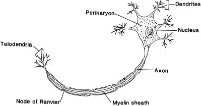
The nervous system comprises many millions of nerve cells, or neurones, which are held together and supported by specialized non-conducting cells known as neuroglia. The major types of neuroglia include astrocytes, oligodendrocytes and microglia. It is the neurones that are responsible for conduction of nerve impulses from one part of the body to another, such as from the central nervous system to the muscles of the speech mechanism to produce the movement of the lips, tongue and so on for speech production. Although there are a number of different types of neurones, most consist of three basic parts: a cell body (also known as a soma or perikaryon) which houses the nucleus of the cell; a variable number of short processes (generally no more than a few millimetres in length) called dendrites (meaning ‘tree-like’) which receive stimuli and conduct nerve impulses; and a single, usually elongated, process called an axon, which in the majority of neurones is surrounded by a segmented fatty insulating sheath called the myelin sheath. A schematic representation of a neurone is shown in Figure 1.2.
Figure 1.1 Basic organization of the nervous system.
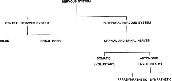
Figure 1.2 Structure of a typical motor neurone.
The cytoplasm of a neurone contains the usual cell organelles (e.g. mitochondria) with the exception of the centrosome. Mature neurones cannot divide or replace themselves because of the lack of a centrosome. In addition to the usual organelles, however, the cytoplasm of nerve cells also contains two organelles unique to neurones: Nissl substance (chromidial substance) and neurofibrils. Seen with the light microscope Nissl substance (bodies) appears as rather large granules widely scattered throughout the cytoplasm of the nerve cell body. Nissl bodies specialize in protein synthesis, thereby providing the protein needs for maintaining and regenerating neurone processes and for renewing chemicals involved in the transmission of nerve impulses from one neurone to another. Seen with the light microscope neurofibrils are tiny tubular structures running through the cell body, axon and dendrites. Although the function of the neurofibrils is uncertain, it has been suggested that they may facilitate the transport of intracellular materials within the neurone. In Alzheimer’s disease, the neurofibrils become abnormally twisted, a feature used in the diagnosis of this condition (see Chapter 6).
In contrast to neurones, neuroglial cells (also often simply referred to as glial cells) contribute to brain function mainly by supporting neuronal functions. Although based on current evidence their role appears to be subordinate to that of the neurones, without glial cells the brain could not function properly. Astrocytes are the most numerous of the glial cells and are widely distributed in the central nervous system. These cells fill spaces between neurones and lie in close proximity to both neurones and capillaries. Evidence suggests that an essential role for astrocytes is the regulation of the chemical content of the extracellular space (e.g. astrocytes envelop synaptic junctions in the brain and thereby restrict the spread of neurotransmitter molecules released by neurones). Further, special proteins found within the membranes of astrocytes may be involved in the removal of many neurotransmitters from the synaptic cleft. In addition to regulating neurotransmitters, astrocytes also regulate the concentration of substances present within the extracellular space that have the potential to interfere with normal functioning of the neurones (e.g. astrocytes regulate the concentration of potassium ions in the extracellular fluid in the brain).
Oligodendrocytes and Schwann cells are two types of glial cells that form the insulation or myelin sheath that surrounds axons in the central and peripheral nervous systems respectively. Oligodendrocytes are only found in the central nervous system (i.e. brain and spinal cord), where they may wrap layers of membrane to form a myelin sheath around several axons. Schwann cells in contrast are located only in the peripheral nervous system, where they myelinate only a single axon. Periodical gaps in the myelin sheath are known as nodes of Ranvier. Nerve impulses travelling down myelinated axons jump from node to node, thereby increasing the speed of transmission, a process known as saltatory conduction.
Other neuroglial cells include ependymal cells and microglia. Ependymal cells provide the lining of fluid-filled cavities within the brain called the ventricles and thereby form a barrier between ventricular fluid and the neuronal substance of the brain. They also form the choroid plexuses, which produce cerebrospinal fluid. Microglia are few in number and small in size and function as phagocytes to remove debris left by dead or degenerating neurones and glial cells.
Synapses and neuroeffector junctions
The axon conducts nerve impulses away from the cell body to the next neurone or to a muscle or gland. The area where two neurones communicate with one another is called a synapse. It represents a region of functional but not anatomical continuity between the axon terminal of one neurone (the pre-synaptic neurone) and the dendrites, cell body or axon of another neurone (the post-synaptic neurone). The synapse is an area where a great degree of control can be exerted over nerve impulses. At the synapse, nerve impulses can be either blocked (inhibited) or facilitated. Axons branch repeatedly, forming anywhere from 1000 to 10 000 synapses. Consequently, there may be thousands of synapses on the surface of a single neurone. When one considers that there are billions of neurones, the complexity of the circuitry of the nervous system is staggering. It has been estimated that the number of synapses in the brain is possibly in the order of 100 trillion. Whether a specific neurone fires is dependent on the summation of the messages it receives from multiple sources.
Structurally, each synapse is made up as follows. As the terminal part of an axon approaches another neurone, it decreases in diameter, loses its myelin (if a myelinated fibre) and divides repeatedly forming small branches, termed telodendria. At the end of each telodendron is a small swelling called a bouton terminal or synaptic knob. The structure of the bouton terminal has been elucidated by electron microscopy. It contains a number of structures, in particular mitochondria and synaptic vesicles. The synaptic vesicles contain a neurotransmitter substance which is released when a nerve impulse arrives at the bouton. There are many kinds of neurotransmitter substance, some of which facilitate (excitatory transmitters) nerve impulse conduction in the post-synaptic neurone while others inhibit (inhibitory transmitters) nerve impulse conduction in the post-synaptic neurone. Some of the more common neurotransmitter substances include acetylcholine, norepinephrine, serotonin, dopamine and gamma aminobutyric acid (GABA).
When released from the synaptic knob the chemical transmitter diffuses across a gap called the synaptic cleft between the bouton and the membrane of the post-synaptic neurone to either excite or inhibit the post-synaptic neurone. As neurotransmitter substance is only located on the pre-synaptic side, a synapse can transmit in only one direction. In addition to the chemical synapses just described, in certain parts of the nervous system electrical synapses or gap junctions are present. In this type of synapse the membranes of the pre- and post-synaptic neurones lie in close proximity to one another and comprise a pathway of low resistance which allows current flow from the pre-synaptic neurone to act upon the post-synaptic neurone.
Neuroeffector junctions are functional contacts between axon terminals and effector cells. Structurally, neuroeffector junctions are similar to synapses with the exception that the post-synaptic structure is not a nerve cell but rather a muscle or gland. We will not concern ourselves greatly with junctions with smooth or cardiac muscles or glands, but will rather concentrate on junctions with skeletal muscles, as this is the type of muscle tissue that comprises the muscles of the speech mechanism.
Figure 1.3 (A) Motor end plate. (B) Close-up of a motor end plate showing the relationship between structures in nerve cell and muscle.
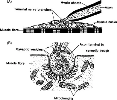
In the case of skeletal muscles, the neuroeffector junction is termed a motor end plate. The structure of a typical motor end plate is shown in Figure 1.3.
Each motor nerve fibre branches at its end to form a complex of branching nerve terminals, each terminal innervating a separate skeletal muscle fibre. A single axon of a motor neurone, therefore, innervates more than one skeletal muscle fibre; the motor neurone plus the muscle fibres it innervates constitute a motor unit. The bouton of each terminal contains synaptic vesicles that contain neurotransmitter substance. The motor neurones running to skeletal muscles use acetylcholine as their transmitter substance. The arrival of a nerve impulse at the bouton causes release of acetylcholine from the vesicles in a similar manner to that for transmission at the synapse, only in this case the transmitter diffuses across the neuromuscular junction to cause contraction of the muscle fibre. In the condition called ‘myasthenia gravis’ there is a failure, possibly as a consequence of antibodies...
Table of contents
- Cover
- Contents
- Title Page
- Copyright
- 1 Neuroanatomical and neuropathological framework of speech and language
- 2 Aphasia syndromes
- 3 Subcortical aphasia syndromes
- 4 Speech-language disorders associated with traumatic brain injury
- 5 Language disorders subsequent to right-hemisphere lesions
- 6 Language disturbances in dementia syndromes
- 7 Language disorders associated with diseases of the cerebral white matter
- 8 Neurological disturbances associated with aphasia
- 9 Dysarthrias associated with upper and lower motor neurone lesions
- 10 Dysarthrias associated with extrapyramidal syndromes
- 11 Dysarthrias associated with lesions in other motor systems
- 12 Acquired childhood speech-language disorders
- Index
Frequently asked questions
Yes, you can cancel anytime from the Subscription tab in your account settings on the Perlego website. Your subscription will stay active until the end of your current billing period. Learn how to cancel your subscription
No, books cannot be downloaded as external files, such as PDFs, for use outside of Perlego. However, you can download books within the Perlego app for offline reading on mobile or tablet. Learn how to download books offline
Perlego offers two plans: Essential and Complete
- Essential is ideal for learners and professionals who enjoy exploring a wide range of subjects. Access the Essential Library with 800,000+ trusted titles and best-sellers across business, personal growth, and the humanities. Includes unlimited reading time and Standard Read Aloud voice.
- Complete: Perfect for advanced learners and researchers needing full, unrestricted access. Unlock 1.4M+ books across hundreds of subjects, including academic and specialized titles. The Complete Plan also includes advanced features like Premium Read Aloud and Research Assistant.
We are an online textbook subscription service, where you can get access to an entire online library for less than the price of a single book per month. With over 1 million books across 990+ topics, we’ve got you covered! Learn about our mission
Look out for the read-aloud symbol on your next book to see if you can listen to it. The read-aloud tool reads text aloud for you, highlighting the text as it is being read. You can pause it, speed it up and slow it down. Learn more about Read Aloud
Yes! You can use the Perlego app on both iOS and Android devices to read anytime, anywhere — even offline. Perfect for commutes or when you’re on the go.
Please note we cannot support devices running on iOS 13 and Android 7 or earlier. Learn more about using the app
Please note we cannot support devices running on iOS 13 and Android 7 or earlier. Learn more about using the app
Yes, you can access Acquired Speech and Language Disorders by Bruce E. Murdoch in PDF and/or ePUB format, as well as other popular books in Medicine & Audiology & Speech Pathology. We have over one million books available in our catalogue for you to explore.