![]()
Chapter 1
Basics
In order to achieve a better understanding of disease processes occurring in different regions of the eye, this section describes the technology currently employed by the histopathologist in the examination of tissue specimens referred by ophthalmologists. It is important to be aware of the range of laboratory services locally available. When there is a suspicion of infection, the relevant specialist (bacteriologist/mycologist/virologist) should be consulted for advice concerning appropriate transport media and therapy. The value of an accurate and concise history cannot be overestimated and good collaboration will be rewarding to both clinicians and laboratory specialists.
Examination of the enucleated eye
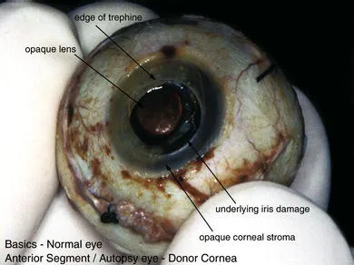
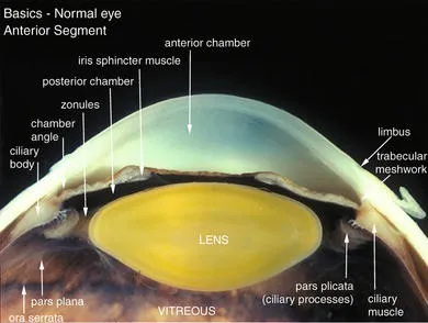
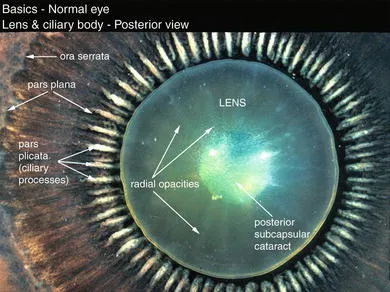
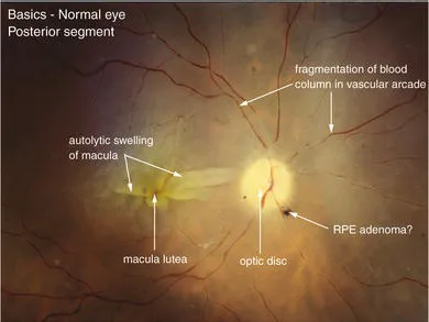
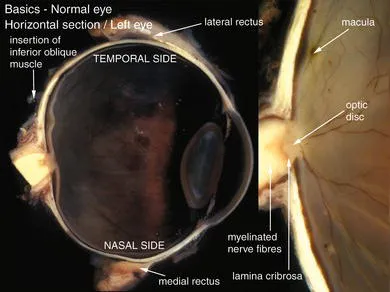
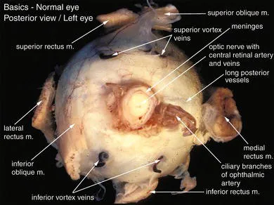
A formalin-fixed enucleated globe bears little resemblance to the in vivo appearance due to opacification of the cornea, lens, vitreous, and retina. Previous intervention, for example removal of keratoplasty tissue, can produce secondary damage to the anterior segment tissues (Figure 1.1). In routine practice, it is unwise to try to cut across the lens because this produces damage to the anterior segment but occasionally a suitable illustration can be provided (Figure 1.2). By dividing the globe in the coronal plane, the pathologist has the advantage of examination of the lens and ciliary body from the posterior aspect (Figure 1.3) and the retina from the anterior aspect (Figure 1.4). For demonstration purposes, it is possible to divide the optic nerve and the lens (Figure 1.5). In general, the globe is divided above the optic nerve and at the edge of the cornea to avoid traumatic artefact to the main axial structures. After paraffin processing, the microtomist cuts into the centre of the eye. The orientation of the extraocular muscles on the posterior aspect of the globe allows the pathologist to identify the side from which the globe was enucleated (Figure 1.6). Orientation of the specimen is vital if the correct plane of cut is to be made.
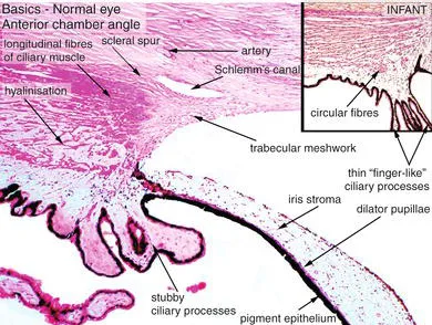
Microscopic features
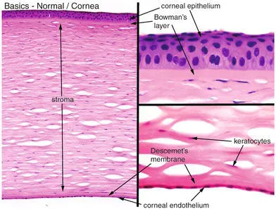
These are described wherever relevant to pathology in the corresponding chapters and are therefore only illustrated briefly in this chapter. The histological features of each of the following tissues are annotated in detail:
- cornea (Figure 1.7)
- chamber angle (Figure 1.8)
- iris (Figures 1.8, 1.9)
- ciliary body (Figures 1.8, 1.10, 1.11)
- lens (Figures 1.9, 1.11)
- retina and choroid (Figure 1.12)
- optic disc (Figure 1.13).
Features for identification of the age of a patient (in this case a child):
- thin Descemet’s membrane
- “finger-like ciliary processes”
- intact, non-hyalinised ciliary muscle
- absence of proliferations in the pars plana epithelium
- absence of sub-RPE (retinal pigment epithelium) deposits (for example drusen).