- English
- ePUB (mobile friendly)
- Available on iOS & Android
eBook - ePub
Practical Guide to Lameness in Horses
About this book
This is an updated, practical version of Dr. Stashak's top selling book Adams' Lameness in Horses, 4th edition. The material is heavily illustrated and provides a hands-on guide to common clinical problems. The authors present important guidelines for decision making and preventive measures. This is a hands-on, authoritative resource that clearly differentiates between important and non-important clinical situations.
Tools to learn more effectively

Saving Books

Keyword Search

Annotating Text

Listen to it instead
Information
CHAPTER ONE
Functional Anatomy*
Anatomy, by nature, is a complex, technical subject. Rather than simplify it too much, we have retained the detail for your future reference.
Anatomic Nomenclature
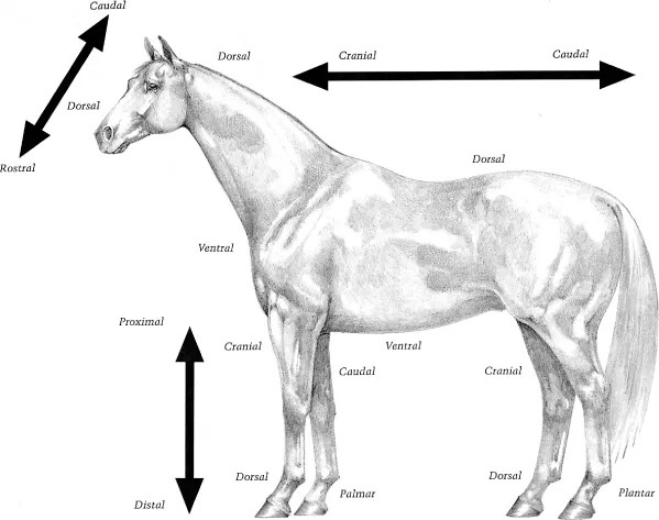
In Figure 1–1, note that the positional adjectives “proximal” and “distal” refer to the limbs and “dorsal” and “ventral” refer to the upper body, head, and neck. “Rostral” is used to indicate the direction toward the nose. With the exception of the eye, the terms “anterior” and “posterior” are not applicable to quadrupeds. “Cranial” and “caudal” apply to the limbs proximal to the knee (antebrachiocarpal radiocarpal) joint and the hock (tarsocrural tibiotarsal) joint. Distal to these joints, “dorsal” and “palmar” (on the forelimb) or “plantar” (on the hindlimb) are the correct terms. The adjective “solar,” is used to designate structures on the palmar (plantar) surface of the coffin bone (distal phalanx) and the ground surface of the hoof.
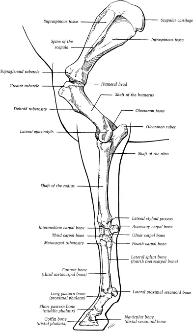
Thoracic Limb (Fig. 1–2)
Digit and Fetlock
The foot and pastern comprise the equine digit. The bones of this region include the coffin bone (also known as [a.k.a.] the distal phalanx, third phalanx, or PIII), the navicular bone (a.k.a. the distal sesamoid), the short pastern bone (a.k.a. the middle phalanx, the second phalanx, or PII), and the long pastern bone (a.k.a. the proximal phalanx, the first phalanx, or PI). The fetlock is the region where the long pastern bone articulates with the cannon bone and the two proximal sesamoid bones.
Foot
The foot consists of the hoof (epidermis) and all it encloses: the corium (dermis), digital cushion, coffin bone, lateral cartilages, coffin joint, distal extremity of the short pastern bone, navicular bone, navicular bursa, several ligaments, tendons of insertion of the common digital extensor and deep digital flexor muscles, blood vessels, and nerves.
The hoof is continuous with the epidermis (outer skin) at the coronet. Here the dermis (inner layer) of the skin is continuous with the dermis (corium) of the hoof. Regions of the corium correspond to the parts of the hoof under which they are located: perioplic corium, coronary corium, laminar corium, corium of the frog, and corium of the sole (Fig. 1–3).
The exterior parts of the hoof protect underlying structures of the foot and dissipate concussive forces when the hoof strikes the ground. Figure 1–4 illustrates the sole, frog, heels, bars, and ground surface of the wall. The ground surface of the forefoot is wider than that of the hindfoot, corresponding to the rounder coffin bone of the forefoot.
The hoof wall extends from the ground surface to the coronary border, where the soft white horn of the periople joins the epidermis of the skin at the coronet. Regions of the wall are the dorsal toe, the medial and lateral quarters, and the heels. From the thick toe, the wall becomes progressively thinner and more elastic toward the heels, where it thickens again at the junction of the bars (the “buttress” of the hoof). The medial wall is usually steeper (more upright) than the lateral wall.
The horn’s tubules are sometimes visible as fine lines on the hoof wall, running from the coronet to the ground (Fig. 1–5). Differential growth rates of the wall from the coronary border toward the ground account for the smooth ridges parallel to the coronary border.
Most of the epidermis is devoid of nerve endings; it is the “insensitive” part of the foot.
Three layers comprise the hoof wall: the stratum tectorium, stratum medium, and stratum internum (see Fig. 1–5). The superficial stratum tectorium is a thin layer of horn extending distally from the periople a variable distance that decreases with age. The bulk of the wall is a stratum medium consisting of horn tubules and intertubular horn. The horn tubules are generated by the germinal layer of the coronary epidermis covering the long papillae of the coronary corium. Intertubular horn is formed in between the projections.
Distal to the coronary groove, approximately 600 primary insensitive (epidermal) laminae interlock with the primary sensitive (dermal) laminae of the laminar corium (Figs. 1–4 and 1–5). Approximately 100 microscopic secondary laminae branch at an angle from each primary lamina, further binding the hoof and corium together (Fig. 1–6).
Some confusion exists concerning the terms “insensitive” and “sensitive” laminae. In the strictest sense, the keratinized parts of the primary epidermal laminae are insensitive; the stratum germinativum, which includes all of the secondary epidermal laminae, and the laminar corium are “sensitive.” Although the terms “epidermal” and “dermal” are far more accurate adjectives, the terms “sensitive” and “insensitive” are still in common use.
FIG. 1–1A. Topographical and directional terms.
Submicroscopic, peglike dermal projections increase the surface of attachment of the sensitive and insensitive structures (dermis and epidermis) of the hoof. This configuration and the blending of the laminar corium with the periosteum of the coffin bone suspend and support the bone, aiding in the dissipation of concussion and the movement of blood.
The wall grows approximately 0.25 inch (6 mm) per month, taking from 9 to 12 months for the toe to grow out. Growth tends to be slower in cold or dry environments.
Stratum medium may be pigmented or nonpigmented. Contrary to popular belief, pigmented hooves are not stronger than nonpigmented hooves.
The slightly concave sole should not bear weight on its ground surface, except near its junction with the white line, but it bears internal weight transmitted from the solar surface of the coffin bone through the solar corium. That portion of the sole at the angle formed by the wall and the bars is the angle of the sole (seat of corn). When the wall is trimmed, the white line is visible where the wall joins the sole. The sensitive corium is immediately internal to the white line that serves as a landmark for determining the position and angle for driving horseshoe nails.
FIG. 1–1B (continued). Topographical and directional terms.
The sole’s horn tubules are oriented vertically, conforming to the direction of the papillae of the solar corium. Intertubular horn binds the tubules together. The relationship of the solar epithelium to the solar corium is responsible for this configuration (Fig. 1–7). Near the ground, the horn tubules curl, accounting for the self-limiting growth of the sole, and cause shedding from the superficial part. Approximately one-third of the sole is water.
FIG. 1–2. Bones of the equine thoracic limb; lateral view of the left limb.
The frog (Fig. 1–4) is a wedge-shaped mass of keratinized epithelium rendered softer than other parts of the hoof by a 50% water content. Merocrine glands deliver their secretions onto the surface superficial to the frog stay (Fig. 1–3). The proximally projecting frog stay contacts the digital cushion.
The corium is rich in elastic fibers, highly vascular, and well supplied with nerves. The arterial supply is from numerous branches radia...
Table of contents
- Cover
- Contents
- Title Page
- Copyright
- Dedication
- Acknowledgments
- Preface
- CHAPTER ONE: Functional Anatomy
- CHAPTER TWO: Conformation and Movement
- CHAPTER THREE: Examination for Lameness
- CHAPTER FOUR: Selected Diseases of Bones, Joints, and Related Structures
- CHAPTER FIVE: Lameness
- CHAPTER SIX: Shoeing for Soundness
- CHAPTER SEVEN: Methods of Therapy
- Index
Frequently asked questions
Yes, you can cancel anytime from the Subscription tab in your account settings on the Perlego website. Your subscription will stay active until the end of your current billing period. Learn how to cancel your subscription
No, books cannot be downloaded as external files, such as PDFs, for use outside of Perlego. However, you can download books within the Perlego app for offline reading on mobile or tablet. Learn how to download books offline
Perlego offers two plans: Essential and Complete
- Essential is ideal for learners and professionals who enjoy exploring a wide range of subjects. Access the Essential Library with 800,000+ trusted titles and best-sellers across business, personal growth, and the humanities. Includes unlimited reading time and Standard Read Aloud voice.
- Complete: Perfect for advanced learners and researchers needing full, unrestricted access. Unlock 1.4M+ books across hundreds of subjects, including academic and specialized titles. The Complete Plan also includes advanced features like Premium Read Aloud and Research Assistant.
We are an online textbook subscription service, where you can get access to an entire online library for less than the price of a single book per month. With over 1 million books across 990+ topics, we’ve got you covered! Learn about our mission
Look out for the read-aloud symbol on your next book to see if you can listen to it. The read-aloud tool reads text aloud for you, highlighting the text as it is being read. You can pause it, speed it up and slow it down. Learn more about Read Aloud
Yes! You can use the Perlego app on both iOS and Android devices to read anytime, anywhere — even offline. Perfect for commutes or when you’re on the go.
Please note we cannot support devices running on iOS 13 and Android 7 or earlier. Learn more about using the app
Please note we cannot support devices running on iOS 13 and Android 7 or earlier. Learn more about using the app
Yes, you can access Practical Guide to Lameness in Horses by Ted S. Stashak,Cherry Hill in PDF and/or ePUB format, as well as other popular books in Medicine & Equine Veterinary Science. We have over one million books available in our catalogue for you to explore.