Introduction to X-Ray Powder Diffractometry
eBook - ePub

Introduction to X-Ray Powder Diffractometry

  1. English
  2. ePUB (mobile friendly)
  3. Available on iOS & Android
eBook - ePub

Introduction to X-Ray Powder Diffractometry

Book details
Book preview
Table of contents
Citations

About This Book

When bombarded with X-rays, solid materials produce distinct scattering patterns similar to fingerprints. X-ray powder diffraction is a technique used to fingerprint solid samples, which are then identified and cataloged for future use-much the way the FBI keeps fingerprints on file. The current database of some 70, 000 material prints has been put to a broad range of uses, from the analysis of moon rocks to testing drugs for purity. Introduction to X-ray Powder Diffractometry fully updates the achievements in the field over the past fifteen years and provides a much-needed explanation of the state-of-the-art techniques involved in characterizing materials. It covers the latest instruments and methods, with an emphasis on the fundamentals of the diffractometer, its components, alignment, calibration, and automation. The first three chapters outline diffraction theory in clear language, accessible to both students and professionals in chemistry, physics, geology, and materials science. The book's middle chapters describe the instrumentation and procedures used in X-ray diffraction, including X-ray sources, X-ray detection, and production of monochromatic radiation. The chapter devoted to instrument design and calibration is followed by an examination of specimen preparation methods, data collection, and reduction. The final two chapters provide in-depth discussions of qualitative and quantitative analysis. While the material is presented in an orderly progression, beginning with basic concepts and moving on to more complex material, each chapter stands on its own and can be studied independently or used as a professional reference. More than 230 illustrations and tables demonstrate techniques and clarify complex material. Self-contained, timely, and user-friendly, Introduction to X-ray Powder Diffractometry is an enormously useful text and professional reference for analytical chemists, physicists, geologists and materials scientists, and upper-level undergraduate and graduate students in materials science and analytical chemistry. X-ray powder diffraction-a technique that has matured significantly in recent years-is used to identify solid samples and determine their composition by analyzing the so-called "fingerprints" they generate when X-rayed. This unique volume fulfills two major roles: it is the first textbook devoted solely to X-ray powder diffractometry, and the first up-to-date treatment of the subject in 20 years. This timely, authoritative volume features:
* Clear, concise descriptions of both theory and practice-including fundamentals of diffraction theory and all aspects of the diffractometer
* A treatment that reflects current trends toward automation, covering the newest instrumentation and automation techniques
* Coverage of all the most common applications, with special emphasis on qualitative and quantitative analysis
* An accessible presentation appropriate for both students and professionals
* More than 230 tables and illustrations Introduction to X-ray Powder Diffractometry, a collaboration between two internationally known and respected experts in the field, provides invaluable guidance to anyone using X-ray powder diffractometers and diffractometry in materials science, ceramics, the pharmaceutical industry, and elsewhere.

Frequently asked questions

Simply head over to the account section in settings and click on “Cancel Subscription” - it’s as simple as that. After you cancel, your membership will stay active for the remainder of the time you’ve paid for. Learn more here.
At the moment all of our mobile-responsive ePub books are available to download via the app. Most of our PDFs are also available to download and we're working on making the final remaining ones downloadable now. Learn more here.
Both plans give you full access to the library and all of Perlego’s features. The only differences are the price and subscription period: With the annual plan you’ll save around 30% compared to 12 months on the monthly plan.
We are an online textbook subscription service, where you can get access to an entire online library for less than the price of a single book per month. With over 1 million books across 1000+ topics, we’ve got you covered! Learn more here.
Look out for the read-aloud symbol on your next book to see if you can listen to it. The read-aloud tool reads text aloud for you, highlighting the text as it is being read. You can pause it, speed it up and slow it down. Learn more here.
Yes, you can access Introduction to X-Ray Powder Diffractometry by Ron Jenkins, Robert Snyder in PDF and/or ePUB format, as well as other popular books in Physical Sciences & Analytic Chemistry. We have over one million books available in our catalogue for you to explore.

Information

Year
2012
ISBN
9781118520925

CHAPTER 1

CHARACTERISTICS OF X-RADIATION

1.1. EARLY DEVELOPMENT OF X-RAY DIFFRACTION

Following the discovery of X-rays by W. C. Röntgen in 1895, three major branches of science have developed from the use of this radiation. The first and oldest of these is X-ray radiography, which makes use of the fact that the relative absorption of X-rays by matter is a function of the average atomic number and density of the matter concerned. From this has developed the whole range of diagnostic methods for medical and industrial use. Early attempts to confirm the dual nature of X-rays, i.e., their particle and wave character, were frustrated by experimental difficulties involved with the handling of the very short wavelengths in question. Not until the classic work of Max von Laue in 1912 was the wave character confirmed by diffraction experiments from a single crystal. From this single experiment has developed the field of X-ray crystallography, of which X-ray powder diffractometry is one important member. X-ray crystallography, using single crystals or powder, is mainly concerned with structure analysis. The third technique, X-ray spectrometry, also has its fundamental roots in the early part of this century, but routine application of X-ray fluorescence spectrometry has only developed over the last 20 to 30 years.
The purpose of this work is to discuss X-ray powder diffractometry. Powder diffractometry is mainly used for the identification of compounds by their diffraction patterns. The first X-ray powder diffractometer was developed in 1935 by Le Galley [1], but, due mainly to the lack of parafocusing conditions, the instrument gave relatively poor intensities. In 1945 Parrish and Gordon [2] developed a Geiger-counter spectrometer1 for the precision cutting of quartz oscillator plates used in frequency control for military communication equipment. At the same time, Friedman [3] was working on X-ray spectrometer techniques at the U.S. Naval Research Laboratory in Washington, DC. The modern parafocusing X-ray powder diffractometer was based on these ideas, and the first commercial equipment was introduced by North American Philips in 1947. The latest versions of the powder diffractometer differ little in their construction and geometry, but considerable advances have been made in detection and counting systems, automation, and in the X-ray tubes themselves.

1.2. ORIGIN OF X-RADIATION

X-rays are relatively short-wavelength, high-energy beams of electromagnetic radiation. When an X-ray beam is viewed as a wave, one can think of it as a sinusoidal oscillating electric field with, at right angles to it, a similarly varying magnetic field changing with time. Another description of X-rays is as particles of energy called photons. All electromagnetic radiation is characterized either by its wave character using its wavelength λ (i.e., the distance between peaks) or its frequency v (the number of peaks that pass a point in unit time) or by means of its photon energy E. The following equations represent the relationships between these quantities:
(1.1)
equation
(1.2)
equation
where c is the speed of light and h is Planck’s constant. The X-ray region is normally considered to be that part of the electromagnetic spectrum lying between 0.1 and 100 Å(1Å = 10-10 m), being bounded by the γ-ray region to the short-wavelength side and the vacuum ultraviolet region to the long-wavelength side. In terms of energy, the X-ray region covers the range from about 0.1 to 100 keV. From a combination of Equations 1.1 and 1.2, it follows that the energy equivalent of an X-ray photon is
(1.3)
equation
Insertion of the appropriate values for the fundamental constants gives
(1.4)
equation
or
(1.5)
equation
where E is in keV and λ in angstroms. As an example the Cu Kα1, Kα2 doublet has an energy of about 8.05 keV, corresponding to a wavelength of 12.398/8.046= 1.541 Å.
In the early days of crystallography there was no standard value for—or way to determine—the wavelength of any particular X-ray photon. A practical definition was made defining wavelength in terms of the cubic lattice parameter of calcite. These units are referred to as kX units and were used in the literature into the 1950s. The angstrom (Å) unit has always been the preferred measure of wavelength and is related to kX (the crystallographic unit) by 1 Å = 1.00025 kX units. Even though the latest recommendation from the International Union of Pure and Applied Chemistry (IUPAC) discourages use of the angstrom and encourages use of the nanometer (nm; 1 × 10⁻⁹ m), the powder diffraction community has fought for retention of the angstrom and this remains the common unit in use in the field today. For this reason, in this book we will use the angstrom unit. The common electron-volt energy unit is also not IUPAC approved in that the standard energy unit is the joule (J), which may be converted by 1 eV = 1.602 × 10⁻¹⁹ J.

1.3. CONTINUOUS RADIATION
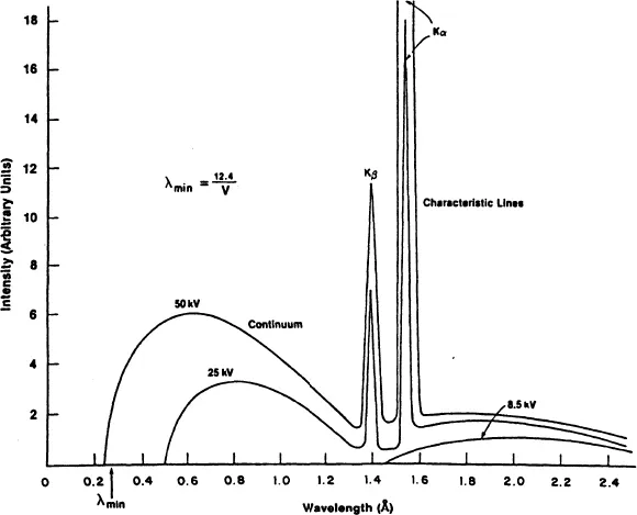
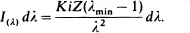
X-radiation arises when matter is irradiated with a beam of high-energy charged particles or photons. When an element is bombarded with electrons the spectrum obtained is similar to that shown in Figure 1.1. The figure illustrates the main features of the spectrum that would be obtained from a copper anode (target) X-ray tube, operated at 8.5, 25, and 50 kV, respectively. It will be seen that the spectrum consists of a broad band of continuous radiation (bremsstrahlung, or white radiation) superimposed on which are discrete wavelengths of varying intensity. The continuous radiation is produced as the impinging high-energy electrons are decelerated by the atomic electrons of the target element. The continuum is typified by a minimum wavelength, λmin, which is related to the maximum accelerating potential V of the electrons. Thus, as follows from Equation 1.5,
Figure 1.1. Continuous and characteristic radiation for copper.
(1.6)
equation
Note from Figure 1.1 that as the operating voltage is increased from 8.5 to 25 to 50 kV, the λmin value shifts to shorter wavelengths and the intensity of the continuum increases. The intensity distribution of the continuum reaches a maximum intensity at a wavelength of about 1.5 to 2 times λmin. The wavelength distribution of the continuum can be expressed quantitatively in terms of the excitation conditions by means of Kramers’ formula [4]:
(1.7)
equation
Kramers’ formula relates the intensity I(λ) from an infinitely thick target of atomic number Z with the applied current i where K is a constant. This expression does not correct for self-absorption by the target, which in practice leads to some modification of the intensity distribution.
It will also be seen from Figure 1.1 that somewhere between X-ray tube potentials of 8.5 and 25 kV sharp lines appear, superimposed on the continuum. These lines were shown by Moseley [5] to be characteristic wavelengths since their v...

Table of contents

  1. Cover
  2. Half Title page
  3. Title page
  4. Copyright page
  5. Dedication
  6. Preface
  7. Cumulative Listing of Volumes in Series
  8. Chapter 1: Characteristics of X-Radiation
  9. Chapter 2: The Crystalline State
  10. Chapter 3: Diffraction Theory
  11. Chapter 4: Sources for the Generation of X-Radiation
  12. Chapter 5: Detectors and Detection Electronics
  13. Chapter 6: Production of Monochromatic Radiation
  14. Chapter 7: Instruments for the Measurement of Powder Patterns
  15. Chapter 8: Alignment and Maintenance of Powder Diffractometers
  16. Chapter 9: Specimen Preparation
  17. Chapter 10: Acquisition of Diffraction Data
  18. Chapter 11: Reduction of Data From Automated Powder Diffractometers
  19. Chapter 12: Qualitative Analysis
  20. Chapter 13: Quantitative Analysis
  21. Appendices
  22. Index