eBook - ePub
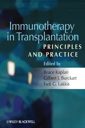
Immunotherapy in Transplantation
Principles and Practice
- English
- ePUB (mobile friendly)
- Available on iOS & Android
eBook - ePub
Immunotherapy in Transplantation
Principles and Practice
About this book
This comprehensive reference source will benefit all transplant specialists working with pharmacologic and biologic agents that modulate the immune system. Compiled by a team of world-renowned editors and contributors covering the fields of transplantation, nephrology, pharmacology, and immunology, the book covers all anti-rejection drugs according to a set template and includes the efficacy of each for specific diseases.
Tools to learn more effectively

Saving Books

Keyword Search

Annotating Text

Listen to it instead
Information
PART 1
Transplantation Immunobiology
CHAPTER 1
The Immune Response to a Transplanted Organ: An Overview
Basic definitions
Organs transplanted between two members of the same species are rejected unless the donor and recipient are genetically indistinguishable (identical twins in the case of humans). Rejection is caused by the recipient’s immune response to foreign elements present on the transplanted organ. These elements are usually proteins that differ between the donor and recipient and are called “alloantigens.” The transplanted organ itself is referred to as the “allograft” and the immune response mounted against it as the “alloimmune response” or “alloimmunity.” The prefix “xeno,” on the other hand, is used to denote the transplantation of organs between members of different species, as in the terms xeno-antigens, xenografts, and xenotransplantation.
The principal players
The T lymphocyte is the principal mediator of the alloimmune response [1, 2]. Experimental animals devoid of T cells do not reject tissue or organ allografts [3, 4]. Similarly, T cell depletion in humans prevents rejection effectively until T cells return to the circulation [5]. T cells cause direct injury to the allograft through a variety of cytotoxic molecules or cause damage indirectly by activating macrophages and other inflammatory cells (Chapter 3). T cells also provide help to B lymphocytes to produce a host of antibodies that recognize alloantigens (“alloantibodies”). Alloantibodies inflict injury on the transplanted organ by activating the complement cascade or by activating macrophages and natural killer cells (Chapter 4). An exception to the T cell requirement for allograft rejection is the rapid rejection of organs transplanted between ABO blood-group-incompatible individuals. In this case, allograft destruction is mediated by preformed anti-ABO antibodies that are produced by B-1 lymphocytes, a subset of B cells that are activated independent of help from T cells. Another potential mechanism of T-cell-independent rejection is graft dysfunction mediated by monocytes. This has been observed in renal transplant recipients after profound T cell depletion [5], but it is unlikely that monocytes lead to full-blown rejection in the absence of T cells or preformed antibodies.
The principal alloantigens recognized by T cells, B cells, and antibodies are the human leukocyte antigens (HLAs). These are cell-surface proteins that are highly variable (polymorphic) between unrelated individuals. Two main classes of HLA proteins have been identified. Class I molecules (HLA-A, -B, and -C) are expressed on all nucleated cells, whereas class II molecules (HLA-DP, -DQ, and -DR) are present on cells of the immune system that process and present foreign proteins to T cells; these are referred to as antigen-presenting cells (APCs) and include B cells, dendritic cells, macrophages, and other phagocytic cells (Chapter 2). In humans, activated T cells and inflamed endothelial cells also express class II molecules. Since HLA inheritance is codominant, any given individual shares one haplotype (one set of alleles) with either biological parent and has a 25 % chance of being HLA-identical (sharing both haplotypes) with a sibling. The chance that two unrelated individuals are HLA-identical is less than 5 %, because of the highly polymorphic nature of the HLA. Although HLA matching between donor and recipient confers long-term survival advantage on grafts [6], it does not in any way obviate the need for immunosuppression. The immune system is, in fact, capable of recognizing any non-HLA protein that differs between the donor and recipient as foreign and of mounting an alloimmune response to it that is sufficient to cause rejection. Non-HLA proteins that trigger an alloimmune response and are targeted during allograft rejection are referred to as “minor histocompatibility antigens” (Chapter 2). It is likely that a large number of minor antigens exist, making it very difficult to match for them.
Types of rejection
Pathologists have traditionally divided allograft rejection into three groups based on the tempo of allograft injury: hyperacute, acute, and chronic. Hyperacute rejection is a very rapid form of rejection that occurs within minutes to hours after transplantation and destroys the allograft in an equally short period of time. It is triggered by preformed anti-ABO or anti-HLA antibodies present in the recipient [7, 8]. Blood typing and clinical cross-matching, whereby preformed anti-HLA antibodies are screened for by mixing recipient serum with donor cells, or more commonly nowadays by sensitive flow-cytometric methods, has virtually eliminated hyperacute rejection. Acute rejection, in contrast, leads to allograft failure over a period of several days rather than minutes or hours. It usually occurs within a few days or weeks after transplantation, but it could happen at much later time points if the immune system is “awakened” by infection or by significant reduction in immunosuppression. Chronic rejection is a slow form of rejection that primarily affects the graft vasculature (or the bronchioles and bile ducts in the case of lung and liver transplants respectively) and causes graft fibrosis. Chronic rejection may become manifest during the first year after transplantation, but more often progresses gradually over several years, eventually leading to the demise of the majority of transplanted organs, with the exception perhaps of liver allografts. Since acute and chronic rejections are caused by T cells, antibodies, or both, it is increasingly common to label rejection by its predominant immunological mechanism, cellular or antibody mediated, in addition to its temporal classification (Chapters 3 and 4). Rejection is also graded according to agreed-upon criteria known collectively as the Banff classification [9]. These are important advances in transplantation pathology, as they often guide the choice of anti-rejection treatment and are used as prognosticators of long-term allograft outcome.
Distinguishing features of the alloimmune response
Although alloimmune responses resemble antimicrobial immune responses in many ways, they are distinguishable by several salient features. These features are highlighted here, as they have direct implications for the development of anti-rejection therapies.
Alloimmune responses are vigorous responses that involve a relatively large proportion of the T cell repertoire
Humans carry a large repertoire of T lymphocytes that recognize and react to virtually any foreign protein with a high degree of specificity. The diversity of T cell reactivity is attributed to the random rearrangement during T cell ontogeny of genes that code for components of the T cell receptor (TCR) for antigen (Chapter 3). The same applies to B cells, leading to an immense variety of antibodies that detect almost any conceivable foreign antigen (Chapter 4). The high specificity of T cells is explained by the fact that TCRs do not recognize whole antigens; instead, they recognize small peptides derived from foreign proteins and presented in the context of HLA molecules on antigen-presenting or infected cells (Chapter 2). This leads to fine molecular specificity in which only a very small proportion of T cells react to a non-self peptide. It is estimated that only 1 in 10 000 or less of all T cells in a human being recognize peptides derived from any given microbe. The small proportion (or precursor frequency) of microbe-specific T cells is nevertheless sufficient to eliminate the infection because of the ability of T lymphocytes to proliferate exponentially (a phenomenon referred to as clonal expansion) before differentiating into effector cells. In sharp contrast, the immune response to an allograft involves anywhere between 1 and 10 % of the T cell repertoire [10, 11] – essentially 10–100 times more than an antimicrobial response. The large-scale participation of T cells in the alloimmune response can be readily demonstrated in the mixed lymphocyte reaction (MLR), a laboratory test in which coculturing recipient peripheral blood mononuclear cells (PBMCs) with donor PBMCs results in conspicuous proliferation of recipient T lymphocytes. Detecting T cell proliferation against microbial antigens, on the other hand, is a much more difficult feat because of the low precursor frequency of microbe-specific lymphocytes. Alloimmune responses, therefore, are especially vigorous responses because of the participation of a significant proportion of T cells with a wide range of specificities. The reasons for this phenomenon, perhaps the dominant obstacle to improving allograft survival without unduly compromising the recipient’s immune system, are explained next.
T cell alloreactivity is cross-reactivity
The immune system has evolved to protect animals against infection. It is not surprising, therefore, that humans and most other vertebrate species are armed with T cells that recognize microbial antigens. Why is it, then, that we also carry a disproportionately large proportion of T cells that react to alloantigens? Based on cellular and molecular studies in humans and experimental animals, it has become evident that TCRs specific for a microbial peptide (presented in the context of self-HLA) are also capable of recognizing allogeneic, non-self HLA [11]. This phenomenon is known as cross-reactivity or heterologous immunity and has been best demonstrated for T cells specific to Epstein–Barr virus (EBV) antigens [12]. The same is likely to be true of T cells specific to other viruses. The inherent ability of developing T cells to bind to HLA molecules also contributes to the high precursor frequency of alloreactive T cells in the mature T cell repertoire [13]. The inherent bias to generate TCRs that “see” HLA is attributed to the fact that T cell education in the thymus and the ultimate development of a mature cellular immune system are dependent on recognition of peptides bound to HLA (Chapter 3). Therefore, alloreactivity is an unintended side effect of an immune system that has evolved to effectively fend off foreign, generally microbial, antigens.
T cell alloreactivity is in large part a memory response, even in naive individuals not previously exposed to alloantigens
The primary immune response to a foreign antigen not previously encountered by the host is mediated by naive T lymphocytes (Chapter 3). Naive T cells specific to the foreign antigen are present at a low precursor frequency, have a relatively high stimulation threshold (e.g., stringent dependence on costimulatory molecules), can only be activated within secondary lymphoid tissues (e.g., the spleen and lymph nodes) [14], and are, therefore, slow to respond. In contrast, the secondary immune response to an antigen previously encountered by an individual (e.g., after vaccination or infection) is mediated by memory T cells and is significantly stronger and faster than a primary response. Antigen-specific memory T cells are long-lived lymphocytes that exist at a greater precursor frequency than their naive counterparts, have a low stimulation threshold and high proliferative capacity, and can be activated within secondary lymphoid tissues or at non-lymphoid sites – for example, the site of infection or in the allograft itself [15]. Memory B cells and plasma cells share some of the properties of memory T cells thus, endowing vaccinated individuals with the ability to rapidly produce high titers of antigen-specific antibodies upon reinfection (Chapter 4). Immunological memory, therefore, provides humans with optimal protection against microbes.
Humans for the most part are not exposed to alloantigens, with the exception of mothers who may have been sensitized to paternal antigens during pregnancy or individuals who had prior transfusions or organ transplants. Yet all humans, including those presumably never exposed to allogeneic cells or tissues, harbor alloreactive memory T cells. Accurate quantitation of alloreactive T cells has demonstrated that approximately 50 % of the alloreactive T cell repertoire in humans is made up of memory T lymphocytes [11, 16, 17]. This finding can again be explained by the phenomenon of cross-reactivity, whereby memory T cells specific to microbial antigens also recognize alloantigens and contribute to the high precursor frequency of alloreactive T cells. Therefore, the extent of one’s alloreactivity is intimately shaped by one’s immunological memory to foreign antigens not necessarily related to the graft.
The distinguishing features of alloimmunity summarized above have important implications for both the immunological monitoring of transplant recipients and the development of anti-rejection therapies. It is becoming increasingly clear that measuring anti-donor memory T cells or donor-specific antibodies either before or after transplantation could predict rejection incidence and graft outcomes [18]. Moreover, T-lymphocyte-depleting agents used to prevent rejection invariably skew T cells that repopulate the host towards memory [19, 20]. These memory T cells arise from antigen-independent, homeostatic proliferation of undepleted naive or memory T cells – a phenomenon known as lymphopenia-induced proliferation [21]. Lymphopenia-induced T cell proliferation is responsible for early and late acute rejection episodes in lymphocyte-depleted transplant recipients and creates an obstacle to minimizing immunosuppression [22]. Another clinical implication of alloreactive memory T cells is that anti-rejection agents that inhibit naive lymphocyte activation or migration are not expected to be as effective as those that suppress both naive and memory lymphocytes. Targeting memory T or B cells, therefore, is desirable but leads to the important conundrum of how to inhibit alloreactivity without compromising beneficial antimicrobial memory. Overcoming this challenge could pave the path towards developing the next generation of immunotherapeutic agents in transplantation.
Immune regulation
The alloimmune response is subject to regulatory mechanisms common to all immune responses. Four principal regulatory mechanisms have been described: activation-induced cell death (AICD), regulation by specialized lymphocyte subsets known as TREG and BREG, anergy, and exhaustion. Thes...
Table of contents
- Cover
- Title page
- Copyright page
- List of Contributors
- Preface
- PART 1: Transplantation Immunobiology
- CHAPTER 1: The Immune Response to a Transplanted Organ: An Overview
- CHAPTER 2: Antigen Presentation in Transplantation
- CHAPTER 3: The T Cell Response to Transplantation Antigens
- CHAPTER 4: The B Cell Response to Transplantation Antigens
- CHAPTER 5: The Innate Response to a Transplanted Organ
- CHAPTER 6: Regulation of the Alloimmune Response
- PART 2: Transplantation Clinical Pharmacology
- CHAPTER 7: Pharmacokinetics
- CHAPTER 8: Therapeutic Drug Monitoring for Immunosuppressive Agents
- CHAPTER 9: Pharmacometrics: Concepts and Applications to Drug Development
- CHAPTER 10: Pharmacogenomics and Organ Transplantation*
- CHAPTER 11: Study Design/Process of Development: Clinical Studies
- PART 3: Agents
- CHAPTER 12: Corticosteroids
- CHAPTER 13: Azathioprine
- CHAPTER 14: Pharmacokinetic and Pharmacodynamic Properties of Mycophenolate
- CHAPTER 15: Cyclosporine: Molecular Action to Clinical Therapeutics
- CHAPTER 16: Tacrolimus
- CHAPTER 17: Inhibitors of Mammalian Target of Rapamycin
- CHAPTER 18: Inhibitors Targeting JAK3*
- CHAPTER 19: Cyclophosphamide
- CHAPTER 20: Application of Antisense Technology in Medicine
- CHAPTER 21: The Role of Ganciclovir in Transplantation
- CHAPTER 22: Transplantation Immunotherapy with Antithymocyte Globulin (ATG)
- CHAPTER 23: The Role of Alemtuzumab in Solid Organ Transplantation
- CHAPTER 24: Rituximab, an Anti-CD20 Monoclonal Antibody
- CHAPTER 25: The Anti-Interleukin 2 Receptor Antibodies
- CHAPTER 26: Infliximab/Anti-TNF
- CHAPTER 27: CTLA4-Ig
- CHAPTER 28: Combination and Adjuvant Therapies to Facilitate the Efficacy of Costimulatory Blockade
- CHAPTER 29: Intravenous Immunoglobulin (IVIG) a Modulator of Immunity and Inflammation with Applications in Solid Organ Transplantation
- Colour plates
- Index
Frequently asked questions
Yes, you can cancel anytime from the Subscription tab in your account settings on the Perlego website. Your subscription will stay active until the end of your current billing period. Learn how to cancel your subscription
No, books cannot be downloaded as external files, such as PDFs, for use outside of Perlego. However, you can download books within the Perlego app for offline reading on mobile or tablet. Learn how to download books offline
Perlego offers two plans: Essential and Complete
- Essential is ideal for learners and professionals who enjoy exploring a wide range of subjects. Access the Essential Library with 800,000+ trusted titles and best-sellers across business, personal growth, and the humanities. Includes unlimited reading time and Standard Read Aloud voice.
- Complete: Perfect for advanced learners and researchers needing full, unrestricted access. Unlock 1.4M+ books across hundreds of subjects, including academic and specialized titles. The Complete Plan also includes advanced features like Premium Read Aloud and Research Assistant.
We are an online textbook subscription service, where you can get access to an entire online library for less than the price of a single book per month. With over 1 million books across 990+ topics, we’ve got you covered! Learn about our mission
Look out for the read-aloud symbol on your next book to see if you can listen to it. The read-aloud tool reads text aloud for you, highlighting the text as it is being read. You can pause it, speed it up and slow it down. Learn more about Read Aloud
Yes! You can use the Perlego app on both iOS and Android devices to read anytime, anywhere — even offline. Perfect for commutes or when you’re on the go.
Please note we cannot support devices running on iOS 13 and Android 7 or earlier. Learn more about using the app
Please note we cannot support devices running on iOS 13 and Android 7 or earlier. Learn more about using the app
Yes, you can access Immunotherapy in Transplantation by Bruce Kaplan, Gilbert J. Burkhart, Fadi G. Lakkis, Bruce Kaplan,Gilbert J. Burkhart,Fadi G. Lakkis in PDF and/or ePUB format, as well as other popular books in Medicine & Public Health, Administration & Care. We have over one million books available in our catalogue for you to explore.