- English
- ePUB (mobile friendly)
- Available on iOS & Android
eBook - ePub
Amplitude Modulation Atomic Force Microscopy
About this book
Filling a gap in the literature, this book features in-depth discussions on amplitude modulation AFM, providing an overview of the theory, instrumental considerations and applications of the technique in both academia and industry. As such, it includes examples from material science, soft condensed matter, molecular biology, and biophysics, among others. The text is written in such a way as to enable readers from different backgrounds and levels of expertise to find the information suitable for their needs.
Tools to learn more effectively

Saving Books

Keyword Search

Annotating Text

Listen to it instead
Information
Edition
11
Introduction
1.1 Historical Perspective
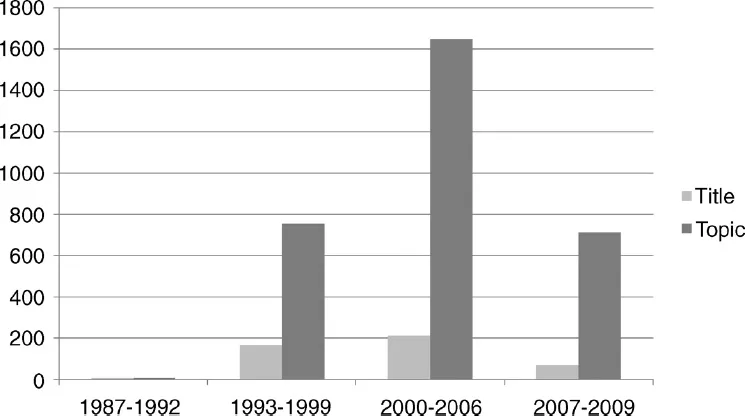
The invention of scanning probe microscopy is considered one of the major advances in materials science since 1950 [1, 2]. Scanning probe microscopy includes a large family of microscopy methods that share two operational elements: the use of a sharp probe (tip) and the feedback mechanism. The feedback loop is characterized by keeping at a constant value the interaction parameter while the probe is scanned across the sample surface. Scanning probe microscopy started with the invention of the scanning tunneling microscope (STM) by Gerd Binnig and Heinrich Rohrer in 1982 [3, 4]. The STM works by detecting the current that flows between a metallic tip situated a few angstroms above a conductive surface when an external voltage is applied. The limitations of the STM to image poorly conducting materials such as biomolecules served as a motivation for Gerd Binnig, Calvin Quate, and Christoph Gerber to invent the atomic force microscope (AFM) in 1986 [5, 6]. The first AFM operated by measuring the static deflection of the probe. This method is called contact mode AFM. One year later Martin, Williams, and Wickramasinghe implemented the dynamic operation in force microscopy [7]. They wanted to use the AFM to measure long-range forces over a distance range of 3–15 nm. They noticed that the amplitude of the tip’s oscillation changed with the tip–surface distance. These changes were related to the gradient of the tip–surface force. At the same time, they proposed to use the amplitude in a feedback loop to get an image of the surface. Such an early start would have anticipated a sudden rise in the number of articles dealing with amplitude modulation AFM. However, it did not happen that way (Figure 1.1). In 1987, the technique was so new that only a handful of groups could master the instrumental and conceptual challenges to design and operate a dynamic AFM [7–11].
Figure 1.1 Histogram of the number of publications per period [104]. In dark gray are the publications that include in the abstract the keywords “tapping mode or tapping force or amplitude modulation force.” In light gray are the publications that include the above key words in the title. The search for the first period was accomplished manually (12 publications). The above figures are merely indicative.
1.2 Evolution Periods and Milestones
The evolution of amplitude modulation AFM is interspersed with several periods where a few topics establish the frontiers of the technique. In some cases, a topic that contributes to shape the technique in a given period could have had its origin in an earlier period. The dates in brackets are indicative.
1.2.1 Early Times (1987–1992)
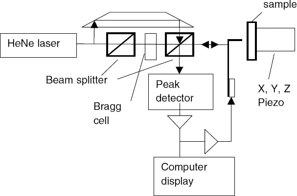
Martin and coworkers recorded the first AFM images obtained in a mode similar or identical to what today is known as amplitude modulation AFM. However, at that time, dynamic AFM was seen as an auxiliary microscopy – its main advantage was its flexibility to be combined with other methods to map magnetic properties [9], electrostatic charges [10], or surface potential differences [11]. The few available dynamic force microscopes were rather complex instruments. For example, the deflection of the lever was measured by using optical interference and the levers were handmade, for example, by bending a tungsten wire.
Figure 1.2 shows the experimental setup of earliest dynamic AFM. The instrument is equipped with an interferometer to measure the lever deflection. The lever is a tungsten needle.
Figure 1.2 Schematic experimental setup of the first dynamic AFM. Notice the use of a bent tungsten wire as the integrated lever–tip system. Adapted from Ref. [7].
1.2.2 Exploration and Expansion (1993–1999)
This period is marked by the rapid expansion and popularization of the technique for surface characterization with nanoscale resolution. The efforts were particularly significant and successful in imaging DNA, proteins and protein–DNA complexes [12–18], and polymer surfaces [19–24]. The progress was sustained by several factors. The implantation of the optical beam deflection method to detect the lever deflection [25, 26] and the fabrication of micromachined cantilevers [27, 28] contributed to the availability of reliable commercial instruments [29]. The operation of tapping mode AFM in liquid [30, 31] made it possible to image dynamic processes, in particular those involving biomolecules with characteristic times in the range of several minutes [32]. The interpretation of the cantilever’s resonance spectra in liquid was simplified by driving the cantilever oscillation with a magnetic force [33]. A significant breakthrough was to implement phase shift measurements simultaneously with topography [34]. This instrumental advance enabled to map compositional variations and changes in material properties in heterogeneous surfaces [35, 36]. In addition, amplitude modulation AFM found new applications by showing its potential for nanopatterning and nanolithography [37, 38].
The collaboration between Paul Hansma and Virgil Elings led to several instrumental and conceptual breakthroughs in probe microscopy. The fabrication of the first wave of short cantilevers is one of them [39]. These cantilevers were initially meant to reduce the thermal noise, but they would have a pivotal role in the development of fast AFM imaging [40].
This period witnessed the earliest attempts to simulate the cantilever–tip dynamics in the presence of an external force [36, 41–44]. Numerical simulations explained the sudden changes, sometimes observed in the amplitude curves in terms of transitions between attractive and repulsive tip–surface interaction regimes [44–46]. Simulations also established that the phase contrast was directly related to energy dissipation processes [47]. This led to the deduction of the first analytical expressions that linked the sine of the phase shift to the energy dissipated by the tip on the sample [48, 49]. Another milestone was the publication of the first comprehensive attempt to describe the dynamics and performance of the microscope in liquid [50].
1.2.3 Cantilever–Tip Dynamics (2000–2006)
Amplitude modulation AFM became so firmly established for nanoscale characterization of surfaces that theoretical contributions devoted to explain its dynamics were initially rejected in the most prestigious physics journals. In fact, during the first two periods, the experimental achievements preceded a comprehensive theoretical understanding of them. Fundamental questions such as the origin of the amplitude reduction, the reconstruction of the force from the observables, or the generation of higher harmonics remained unsatisfactorily addressed. It became evident that the lack of a theory was limiting the evolution of the technique. A significant number of publications were devoted to explain the nonlinear character of the cantilever dynamics [51–54], the generation [55, 56] and the use of higher harmonics, or modes to map material properties [57–59]. A remarkable result was the method developed by Stark and coworkers to obtain the time-resolved force by integrating the higher harmonics of the oscillation [58]. The spatial reconstruction of the force from the amplitude and phase shift curves was also accomplished [60, 61].
Two experimental results highlighted the molecular resolution capabilities of the instrument. Muller and coworkers provided images of a purple membrane surface with a lateral resolution of 1.1 nm [62], a value almost identical to the one reported by the same authors with contact AFM. Later, Klinov and Magonov dispelled the myth that molecular resolution was incompatible with operation in air by imaging a polymer crystal with a resolution of about 0.4 nm [63].
Three instrumental developments underlined the vitality of the technique. First, the promise to observe dynamic processes in real time was eventually fulfilled by designing a microscope with a frame rate of 12.5 s−1 [64]. Second, the observation that the oscillation is asymmetric in liquid led to combine topography and molecular recognition imaging, in a single pass, [65, 66]. Third, the simultaneous excitation of the first two cantilever modes provided a way to separate topography from compositional contrast and to enhance spatial resolution while minimizing tip–surface forces [67, 68].
1.2.4 Multifrequency AFM (2007 to Present)
Several experimental schemes are being proposed to increase the capacities of the instruments to measure material properties while aiming molecular spatial resolution. These methods are based on the simultaneous detection of several harmonics and/or modes of the tip oscillation. In some cases, such as in bimodal AFM, two modes of the cantilever are externally excited [69], while in other cases, for example, it happens in liquid, the higher modes or harmonics are activated by the tip–surface interaction [70, 71]. In other cases, the use of a special cantilever where the tip is offset at one edge of the cantilever enables to measure time-varying forces [72, 73]. Other approaches are exploring the cantilever response when the excitation involves a discrete series [74] or a band of frequencies [75].
The theoretical efforts are focused on two different directions. First, several aspects of the cantilever dynamics in liquid such as the origin of the asymmetry observed in the oscillation, the generation of higher harmonics, the interplay between damping and added mass effects, or the influence of the excitation mode on the dynamics are being addressed [76–81]. Second, the operation of the microscope in various multifrequency regimes is under investigation [80, 82].
1.3 Tapping Mode or Amplitude Modulation Force Microscopy?
Amplitude modulation AFM suffers from a problem of terminology. This is a common issue with scanning probe techniques where standardization is coming rather slowly. Different terms are being used to describe the same method or technique. This anomaly can be partially explained by the vitality of the technique that every now and then surprises with new developments. The first publication describing tapping mode AFM operation [19] makes a point in distinguishing tapping mode from the noncontact method proposed by Martin et al. [7]. Elings and coworkers emphasized that tapping mode AFM operated with amplitudes ranging between 20 and 100 nm, while amplitudes of 5 nm or less were required for noncontact operation. It was claimed that large amplitude values were needed to overcome the adhesion forces and reach stable imaging conditions. However, an analysis based on the equation of motion and the feedback mechanism shows that there are no grounds to support that distinction [83, 84]. The value of the free amplitude is certainly one of the factors that contributes to establishing the operation regime, attractive (usually noncontact) versus repulsive (intermittent contact). But there are other relevant factors such as the radius, the cantilever’s force constant, or the surface energy. Furthermore, the attractive and the repulsive regimes are the solutions of the same equation of motion [46]. Nonlinear dynamics studies have shown that these solutions do coexist under the same operational parameters [52, 83]. The alternation between stable and unstable imaging while the set point amplitude is reduced represents a direct experimental evidence of the coexistence of solutions [52]. Furthermore, the coexistence of solutions is observed for amplitude values ranging from 5 to 25 nm. In fact, the technique is characterized by the use of the amplitude as the feedback parameter not by the values of the amplitude.
In this sense, amplitude modulation AFM seems a more appropriate name for the technique. Furthermore, this term helps us to rationalize the separation of dynamic AFM methods into two modes or techniques, frequency and amplitude modulation, according to the parameter used to establish the feedback mechanism [84, 85]. Acceptable variations are amplitude-modulated or amplitude mode AFM. Nonetheless, on a few occasions I will also use the tapping mode AFM term. The latter is both a tribute to its widespread use and a means to emphasize that we are dealing with the same experimental technique.
1.4 Other Dynamic AFM Methods
Nanoscience sets the scenario for an intense competition among the different microscopy techniques. In the field of force microscopy, there are two major dynamic modes that, on a few occasions, might be seen as competitors. The other technique uses the frequency as the feedback parameter and it is rightly called frequency modulation AFM.
1.4.1 Frequency Modulation AFM
In frequency modulation AFM, the feedback parameter is a frequency shift between the resonant frequency far from the sample surface and the resonant frequency closer to the surface [84, 86, 87]. The resonant frequency depends on the forces acting between the tip and the sample surface. The spatial dependence of the frequency shift, the difference between the actual resonant frequency and that of the free lever, is the source of contrast. An image is formed by profiling the surface topography with a constant frequency shift. FM-AFM has been primarily used to obtain atomic resolution images of semiconductor, metals, insulators, or adsorbates in ultrahigh vacuum [88–94]. The ability to measure the tip–surface force with great accuracy has led to several approaches to identify atoms [95, 96]. Initially, FM-AFM operation was restricted to ultrahigh-vacuum conditions; however, recent results show that molecular resolution imaging can also be achieved in liquid [97–100].
1.4.2 Amplitude Modulation versus Frequency Modulation AFM
Amplitude and frequency modulation AFM are both sophisticated instruments with atomic and molecular resolution capabilities. Mostly, they are seen as complementary dynamic AFM methods because they operate in different environments. However, FM-AFM has also shown its capability to operate under air or liquid environments. This makes it pertinent to comment on the advantages of one method with respect to the other. Let us start by stating that there are no quantitative studies showing that one method has an intrinsic higher signal-to-noise ratio than the other. Some direct comparisons at nanoscale resolution have been attempted by imaging the same sample with the same instrument [101, 102]. However, the results seem to reflect more the skill of the microscopist with a given technique than an intrinsic difference between them. More meaningful information could be extracted by comparing molecular resolution images obtained on the same surface. The cytoplasmic side of purple membrane has been imaged by both techniques [100, 103]. In this case, both methods produce images of similar spatial resolution. However, the versatility of amplitude modulation AFM in achieving molecular resolution while having the possibility of scanning μm2 regions and coping with the presence of sudden height changes is still no mach for frequency modulation AFM instruments.
2
Instrumental and Conceptual Aspects
2.1 Introduction
This chapter introduces the main concepts and elements needed to operate an amplitude modulation AFM (AM-AFM). Expressio...
Table of contents
- Cover
- Title
- Copyright
- Author
- Preface
- Annotation List
- 1: Introduction
- 2: Instrumental and Conceptual Aspects
- 3: Tip–Surface Interaction Forces
- 4: Theory of Amplitude Modulation AFM
- 5: Advanced Theory of Amplitude Modulation AFM
- 6: Amplitude Modulation AFM in Liquid
- 7: Phase Imaging Atomic Force Microscopy
- 8: Resolution, Noise, and Sensitivity
- 9: Multifrequency Atomic Force Microscopy
- 10: Beyond Topographic Imaging
- References
- Index
Frequently asked questions
Yes, you can cancel anytime from the Subscription tab in your account settings on the Perlego website. Your subscription will stay active until the end of your current billing period. Learn how to cancel your subscription
No, books cannot be downloaded as external files, such as PDFs, for use outside of Perlego. However, you can download books within the Perlego app for offline reading on mobile or tablet. Learn how to download books offline
Perlego offers two plans: Essential and Complete
- Essential is ideal for learners and professionals who enjoy exploring a wide range of subjects. Access the Essential Library with 800,000+ trusted titles and best-sellers across business, personal growth, and the humanities. Includes unlimited reading time and Standard Read Aloud voice.
- Complete: Perfect for advanced learners and researchers needing full, unrestricted access. Unlock 1.4M+ books across hundreds of subjects, including academic and specialized titles. The Complete Plan also includes advanced features like Premium Read Aloud and Research Assistant.
We are an online textbook subscription service, where you can get access to an entire online library for less than the price of a single book per month. With over 1 million books across 990+ topics, we’ve got you covered! Learn about our mission
Look out for the read-aloud symbol on your next book to see if you can listen to it. The read-aloud tool reads text aloud for you, highlighting the text as it is being read. You can pause it, speed it up and slow it down. Learn more about Read Aloud
Yes! You can use the Perlego app on both iOS and Android devices to read anytime, anywhere — even offline. Perfect for commutes or when you’re on the go.
Please note we cannot support devices running on iOS 13 and Android 7 or earlier. Learn more about using the app
Please note we cannot support devices running on iOS 13 and Android 7 or earlier. Learn more about using the app
Yes, you can access Amplitude Modulation Atomic Force Microscopy by Ricardo García in PDF and/or ePUB format, as well as other popular books in Technology & Engineering & Electrical Engineering & Telecommunications. We have over one million books available in our catalogue for you to explore.