This book begins with a concise review of the basic science of tissues and then moves into diagnosis, treatment planning, and surgical procedures in endodontics, with an emphasis on the use of enhanced magnification, ultrasonic tips, microinstruments, newer root-end filling materials, and CBCT. Chapters on the maxillary sinus and its relation to surgical endodontics, soft and hard tissue healing, and adjunctive surgical procedures and considerations such as management of procedural accidents, resorption, root amputation, hemisection, replantation, transplantation, crown lengthening, grafting materials, and pharmacology are followed by an assessment of the outcomes of surgical endodontics based on current evidence. The accompanying DVDs present valuable videos demonstrating many of the procedures. These features provide the reader with a textbook that is concise, current, and easy to follow in an interactive manner. Written by a team of leading authorities and richly illustrated, this new compendium of state-of-the-art knowledge and protocols is essential reading for practicing endodontists and residents alike.
eBook - ePub
The Art and Science of Contemporary Surgical Endodontics
- 336 pages
- English
- ePUB (mobile friendly)
- Available on iOS & Android
eBook - ePub
The Art and Science of Contemporary Surgical Endodontics
About this book
Trusted by 375,005 students
Access to over 1.5 million titles for a fair monthly price.
Study more efficiently using our study tools.
Information

The oral environment is a complex region formed of a mixture of hard and soft tissues. Its functions include chewing, swallowing, and speech, as well as acting as an accessory airway. Maintenance of a healthy dentition is imperative to the overall well-being of the individual and proper functioning of the alimentary system. An in-depth understanding of the structure and function of the oral apparatus is required to provide proper care to oral structures and tissues.
In addition to posing the risk of damaging parts of the tooth, endodontic procedures also risk damaging tissues and anatomical structures surrounding the tooth root.1 It is therefore essential to have a thorough understanding of the anatomy of the jaws and in particular the parts of these bones housing neurovascular structures or pneumatic spaces. In this chapter, we examine the anatomy of the maxillary sinus, the mandibular canal with its branches (the mental canal/foramen and the incisive canal), and the incisive canal and palatine foramina of the maxilla, as well as their relationships to the roots of teeth. But first we examine the anatomy of the oral region in general.
The Bony Framework
Maxilla
The maxilla forms much of the midportion of the face, the borders of the nasal aperture, part of the margin of the orbits, and most of the hard palate and the support for the upper lip and teeth (Fig 1-1). Branches from the maxillary artery supply most of the maxillary region, and sensory innervation is provided by the maxillary division of the trigeminal nerve, designated as cranial nerve V2.
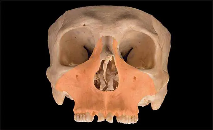
Fig 1-1 The maxilla shown from an anterior angulation. Borders of the maxilla include the floor of the orbit, the zygomatic bone, and the lateral borders of the nasal cavity. The alveolar process forms the inferior boundary.
Associated with the nasal cavity are four sets of paranasal sinuses, found in the frontal, maxillary, sphenoid, and ethmoid bones. The largest of the paranasal sinuses, the maxillary sinus, is housed in the body of the maxilla (Fig 1-2). This pneumatic space is roughly pyramid shaped, with the base of the pyramid formed by the medial wall of the sinus, which is also the lateral wall of the nasal cavity. The medial wall of the sinus is actually formed by parts of five bones—the maxilla, the lacrimal bone, the inferior nasal concha, the perpendicular plate of the palatine bone, and the uncinate process of the ethmoid bone. The ostium of the sinus drains to the middle meatus, the space inferior to the middle nasal concha, on the lateral nasal wall (Fig 1-3). The posterior wall of the sinus faces the maxillary tuberosity, the roof forms the floor of the orbit, and the floor of the sinus extends inferiorly into the alveolar ridge of the maxilla, most commonly in the area of the second premolar and first and second molars. Innervation of the mucosa lining the maxillary sinus is provided by the posterior, middle, and anterior superior alveolar nerves and the infraorbital nerve, all branches of V2. The blood supply is primarily from branches of the maxillary artery accompanying these nerve branches, as well as the descending palatine artery, which accompanies the greater and lesser palatine nerves, and sometimes the posterior superior alveolar artery. During endodontic surgery, it is important to be aware of the position of the posterior superior alveolar nerve to avoid damaging it.
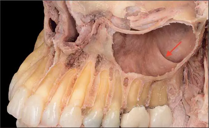
Fig 1-2 The maxillary sinus is housed in the body of the maxilla. This pneumatic space is roughly pyramid shaped, with the base of the pyramid formed by the medial wall of the sinus, which is also the lateral wall of the nasal cavity. Note the presence of a sinus septum (arrow).
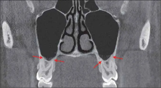
Fig 1-3 A coronal section of the maxillary sinus using cone beam computed tomography (CBCT) imaging. Note the close proximity of the root tips to the floor of the sinus (red arrows).
The maxillary sinus, like all the paranasal sinuses, is lined by respiratory mucosa, comprising pseudostratified ciliated columnar epithelium with goblet cells overlying a rather thin lamina propria that adheres to the periosteum covering underlying bone (mucoperiosteum). The mucosa covering the floor of the sinus—the sinus membrane—is often somewhat thickened and is sometimes referred to as the Schneiderian membrane clinically (Figs 1-4 and 1-5).
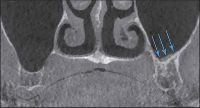
Fig 1-4 Coronal CBCT image of a slightly thickened sinus membrane (arrows).
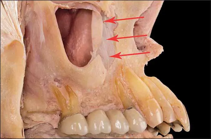
Fig 1-5 The mucosa covering the floor and walls of the sinus is sometimes referred to as the Schneiderian membrane clinically. The arrows point to a section left during the dissection. Notice its thin, delicate nature.
The alveolar process or ridge is the portion of the maxilla that houses the roots of the teeth. The cortical plate forming the outer walls of this ridge is relatively thin, allowing the infiltration of anesthetics. Below the midpoint of the inferior orbital rim, an infraorbital foramen provides passage for the infraorbital nerve, a continuation of the maxillary nerve (V2), along with infraorbital vessels. A canine eminence shows the location of the root of the canine tooth. Medial to this eminence is an incisive fossa, and lateral to the eminence is a canine fossa (Fig 1-6).
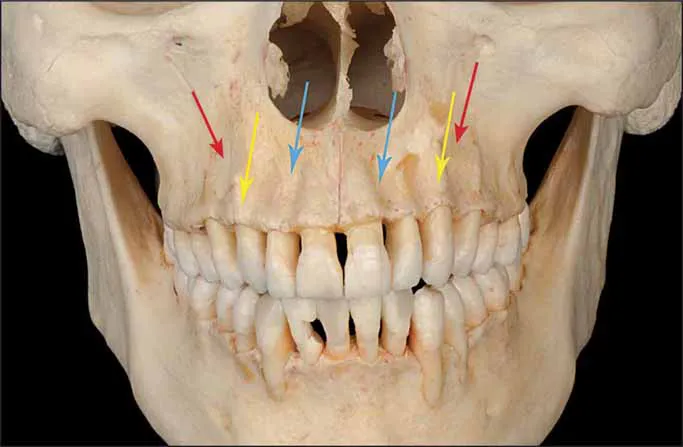
Fig 1-6 The canine eminence (yellow arrows) shows the location of the root of the canine tooth. Medial to this eminence is an incisive fossa (blue arrows), and lateral to the eminence is a canine fossa (red arrows).
The hard palate is formed primarily by the two lateral palatine processes of the maxilla, which fuse in the midline to form the intermaxillary, or median palatine, suture. Two transverse palatine sutures separate the posterior borders of the palatine processes of the maxilla from the horizontal plates of the palatine bones, which form the posterior third of the hard palate. These sutures are sometimes incomplete laterally, forming greater palatine foramina that transmit the greater palatine nerves and vessels. Posterior to the greater palatine foramina are smaller lesser palatine foramina, located within the pyramidal processes of the palatine bones and transmitting the lesser palatine nerves and vessels. Just posterior to the maxillary central incisors lies the incisive fossa, into which open incisive canals by way of incisive foramina, transmitting the nasopalatine nerves and sphenopalatine vessels from the nasal cavity (Fig 1-7).

Fig 1-7 (a and b) Remarkable features of the hard palate include the intermaxillary suture (blue arrows), greater palatine foramina (red arrows), and lesser palatine foramina (green arrows). Note the supplemental foramina (yellow arrows) in the posterior region (b).
The oral portion of the maxilla is covered by mucosa. The buccal or vestibular surface of the maxilla is c...
Table of contents
- Cover
- Title Page
- Copyright Page
- Contents
- Foreword
- Preface
- Contributors
- List of Videos
- 1 Anatomical Zones in Endodontic Surgery
- 2 Histology of Tissues Involved in Surgical Endodontics
- 3 Bone Physiology and Metabolism in Endodontic Surgery
- 4 Radiolucent Periapical Pathosis
- 5 Diagnosis and Treatment Planning
- 6 Cone Beam Computed Tomography in Treatment Planning of Periapical Surgery
- 7 Magnification and Illumination in Apical Surgery
- 8 Local Anesthesia and Hemostasis
- 9 Soft Tissue Management
- 10 Apical Microsurgery: Application of Armamentaria, Materials, and Methods
- 11 Root-End Filling Materials
- 12 Surgical Endodontics and the Maxillary Sinus
- 13 Suturing and Postoperative Instructions
- 14 Wound Healing
- 15 Adjunctive Procedures
- 16 Pharmacology in Surgical Endodontics
- 17 Outcomes of Endodontic Surgery
- Index
Frequently asked questions
Yes, you can cancel anytime from the Subscription tab in your account settings on the Perlego website. Your subscription will stay active until the end of your current billing period. Learn how to cancel your subscription
No, books cannot be downloaded as external files, such as PDFs, for use outside of Perlego. However, you can download books within the Perlego app for offline reading on mobile or tablet. Learn how to download books offline
Perlego offers two plans: Essential and Complete
- Essential is ideal for learners and professionals who enjoy exploring a wide range of subjects. Access the Essential Library with 800,000+ trusted titles and best-sellers across business, personal growth, and the humanities. Includes unlimited reading time and Standard Read Aloud voice.
- Complete: Perfect for advanced learners and researchers needing full, unrestricted access. Unlock 1.5M+ books across hundreds of subjects, including academic and specialized titles. The Complete Plan also includes advanced features like Premium Read Aloud and Research Assistant.
We are an online textbook subscription service, where you can get access to an entire online library for less than the price of a single book per month. With over 1.5 million books across 990+ topics, we’ve got you covered! Learn about our mission
Look out for the read-aloud symbol on your next book to see if you can listen to it. The read-aloud tool reads text aloud for you, highlighting the text as it is being read. You can pause it, speed it up and slow it down. Learn more about Read Aloud
Yes! You can use the Perlego app on both iOS and Android devices to read anytime, anywhere — even offline. Perfect for commutes or when you’re on the go.
Please note we cannot support devices running on iOS 13 and Android 7 or earlier. Learn more about using the app
Please note we cannot support devices running on iOS 13 and Android 7 or earlier. Learn more about using the app
Yes, you can access The Art and Science of Contemporary Surgical Endodontics by Mahmoud Torabinejad,Richard Rubinstein in PDF and/or ePUB format, as well as other popular books in Medicine & Dentistry. We have over 1.5 million books available in our catalogue for you to explore.