- 170 pages
- English
- ePUB (mobile friendly)
- Available on iOS & Android
eBook - ePub
Physical Sensors for Biomedical Applications
About this book
The material in this book is based upon a two-day workshop on solid state physical sensors for biomedical applications held in Huron, Ohio, December 8-9, 1977. The individual sections of the book are based upon presentations made by the authors at the workshop. Each presentation was transcribed and given to the authors for revision. Also, transcribed, are the discussions had following each presentation.
Tools to learn more effectively

Saving Books

Keyword Search

Annotating Text

Listen to it instead
Information
Applications of Physical Sensors in Life Sciences
Chapter 7
The Application of Physical Sensors to Studies of the Cardiovascular System
Ernest P. McCutcheon
Table of Contents
- I. Introduction
- II. Force, Dimensions, and Pressure
- A. Force and Dimensions: Isolated Tissue
- B. Force and Dimension: Intact Ventricle
- C. Force and Dimension: In Situ Veins and Arteries
- D. Pressure
- III. Volume, Flow, and Motion
- A. Volume: Single Dimension
- B. Volume: Area
- C. Flow
- D. Motion
- IV. Biopotentials
- V. Comments on Device Criteria
- VI. Conclusions
- References
I. Introduction
The purpose of this presentation is to initiate and stimulate discussion of the physical sensors used to obtain significant information on cardiovascular function. It is not an encyclopedic review of all possible applications to this area, since virtually every type or class of physical sensor has been used or has the potential for use in cardiovascular studies. It is intended to provide a representative example of the broad applications of physical sensors in a specific specialty area. My viewpoint is that of the user community rather than the instrumentation design engineer. Examples have been chosen to illustrate particular classes of problems and to demonstrate uncertainties created by shortcomings of existing sensing techniques. Emphasis is on the more pragmatic and realizable rather than the theoretical and optimal. Human applications have received considerable emphasis in earlier chapters, and for that reason as well as my personal research orientation, applications in animal studies will be stressed.
In every case, certain background considerations always apply even if they are not mentioned explicitly. Factors to be kept in mind affecting the choice of sensors and implementation of sensor systems include (1) the degree to which the characteristics of the variable are static or dynamic, (2) whether the structure or organ sampled is isolated and perfused in a bath, or within the intact organism, (3) the extent to which the required approach is noninvasive or invasive and implantable, (4) the length of time required for the measurement, ranging from brief and acute to long-term and chronic, and (5) whether the sensor output is accessed through a directly wired connection or transmitted through a telemetry link. Such considerations always impact the measurement process and illustrate the broad range of factors affecting applications of physical sensors in analysis of cardiovascular function.
The selected examples of cardiovascular measurements have been grouped into three primary areas. First is force and its spatial or structural distribution as tension (force/length), pressure and stress (force/area), and the proportional displacement of strain (Δ length/initial length). Second is the determination of volumes and the movement of fluids and structures, an aspect also including sound. The third area includes the biopotentials and electrical activity, and the thermal processes underlying all cardiovascular function, about which very little will be said in this paper since the emphasis is on sampling of physical-mechanical events.
II. Force, Dimensions, and Pressure
A. Force and Dimensions: Isolated Tissue
Let us first consider an example illustrating the class of an isolated tissue specimen in a comparatively highly controlled environment, a configuration often taken for granted (Figure I).1 This approach is particularly suitable for the determination of force, tension, and stress/strain properties. This is a very significant and fundamental technique for obtaining information about cardiovascular function, and perfused segments of vessels or heart muscle are frequently studied in this manner.1-7 The configuration illustrated in Figure 1 is for the study of papillary muscle.1 One of the notable features is its horizontal orientation, technically somewhat easier to manage than a vertical system. The force transducer is incorporated in the short, rigid lever arm. A muscle lever with clamp (not shown) is attached to the transducer mounting arm to hold one end of the specimen. The other clamp (shown) holds the opposite end of the tissue sample, and a micrometer adjusts the overall specimen length. The pair of clamps holds the sample in the isometric state so that it does not shorten with contraction. Thus, the transducing system must have very low compliance. Frequently, such systems have used a deflecting beam with an attached strain gauge for measurement of the contraction-generated force. A special feature of the device of Figure 1 is the linear semiconductor element (Pitran transistor) incorporated in the fixed transducer mounting arm to provide a highly sensitive detector, A very stable system with a capacitive transducer has also been described.2 One of the nicest things about this bath preparation is that the desired dimensions such as length, circumference, and cross-sectional areas can be determined easily by a variety of techniques, from rulers and calipers3-5 to optical, noncontacting approaches such as those described in the Workshop paper by Christensen. Results of such experiments have been valuable in documenting the characteristic exponential stress-strain relationship of cardiovascular structures and the linearity of the elastic modulus vs. stress. Interestingly, a papillary muscle in a configuration of this general type was thought initially to contract homogeneously, but on closer examination, the external fibers are observed to shorten while those in the central core lengthen, so that even isolated samples can be very complicated systems.6,7 Testing of vessel pieces under similar conditions has quantified differences between veins and arteries, and the increased passive arterial stiffness and other changes with age and atherosclerosis.8,9
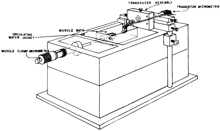
FIGURE 1. Temperature controlled bath for study of isolated tissue specimens. The force transducer (Pitran transistor) is mounted in the short, rigid lever arm. A muscle lever with clamp (not shown) secures one end of the specimen and is placed so that its projecting bead activates the transducer. Th...
Table of contents
- Cover
- Title
- Copyright
- PREFACE
- THE EDITORS
- CONTRIBUTORS
- Contents
- Basic Physics of Physical Sensors
- State of the Art
- Applications of Physical Sensors in Life Sciences
- Index
Frequently asked questions
Yes, you can cancel anytime from the Subscription tab in your account settings on the Perlego website. Your subscription will stay active until the end of your current billing period. Learn how to cancel your subscription
No, books cannot be downloaded as external files, such as PDFs, for use outside of Perlego. However, you can download books within the Perlego app for offline reading on mobile or tablet. Learn how to download books offline
Perlego offers two plans: Essential and Complete
- Essential is ideal for learners and professionals who enjoy exploring a wide range of subjects. Access the Essential Library with 800,000+ trusted titles and best-sellers across business, personal growth, and the humanities. Includes unlimited reading time and Standard Read Aloud voice.
- Complete: Perfect for advanced learners and researchers needing full, unrestricted access. Unlock 1.4M+ books across hundreds of subjects, including academic and specialized titles. The Complete Plan also includes advanced features like Premium Read Aloud and Research Assistant.
We are an online textbook subscription service, where you can get access to an entire online library for less than the price of a single book per month. With over 1 million books across 990+ topics, we’ve got you covered! Learn about our mission
Look out for the read-aloud symbol on your next book to see if you can listen to it. The read-aloud tool reads text aloud for you, highlighting the text as it is being read. You can pause it, speed it up and slow it down. Learn more about Read Aloud
Yes! You can use the Perlego app on both iOS and Android devices to read anytime, anywhere — even offline. Perfect for commutes or when you’re on the go.
Please note we cannot support devices running on iOS 13 and Android 7 or earlier. Learn more about using the app
Please note we cannot support devices running on iOS 13 and Android 7 or earlier. Learn more about using the app
Yes, you can access Physical Sensors for Biomedical Applications by Michael R. Neuman in PDF and/or ePUB format, as well as other popular books in Physical Sciences & Biology. We have over one million books available in our catalogue for you to explore.