eBook - ePub
Organization of the Extracellular Matrix
A Polarization Microscopic Approach
- 295 pages
- English
- ePUB (mobile friendly)
- Available on iOS & Android
eBook - ePub
About this book
This book describes analysis techniques and results of topics such as physical backgrounds, chemical backgrounds, and principal methods of topo-optical reactions used in ultrastructure research of the ECM; orientation patterns of GAGs and collagen in different tissues/cartilage, cornea, kidney basement membranes, and skin; factors involved in the formation of submicroscopically ordered matrix structure; and cell-matrix interactions, including cytoskeleton-cell-membrane-matrix relationships. A summarization of the advantages and limitations of polarization microscopy compared to electron microscopy in ultracellular research is also included. Cell biologists, histologists, pathologists, and biochemists in connective tissue research will find this book to be an invaluable reference tool.
Trusted by 375,005 students
Access to over 1.5 million titles for a fair monthly price.
Study more efficiently using our study tools.
Information
Chapter 1
A SHORT OVERVIEW OF MAJOR CONSTITUENTS OF THE EXTRACELLULAR MATRIX
TABLE OF CONTENTS
I. | Introduction | |
II. | Major Constituents of the Matrix | |
A. | Collagen | |
B. | Elastin | |
C. | Proteoglycans | |
D. | Noncollagenous Proteins | |
III. | Receptors Involved in Cell-Matrix Interactions | |
References | ||
I. INTRODUCTION
The extracellular matrix (ECM) constitutes a significant part of the multicellular animal organism. Except blood, there are no tissues without an ECM. In connective tissue the matrix is abundant, while its bulk is much less in other tissues where sometimes special techniques are needed for its detection. The matrix molecules are produced and degraded by cells.
After deposition the ECM, or some of its constituents, influence a number of functions of contacting cells. The cell-matrix interactions are generally mediated by specific receptors in the cell membrane. The ECM can control the metabolism, growth, differentiation, migration, and adhesion of cells, the cytoskeletal organization and cell shape, and the organization of the tissues1, 2, 3, 4, 5 (Figure 1). There are experimental data suggesting that ECM exerts an influence on gene expression via transmembrane proteins and cytoskeletal compartments which are associated with polyribosomes as well as the nuclear matrix.6 According to this theory, the cytoskeletal association with polyribosomes may affect the rate of protein sythesis, while the matrix-cell membrane-cytoskeleton-nuclear matrix interaction could affect mRNA processing and rates of transcription.
Thus, the ECM is not a simple and passive structural scaffold. Multicellular animals can function only because single cells learned to synthesize, and maintain macromolecular connections between them, and they have become dependent upon the matrix they made.7 This has been realized in the past decade due to the large amounts of new data accumulated about the biosynthesis, molecular anatomy, functions, and interactions of the ECM constituents. It is now, therefore, generally accepted that the interest of cell biology extends beyond the cell membrane.
There are different major trends in the contemporary ECM research. One of them is — the most classical — the biochemical approach with the aim of a detailed description of the matrix constituents and their interactions. The second and tremendously increasing field is the molecular biology of the ECM.8 The third trend — which is rapidly developing — is the study of the role of the ECM in diseases.9, 10, 11 A fourth direction is to get new data about the ECM structure in different normal and pathological tissues. In this respect, immunohistochemistry has contributed much to our present knowledge.12 Other morphological techniques like electron microscopy and polarization microscopy, however, are also indispensable tools of ECM research. As this volume is dedicated to a morphological approach of the ECM organization, it is beyond the scope of the present work to give a detailed description of the ECM constituents. This chapter shortly summarizes some basic informations about the major macromolecular components of the ECM, other details are found in the references given as well as in the relevant chapters of this volume.
II. MAJOR CONSTITUENTS OF THE MATRIX
A. COLLAGEN
Collagen is the most ubiquitous protein in animal kingdom. It is a heterogenous family of proteins, but the members share common features both in primary structure and conformation, e.g., the repeating Gly-X-Y triplet in the polypeptide chain, and the presence of hydroxyproline and hydroxylysine.
Today, 14 collagens are distinguished. They differ genetically, chemically, and immunologically.13, 14, 15, 16 There are 5 of the collagen types which are known to be involved in the formation of crossbanded fibrils (types I, II, III, V, XI), but other collagens (e.g., type IX) may also be associated with the fibrillar surface. The collagen fibrils and fibers represent systems where the degree of the orientation of the macromolecular constituents is the highest in the ECM. Some collagen types are tissue-specific: types II, IX, X and XI are round in cartilage (and also in vitreous body and notochord) (see Chapter 4). Collagen type I molecules form the thick, interstitial collagen fibers (in skin, bone, tendon, etc.), while type III is present in a wide variety of tissues, generally associated with type I collagen, forming thinner fibrils (“reticulin” fibers of the classical histology). Type IV collagen is specific to basement membranes, they form an orientad micellar network but not fibrils (see Chapter 7). Type V collagen shows a wide distribution. It is generally found in the pericellular matrix in form of small fibrils, and, therefore, it is frequently named as pericellular collagen and considered as a part of the exocytoskeleton.13,17 Collagen type VI is present in a wide variety of tissues as interstitial collagen. Type VIII collagen is secreted by endothelial cells. Type XII collagen is expressed in embryonic tendon, but its role is still unknown.
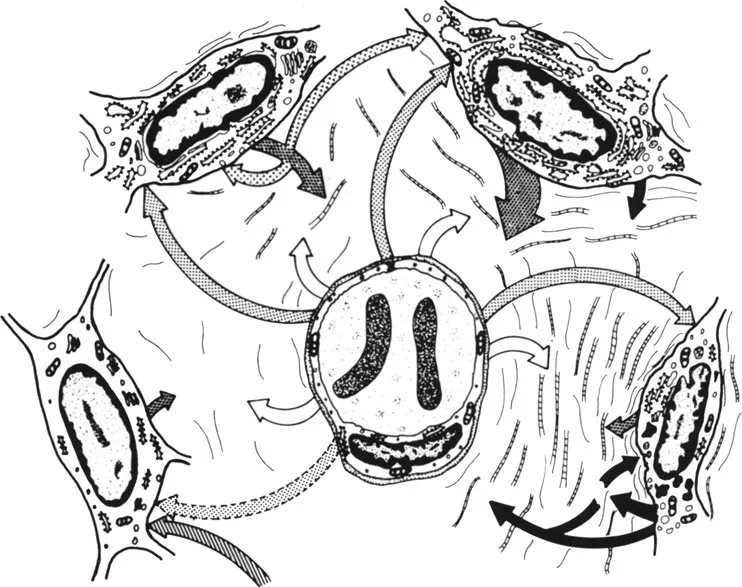
FIGURE 1. A scheme illustrating some selected aspects of cell-matrix interactions. As an example, a portion of a connective tissue is depicted. In the center of the diagram, a blood capillary can be seen. Around the capillary, cells of varying degree of differentiation are shown (from left to right: undifferentiated, differentiating, mature and aging cell). The space between the cells and the capillary is occupied by the extracellular matrix (the fibrillar components are masked by a few fine lines). Empty arrows: anabolic transport; dotted arrows: humoral factors regulating cell functions; arrows filled with circles: secretion of matrix constituents; arrows filled with triangles: effects of matrix on cell functions; dense arrows: matrix degradation and catabolic transport. (From Módis, L., Acta Biol. Acad. Sci. Hung., 29, 197, 1978. With permission.)4
Collagen may be involved in different interactions. Collagen molecules of the same type can be associated with each other forming fibrils or micelles (e.g., type I fibrils and type IV micelles). Different collagen types can also be bound with each other (e.g., in collagen fibrils of the hyaline cartilage18). Type VII — as a major part of anchoring fibrils-interconnects basement lamina to the underlying connective tissue matrix.19 Different collagen types may be attached to the cell membrane via collagen receptors or by nectin molecules (fibronectin, chondronectin, laminin, see later). Proteoglycans also interact with collagen, predominantly by electrostatic forces but more specific interactions may also occur.20 Considering these multisided interactions, collagens can be regarded as key molecules of the extracellular matrix organization.
B. ELASTIN
The elastic behavior of some tissues (lung, artery wall, skin) is mainly due to the presence of elastic fibers in the ECM. The morphological appearance of elastic fibers is simple, the mature fibers appear as homogeneous structures under electron microscope. They have been, therefore, considered to possess an amorphous structure. Polarization microscopy revealed that elastic fibers have submicroscopically oriented constituents.21 These may correspond to the microfibrils of 10 to 12 n...
Table of contents
- Cover
- Title Page
- Copyright Page
- Dedication
- Table of Contents
- Chapter 1 A Short Overview of Major Constituents of the Extracellular Matrix
- Chapter 2 Physical Backgrounds of Polarization Microscopy
- Chapter 3 Topo-Optical Reactions Used in Polarization Microscopic Ultrastructure Research
- Chapter 4 Structure of the Hyaline Cartilage Matrix
- Chapter 5 The Organic Bone Matrix
- Chapter 6 Structure of the Corneal Stroma
- Chapter 7 Structure of Kidney Basement Membranes
- Chapter 8 Experimental Reconstruction of the Oriented Glycosaminoglycan Structure of the Extracellular Matrix
- Chapter 9 Factors Involved in Formation and Maintenance of Oriented Microstructure of Matrix Constituents
- Chapter 10 Polarization Microscopy of Amyloid
- Chapter 11 Correlation Between Polarization and Electron Microscopic Findings in Ultrastructure Research of Proteoglycans
- Chapter 12 Methodological Appendix
- Index
Frequently asked questions
Yes, you can cancel anytime from the Subscription tab in your account settings on the Perlego website. Your subscription will stay active until the end of your current billing period. Learn how to cancel your subscription
No, books cannot be downloaded as external files, such as PDFs, for use outside of Perlego. However, you can download books within the Perlego app for offline reading on mobile or tablet. Learn how to download books offline
Perlego offers two plans: Essential and Complete
- Essential is ideal for learners and professionals who enjoy exploring a wide range of subjects. Access the Essential Library with 800,000+ trusted titles and best-sellers across business, personal growth, and the humanities. Includes unlimited reading time and Standard Read Aloud voice.
- Complete: Perfect for advanced learners and researchers needing full, unrestricted access. Unlock 1.5M+ books across hundreds of subjects, including academic and specialized titles. The Complete Plan also includes advanced features like Premium Read Aloud and Research Assistant.
We are an online textbook subscription service, where you can get access to an entire online library for less than the price of a single book per month. With over 1.5 million books across 990+ topics, we’ve got you covered! Learn about our mission
Look out for the read-aloud symbol on your next book to see if you can listen to it. The read-aloud tool reads text aloud for you, highlighting the text as it is being read. You can pause it, speed it up and slow it down. Learn more about Read Aloud
Yes! You can use the Perlego app on both iOS and Android devices to read anytime, anywhere — even offline. Perfect for commutes or when you’re on the go.
Please note we cannot support devices running on iOS 13 and Android 7 or earlier. Learn more about using the app
Please note we cannot support devices running on iOS 13 and Android 7 or earlier. Learn more about using the app
Yes, you can access Organization of the Extracellular Matrix by Laszlo Modis in PDF and/or ePUB format, as well as other popular books in Biological Sciences & Biology. We have over 1.5 million books available in our catalogue for you to explore.