- 125 pages
- English
- ePUB (mobile friendly)
- Available on iOS & Android
eBook - ePub
Imaging Atlas of the Normal Gallbladder and Its Variants
About this book
An in-depth knowledge of the wide spectrum of normal gallbladder appearances is vital to appropriate clinical workup and the correct diagnosis of patients with upper abdominal symptoms. This book covers the range of appearances of the normal gallbladder and its variants, including discussions of anatomy, embryology, and imaging techniques to promote a better understanding of the subject. Anomalies of number, location, and form are each addressed in separate chapters, and discussions of imaging artifacts of the gallbladder and pseudolesions, which can mimic gallbladder disease, are also included. A short chapter on the appearance of the fetal gallbladder is included to round out this comprehensive volume.
Trusted by 375,005 students
Access to over 1.5 million titles for a fair monthly price.
Study more efficiently using our study tools.
Information
Chapte 1
INTRODUCTION
Jon W. Meilstrup
It is not surprising that the gallbladder, a small sac-like structure which serves as the biliary reservoir, has been the source of so much discussion in recent years. Fifteen million Americans have gallstones,1 many of which are asymptomatic.2 With this potential cause of significant morbidity, it is no wonder there is interest in the gallbladder and the diagnosis of gall-bladder disease.
Since 1900, when gallstones were first radiographically detected, continual improvement has taken place in gallbladder imaging.3 The development of contrast media led to oral cholecystography, and with additional technological advances, sonography became available. While they have limited roles in specifically imaging the gallbladder, computed tomography (CT) and magnetic resonance imaging (MRI) are frequently used to evaluate the abdomen, including the gallbladder. Recently, percutaneous intraluminal gallbladder ultrasound has even been reported as a diagnostic tool.4 New therapies for gallbladder disease, such as chemical dissolution of gallstones, lithotripsy, laparoscopic cholecystectomy, and percutaneous cholecystostomy and drainage attest to the continued need for appropriate imaging and diagnosis in gallbladder disease. Vital to current clinical workup and correct diagnosis is a knowledge of the wide array of normal gallbladder appearances, variants, mimics, and artifacts.
This volume deals with the range of appearances of the normal gall-bladder and its variants. A discussion of anatomy, embryology, and imaging techniques is included for a better understanding of the normal gallbladder. Variants are grouped into three categories: anomalies of number, location, and form,5 each addressed in a separate chapter. Discussions of imaging artifacts of the gallbladder and pseudolesions which can mimic gallbladder disease are also included. A short chapter on the appearance of the fetal gallbladder is provided for completeness. Biliary duct variants are not treated here, except where they relate to a specific gallbladder entity.
Because sonography and CT are so frequently used to evaluate the gall-bladder and upper abdomen, variants seen with these modalities are emphasized throughout this volume. However, other imaging modalities such as oral cholecystography, endoscopic retrograde cholecystopancreatography (ERCP), scintigraphy, and MRI are also included.
The case examples used in this volume have been largely collected by the editor and contributors because of their particular fascination with the variable appearance of the normal gallbladder. As various imaging modalities have developed, we have been excited to see the gallbladder displayed to new and different advantages. When viewing gallbladder imaging studies, especially when an unusual variant is evident, each image must be carefully evaluated against a backdrop of knowledge of what is normal and what is not. We believe that “. . . improvement in the accuracy of differential diagnosis resulting from the more effective use of normal findings should improve patient care by aiding in the process of making sound decisions.”6
To this end, it is hoped that the reader finds a similar interest in the many gallbladder variants that inevitably will be seen in clinical practice, and that this small volume will help the reader in making an appropriate clinical diagnosis when imaging the normal gallbladder.
REFERENCES
1. Kaiser, M. H., Cholelithiasis: clinical aspects, in Bockus Gastroenterology, 4th ed., Berk, J. E., Haubrich, W. S., Kaiser, M. H., Roth, J. L. A., and Schaffner, F., Eds., W. B. Saunders, Philadelphia, 1985, 3619.
2. Hopper, K. D., Landis, J. R., Meilstrup, J. W., McCauslin, M.A., and Sechtin, A. G., The prevalence of asymptomatic gallstones in the general population, Invest. Radiol., 26, 939, 1991.
3. Feld, R., Kurtz, A. B., and Zeman, R. K., Imaging the gallbladder: a historical perspective, AJR, 156, 737, 1991.
4. van Sonnenberg, E., D’Agostino, H. B., Sanchez, R. L., Goodacre, B. B., Esch, O. G., Easter, D. E., and Gosink, B. B., Percutaneous intraluminal US in the gallbladder and bile ducts, Radiology, 182, 693, 1992.
5. Lockwood, B. C., Congenital anomalies of the gallbladder, JAMA, 136, 678, 1948.
6. Gorry, G. A., Pauker, S. G., and Schwartz, W. B., The diagnostic importance of the normal finding, N. Engl. J. Med., 298, 486, 1978.
Chapter 2
NORMAL GALLBLADDER ANATOMY AND IMAGING
Simon Westacott and Rickhesvar Mahraj
TABLE OF CONTENTS
- Introduction
- Normal Anatomy
- Connections and Relations
- Vascular Supply
- Histology
- Imaging the Normal Gallbladder
- Oral Cholecystography
- Sonography
- Scintigraphy
- Computed Tomography
- Magnetic Resonance Imaging
References
I. Introduction
This chapter defines the most common arrangement of the gross anatomy of the gallbladder, including its relations and vascular anatomy, gives a short account of its microscopic structure, and describes the appearance of these structures when imaged by different modalities.
II. Normal Anatomy
The gallbladder is a pear-shaped, hollow viscus (Figure 1) that is connected to the extrahepatic biliary tree by the cystic duct. Bile from the hepatocytes is stored and concentrated in the gallbladder and ultimately ejected into the gastrointestinal (GI) tract, under the influence of the parasympathetic nervous system and circulating hormones, notably cholecystokinin. There is considerable variation in dimensions; approximate length and diameter of the fully distended gallbladder are l 0 and 4 cm, respectively. The gallbladder’s capacity is up to 45 ml.1
The gallbladder is divided into a fundus, body, and neck. The fundus is a hemispherical ending which forms the anteroinferior margin. The neck forms the posterosuperior limit of the gallbladder and connects it to the cystic duct. The gallbladder lumen is widest at the junction of the body and the fundus and tapers toward the neck; the most rapidly tapering segment is called the infundibulum. Hartmann’s pouch is a pathological saccular expansion of the neck and not a feature of normal anatomy.2
The gallbladder is located in the gallbladder fossa,3 a concavity of the inferior surface of the liver which generally marks the caudal limit of the interlobar fissure* separating the right and left hepatic lobes. The body and neck are loosely attached to the liver by connective tissue, which is continuous with the connective tissue of the interlobar fissure. Peritoneum covers the fundus, which frequently projects beyond the inferior margin of the liver, and those parts of the gallbladder not directly adherent to the liver. The degree of contact between the gallbladder and the liver is highly variable: in some individuals the gallbladder is invaginated into the interlobar fissure; conversely, it may be entirely enveloped by peritoneum, in which case it is attached to the liver by a mesentery.
The gallbladder fossa is continuous posterosuperiorly with the porta hepatis, a transverse groove in the inferior surface of the liver traversed by the hepatic artery, portal vein, and common bile duct. A reflection of the peritoneum which covers the inferior surface of the liver forms a sleeve around these structures known as the hepatoduodenal ligament (free edge of the lesser omentum). Occasionally, a fold of peritoneum may connect the hepatoduodenal ligament to the peritoneal reflections around the gallbladder, forming the cholecystoduodenal ligament.
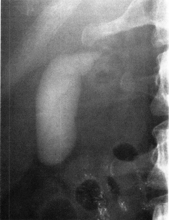
Figure 1 Oral cholecystogram demonstrating normal opacitication of the gallbladder. Unabsorbed contrast remains in the transverse colon.
A. Connections and Relations
The gallbladder is connected to the biliary tree by the cystic duct, which joins the common hepatic duct to form the common bile duct. Anteriorly, the fundus of the gallbladder is related to the parietal peritoneum of the anterior abdominal wall; its surface marking is the junction of the costal margin with the lateral border of the rectus abdominis muscle. Anterosuperiorly, the body of the gallbladder is in close contact with the liver. Posteroinferiorly, the gallbladder is related to the second part of the duodenum and the transverse colon and, occasionally, to the inferior vena cava (Figure 2).
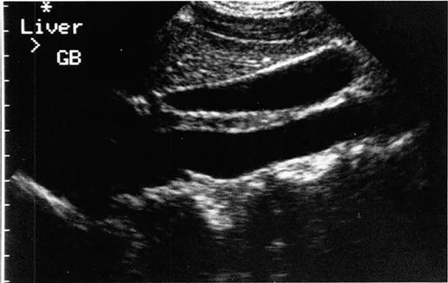
FIGURE 2 Longitudinal sonogram through a normal gallbladder.
B. Vascular Supply
The arterial supply of the gallbladder emanates from the cystic artery, most commonly a branch of the right hepatic artery. Venous drainage is by multiple, small, unnamed vessels into the portal venous system. Lymphatic drainage is to nodes in the porta hepatis and subsequently to preaortic nodes near the celiac axis.
C. Histology
The gallbladder wall has three distinct layers. The outermost (serosal) layer is the peritoneum, as described above. The serosa is deficient in the areas in which the gallbladder is in contact with the liver, where it is replaced by connective tissue. Microscop...
Table of contents
- Cover Page
- Halftitle Page
- Title Page
- Copyright Page
- Content Page
- Chapter 1 Introduction
- Chapter 2 Anatomy and Imaging of the Normal Gallbladder
- Chapter 3 Embryology of the Gallbladder
- Chapter 4 The Fetal Gallbladder
- Chapter 5 Variations of Location
- Chapter 6 Variations of Number
- Chapter 7 Variations of Form
- Chapter 8 Artifacts
- Chapter 9 Pseudolesions
- Index
Frequently asked questions
Yes, you can cancel anytime from the Subscription tab in your account settings on the Perlego website. Your subscription will stay active until the end of your current billing period. Learn how to cancel your subscription
No, books cannot be downloaded as external files, such as PDFs, for use outside of Perlego. However, you can download books within the Perlego app for offline reading on mobile or tablet. Learn how to download books offline
Perlego offers two plans: Essential and Complete
- Essential is ideal for learners and professionals who enjoy exploring a wide range of subjects. Access the Essential Library with 800,000+ trusted titles and best-sellers across business, personal growth, and the humanities. Includes unlimited reading time and Standard Read Aloud voice.
- Complete: Perfect for advanced learners and researchers needing full, unrestricted access. Unlock 1.5M+ books across hundreds of subjects, including academic and specialized titles. The Complete Plan also includes advanced features like Premium Read Aloud and Research Assistant.
We are an online textbook subscription service, where you can get access to an entire online library for less than the price of a single book per month. With over 1.5 million books across 990+ topics, we’ve got you covered! Learn about our mission
Look out for the read-aloud symbol on your next book to see if you can listen to it. The read-aloud tool reads text aloud for you, highlighting the text as it is being read. You can pause it, speed it up and slow it down. Learn more about Read Aloud
Yes! You can use the Perlego app on both iOS and Android devices to read anytime, anywhere — even offline. Perfect for commutes or when you’re on the go.
Please note we cannot support devices running on iOS 13 and Android 7 or earlier. Learn more about using the app
Please note we cannot support devices running on iOS 13 and Android 7 or earlier. Learn more about using the app
Yes, you can access Imaging Atlas of the Normal Gallbladder and Its Variants by J.W. Meilstrup in PDF and/or ePUB format, as well as other popular books in Law & Forensic Science. We have over 1.5 million books available in our catalogue for you to explore.