- 392 pages
- English
- ePUB (mobile friendly)
- Available on iOS & Android
eBook - ePub
Diagnosis of Plant Virus Diseases
About this book
Diagnosis of Plant Virus Diseases presents a comprehensive summary of methods currently available for the diagnosis of plant diseases caused by viruses and viroids. Up-to-date literature references are provided, brief accounts of the basis for particular methods are included, and detailed protocols are presented. Procedures discussed include the use of host plants, electron microscopy of in vitro preparations, serological procedures (especially forms of ELISA, monoclonal antibodies, serological specific electron microscopy, and immunoblotting), and nucleic acid hybridization procedures. Strategies are outlined for implicating virus-like pathogens as causes of diseases of unknown etiology, and problems involved in identifying complexes of transmission-dependent and helper viruses are discussed. The book will be extremely useful for phytopathologists, plant virologists, and research students and workers in plant virology laboratories and diagnostic plant pathology laboratories.
Trusted by 375,005 students
Access to over 1.5 million titles for a fair monthly price.
Study more efficiently using our study tools.
Information
Chapter 1
OVERVIEW
R. E. F. Matthews
Table of Contents
- I. Introduction
- II. Classification as a Framework for Diagnosis
- III. An Overview of Techniques
- IV. Databases as Aids in Diagnosis
- V. Koch’s Postulates in Relation to Diagnosis of Plant Virus Diseases
- VI. Agents Causing Vims-Like Diseases
- VII. Virus Diseases with More than One Causal Agent
- A. Two or More Strains of the Same Virus
- B. Chance Infections with Two or More Unrelated Viruses
- C. A Virus and a Satellite RNA
- D. A Virus and Some Noninfectious Agent
- E. Two Unrelated Viruses in a Fixed Relationship
- VIII. Summary
- References
I. Introduction
Diseases due to plant viruses cause losses estimated at about $15 billion per year worldwide in both agricultural and horticultural crops. For various reasons, such losses may be particularly important in tropical and subtropical regions. In attempts to control these diseases, the first essential step is to establish the identity of the virus causing the disease and to determine its properties. Over recent years, this task has been made substantially less difficult by two kinds of development.
First, the physical, genetic, and biological properties of many viruses have been sufficiently well described for the International Committee on Taxonomy of Viruses (ICTV) to be able to arrange them in families and groups of viruses with similar properties, thus providing an overall framework for virus identification.
The second major factor facilitating diagnosis has been the development of improved technologies for virus identification. Refined serological and electron microscopic procedures, and the development of widely applicable methods using nucleic acid hybridization have become particularly important.
II. Classification as a Framework for Diagnosis
The current internationally approved classification and nomenclature of viruses is given by Francki et al.1 This publication forms a useful companion to this volume for those involved in diagnosis. Here, I will give a brief overview of the current classification. Figure 1 illustrates in outline drawings of particle morphology the 35 groups and families of plant viruses that have been delineated and approved by the ICTV.
This classification provides an excellent framework for diagnosis. There is now a reasonably high probability that a virus causing a disease found in a new crop, or from a new region, will belong in one of these 35 families or groups; and that it is likely to be a strain of a virus that has already been extensively studied. There have been 394 viruses allocated to these taxa, with a further 320 being given a provisional allocation (Table 1).
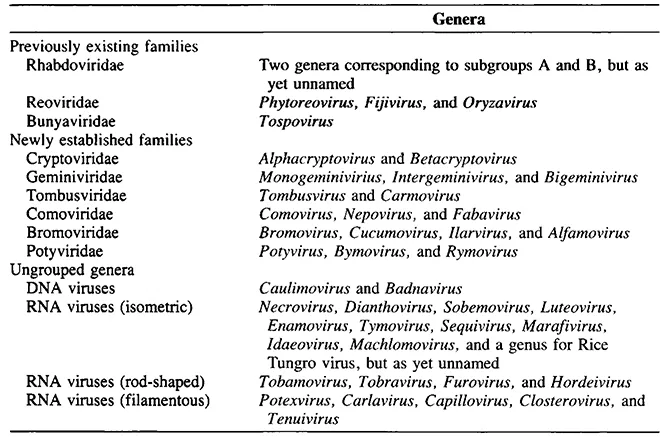
The Plant Virus Subcommittee of the ICTV is moving to adopt the familygenus-species classification for all plant viruses. Current proposals for families and genera already approved by the Executive Committee of the ICTV are summarized in Table 2. It is almost certain that these proposals will be adopted at the next meeting of the ICTV in 1993.
III. An Overview of Techniques
The main techniques currently available for the diagnosis of virus diseases in plants are dealt with in the 12 chapters which follow. Inoculation to the original host species or cultivar (Chapter 2) is necessary to demonstrate that the virus identified by other means is in fact the cause of the disease in question (see Section V). Thus, positive diagnosis will almost always involve the application of two or more of the available techniques.
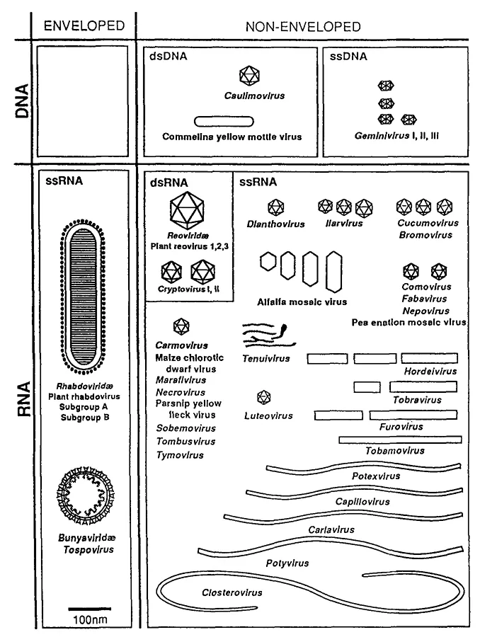
FIGURE 1. The families and groups of plant viruses. Outline diagrams drawn approximately to scale. (From Francki, R. I. B. et al., Arch. Virol. Suppl., 2, 450, 1991. With permission.)
A knowledge of the size, shape, and overall properties of the virus particle are usually important for placing a virus in its correct family or group. Electron microscopy is the most direct method now used for determining size and shape. This technique applied to virus preparations is detailed in Chapter 8. Viruses with large particles, particularly those with membranes, can be characterized in thin sections of infected tissue. I have not included a chapter on thin-sectioning techniques because many good accounts of these methods are available elsewhere.4
Table 1. The 35 Families and Groups of Plant Viruses Approved by the ICTVa
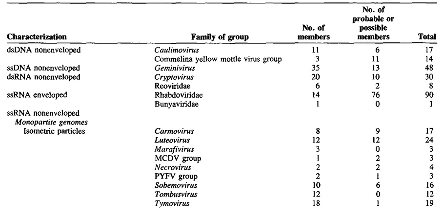
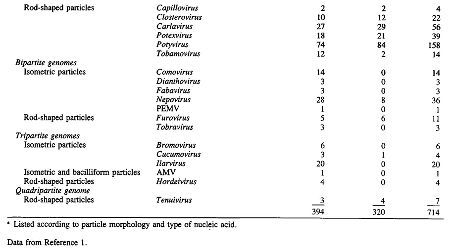
Table 2. Provisional Classification of Plant Viruses in Families and Genera
The value of particle morphology for placing a virus in its correct group or family varies significantly for different kinds of virus. Table 3 summarizes some properties of seven virus groups and families which may be quite readily distinguished by particle morphology.
Table 4 lists some properties of the rod-shaped plant viruses. Members of some of these groups, such as tobamoviruses, tobraviruses, and tenuiviruses can usually be placed tentatively in their correct group on particle morphology alone. For some others this would be much more difficult.
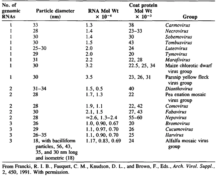
Table 5 details the 18 plant virus groups with isometric particles. With the exception of alfalfa mosaic virus, which has bacilliform particles as well as isometric, these groups may be difficult or impossible to distinguish on particle morphology alone. Most have particle diameters around 30 nm. Furthermore, different treatments may significantly affect the apparent particle diameter of an icosahedral virus.5 Differences in surface morphology can be observed in negatively stained preparations between some of the virus groups listed in Table 5, provided great care is taken with the preparation procedure.6
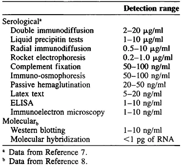
Two kinds of techniques of wide applicability in virus diagnosis have now emerged. These are serological procedures, especially various forms of enzyme-linked immunosorbent assay (ELISA) (Chapter 7) and nucleic acid hybridization (Chapter 9). Table 6 summarizes the relative sensitivity of some of these procedures for the detection of plant viruses.
Table 3. Seven Virus Groups and Families with Genomes Other Than ss Positive Sense RNA
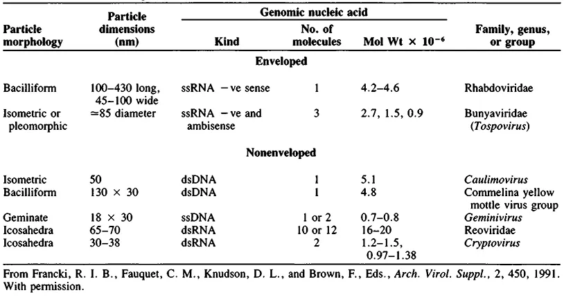
Table 4. Ten Plant Virus Groups with ss Positive Sense RNA and Rod-Shaped Particles
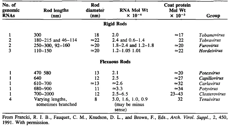
Table 5. Eighteen Plant Virus Groups with ss Positive Sense RNA and Isometric Particles
Besides the size and morphology of the virus particle as revealed by electron microscopy, various other physical properties of the virus particle have in the past been used in diagnosis. In the early years of plant virus research three so-called physical properties feature were commonly reported. These were the thermal death point, dilution end point, and longevity in vitro, all based on measurements of infectivity of the virus, usually in crude extracts. These properties have been shown to be quite unreliable as diagnostic criteria and are now rarely used.9 Three more precisely determined physical properties have been reported for many viruses. These are (1) density of the virus measured in water, sucrose, or CsCl solutions, (2) sedimentation coefficient and diffusion coefficient, and (3) electrophoretic mobility of the virus. These properties can be somewhat laborious to determine if accurate numbers are required. Furthermore, while they may provide useful additional data for placing a new virus in its correct family or group, these properties do not often distinguish between individual viruses within a family or group. To describe adequately and identify a virus it is usually necessary to isolate the virus in reasonably purified form. The procedure that can be used for successful isolation will depend to a large extent on the properties of the virus particle and the concentration it reaches in the host plant. Thus, a method that is successful for purification of a virus may sometimes itself provide a useful taxonomic guideline (Chapter 6).
Table 6. Sensitivity of Some Serological and Molecular Techniques for Plant Virus Detection and Identification
Besides information on the host range of a virus and the macroscopic disease symptoms it causes (Chapter 2), host plants can be used to establish other biological properties which may be useful in diagnosis. The ways in which a virus is transmitted can give clues as to identity. These include: experimental transmission, for example by mechanical inoculation, and transmission through s...
Table of contents
- Cover
- Title Page
- Copyright Page
- Preface
- The Editor
- Contributors
- Table of Contents
- Chapter 1 Overview
- Chapter 2 Host Plants in Diagnosis
- Chapter 3 Experimental Transmission of Viruses in Diagnosis
- Chapter 4 Virus Transmission Through Soil and By Soil-Inhabiting Organisms in Diagnosis
- Chapter 5 Inclusions in Diagnosing Plant Virus Diseases
- Chapter 6 Virus Purification in Relation to Diagnosis
- Chapter 7 Serological Procedures
- Chapter 8 Electron Microscopy of In Vitro Preparations
- Chapter 9 Nucleic Acid Hybridization Procedures
- Chapter 10 dsRNA in Diagnosis
- Chapter 11 Diagnostic Methods Applicable to Viroids
- Chapter 12 Strategies for Implicating Virus-Like Pathogens as the Cause of Diseases of Unknown Etiology
- Chapter 13 Complexes of Transmission-Dependent and Helper Viruses
- Index
Frequently asked questions
Yes, you can cancel anytime from the Subscription tab in your account settings on the Perlego website. Your subscription will stay active until the end of your current billing period. Learn how to cancel your subscription
No, books cannot be downloaded as external files, such as PDFs, for use outside of Perlego. However, you can download books within the Perlego app for offline reading on mobile or tablet. Learn how to download books offline
Perlego offers two plans: Essential and Complete
- Essential is ideal for learners and professionals who enjoy exploring a wide range of subjects. Access the Essential Library with 800,000+ trusted titles and best-sellers across business, personal growth, and the humanities. Includes unlimited reading time and Standard Read Aloud voice.
- Complete: Perfect for advanced learners and researchers needing full, unrestricted access. Unlock 1.5M+ books across hundreds of subjects, including academic and specialized titles. The Complete Plan also includes advanced features like Premium Read Aloud and Research Assistant.
We are an online textbook subscription service, where you can get access to an entire online library for less than the price of a single book per month. With over 1.5 million books across 990+ topics, we’ve got you covered! Learn about our mission
Look out for the read-aloud symbol on your next book to see if you can listen to it. The read-aloud tool reads text aloud for you, highlighting the text as it is being read. You can pause it, speed it up and slow it down. Learn more about Read Aloud
Yes! You can use the Perlego app on both iOS and Android devices to read anytime, anywhere — even offline. Perfect for commutes or when you’re on the go.
Please note we cannot support devices running on iOS 13 and Android 7 or earlier. Learn more about using the app
Please note we cannot support devices running on iOS 13 and Android 7 or earlier. Learn more about using the app
Yes, you can access Diagnosis of Plant Virus Diseases by R. E. F. Matthews in PDF and/or ePUB format, as well as other popular books in Biological Sciences & Biology. We have over 1.5 million books available in our catalogue for you to explore.