eBook - ePub
Laboratory Manual for Classification and Morphology of Rumen Ciliate Protozoa
- 128 pages
- English
- ePUB (mobile friendly)
- Available on iOS & Android
eBook - ePub
Laboratory Manual for Classification and Morphology of Rumen Ciliate Protozoa
About this book
The only rumen protozoa lab guide featuring line drawings created by a leading scientist in the field
Laboratory Manual for Classification and Morphology of Rumen Ciliate Protozoa is a unique lab guide for learning how to count and identify rumen protozoa. In this guide, Professor Dehority has created line drawings of rumen protozoa that emphasize morphological features and size measurements. The book also provides keys for identifying genera and species, and it contains classifications and descriptions of the different orders and families of rumen ciliate protozoa. Procedures for counting rumen protozoa and identifying individual species are included as well.
Laboratory Manual for Classification and Morphology of Rumen Ciliate Protozoa will be an excellent identification guide for protozoologists, microbiologists, dairy scientists, and any researcher or student working with rumen protozoa.
Trusted by 375,005 students
Access to over 1.5 million titles for a fair monthly price.
Study more efficiently using our study tools.
Information
Topic
MedicineSubtopic
Veterinary MedicineFamily Ophryoscolecidae
Morphological Orientation
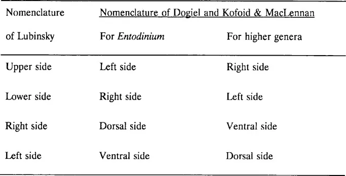
Considerable discrepancy has arisen over the terminology used to describe the different cell surfaces in the family Ophryoscolecidae. When asymmetrical organisms are not oriented in relation to the force of gravity, describing body sides as “dorsal” and “ventral” is confusing. The system used in this book has been proposed by Lubinsky (1958), and is presented in detail on the next page. The relationship between this system of terminology and the nomenclature of Dogiel (1927) and Kofoid and MacLennan (1930) are shown in the following table:
Subfamilies of Ophryoscolecidae
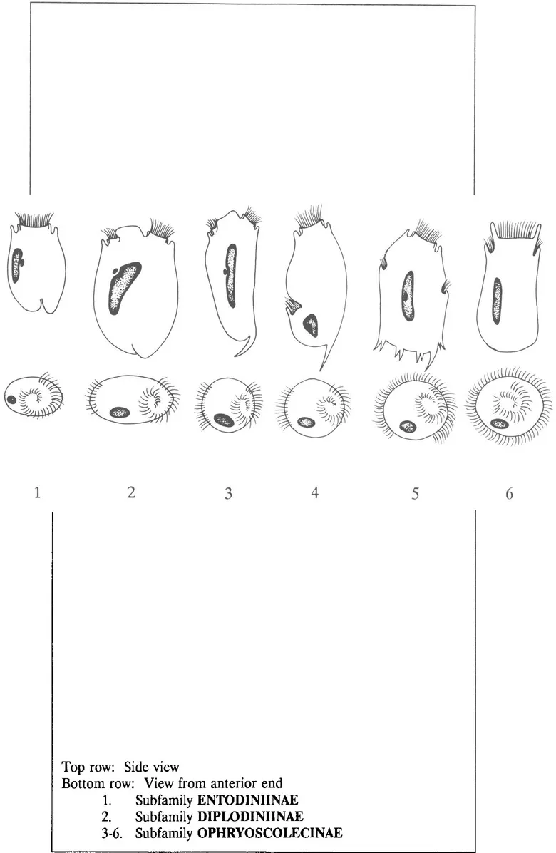
Subfamily ENTODINIINAE Lubinsky, 1957 - One ciliary zone; one contractile vacuole; in side view, the macronucleus lies between the micronucleus and nearest body side (Fig. 9-1).
Subfamily DIPLODINIINAE Lubinsky, 1957 - Two ciliary zones located in one transverse plane at anterior end of cell; two or more contractile vacuoles; in side view, micronucleus lies between macronucleus and nearest body side; skeletal plates absent or present; body more or less flattened (Fig. 9-2).
Subfamily OPHRYOSCOLECINAE Lubinsky, 1957 - Two ciliary zones, located in different transverse planes; two or more contractile vacuoles; in side view the micronucleus situated between the macronucleus and nearest body side; skeletal plates present; body more or less cylindrical (Fig. 9-3,4,5,6).
Figure 9 Location of ciliary zones in the family OPHRYOSCOLECIDAE (redrawn from Latteur, 1966 and Hungate, 1978).
Subfamily Entodiniinae
Entodinium is the only genus in this subfamily which commonly occurs in the rumen. General features used to classify rumen protozoa into the genus Entodinium and species within the genus are as follows (also see Fig. 10).
- The presence of a single oral (adorai) ciliary zone.
- Lack of skeletal plates.
- Position of macronucleus - lies between micronucleus and closest body side.
- Position of the contractile vacuole.
- Body length - Body length is designated as the distance between the anterior pole of the body and the anal opening.
- Body width.
- Ratio of body length to body width (L/W).
- Length of macronucleus - The distance, on a straight line, between the anterior and posterior tips.
- Shape of macronucleus.
- Location of micronucleus.
- Overall shape of cell.
Figure 10 Cell morphology of protozoa in the genus Entodinium
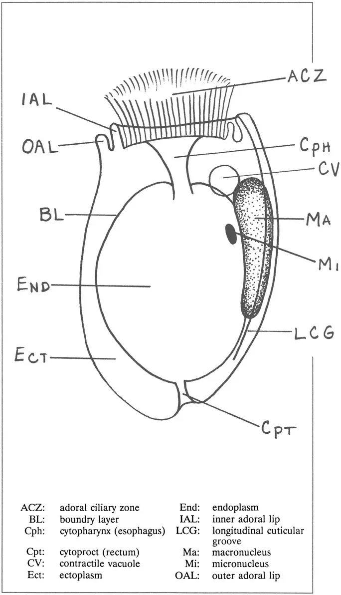
Figure 11 Variation in caudal spination within an Entodinium species (redraw after Lubinsky, 1957)
Key for Commonly Occurring Species of Entodinium
- With one or more caudal spine-like projections.................... 2Without caudal spine-like projections.................... 7
- Single heavy spine on posterior left side; contractile vacuole directly anterior of macronucleus.................... Entodinium rostratumOne long spine on posterior right side; one or more lobes on posterior left side.................... 3
- Contractile vacuole adjacent to anterior left edge of macronucleus.................... 4Contractile vacuole near middle of body to the left of triangular groove on right side of upper surface.................... 5Contractile vacuole near middle of body, to the left of distinct narrow groove on right side of upper surface, which extends anteriorly to outer adorai lip.................... 6
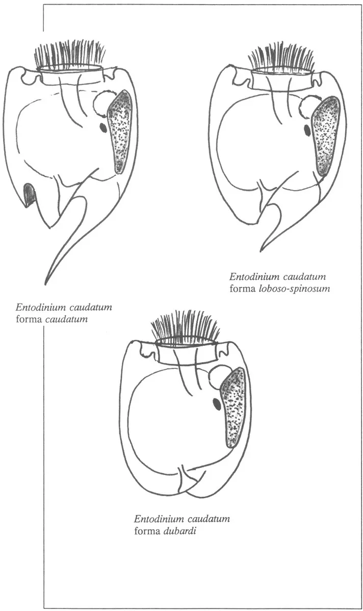
- A pointed to slightly rounded lobe on posterior left side .................... Entodinium caudatum forma loboso-spinosumPointed to slightly rounded lobes on both upper and lower posterior left side.................... Entodinium caudatum forma caudatum
- A pointed to slightly rounded lobe on posterior left side ..........
Table of contents
- Cover
- Title Page
- Copyright Page
- Contents
- Preface
- Classification of Rumen Ciliate Protozoa
- Key for Identifying Genera of Rumen Protozoa
- Rumen Protozoa in the Orders Prostomatida (Family Buetschliidae) and Trichostomatida (Families Isotrichidae and Blepharocorythidae)
- Rumen Protozoa in the Order Entodiniomorphida
- Family Ophryoscolecidae
- Procedure for Counting Total Protozoan Numbers in Rumen Contents
Frequently asked questions
Yes, you can cancel anytime from the Subscription tab in your account settings on the Perlego website. Your subscription will stay active until the end of your current billing period. Learn how to cancel your subscription
No, books cannot be downloaded as external files, such as PDFs, for use outside of Perlego. However, you can download books within the Perlego app for offline reading on mobile or tablet. Learn how to download books offline
Perlego offers two plans: Essential and Complete
- Essential is ideal for learners and professionals who enjoy exploring a wide range of subjects. Access the Essential Library with 800,000+ trusted titles and best-sellers across business, personal growth, and the humanities. Includes unlimited reading time and Standard Read Aloud voice.
- Complete: Perfect for advanced learners and researchers needing full, unrestricted access. Unlock 1.5M+ books across hundreds of subjects, including academic and specialized titles. The Complete Plan also includes advanced features like Premium Read Aloud and Research Assistant.
We are an online textbook subscription service, where you can get access to an entire online library for less than the price of a single book per month. With over 1.5 million books across 990+ topics, we’ve got you covered! Learn about our mission
Look out for the read-aloud symbol on your next book to see if you can listen to it. The read-aloud tool reads text aloud for you, highlighting the text as it is being read. You can pause it, speed it up and slow it down. Learn more about Read Aloud
Yes! You can use the Perlego app on both iOS and Android devices to read anytime, anywhere — even offline. Perfect for commutes or when you’re on the go.
Please note we cannot support devices running on iOS 13 and Android 7 or earlier. Learn more about using the app
Please note we cannot support devices running on iOS 13 and Android 7 or earlier. Learn more about using the app
Yes, you can access Laboratory Manual for Classification and Morphology of Rumen Ciliate Protozoa by B.A. Dehority in PDF and/or ePUB format, as well as other popular books in Medicine & Veterinary Medicine. We have over 1.5 million books available in our catalogue for you to explore.