This book provides a first authoritative text on radiochromic film, covering the basic principles, technology advances, practical methods, and applications. It focuses on practical uses of radiochromic film in radiation dosimetry for diagnostic x-rays, brachytherapy, radiosurgery, external beam therapies (photon, electron, protons), stereotactic body radiotherapy, intensity-modulated radiotherapy, and other emerging radiation technologies. The expert authors address basic concepts, advantages, and the main applications including kilovoltage, brachytherapy, megavoltage, electron beam, proton beam, skin dose, in vivo dosimetry, postal and clinical trial dosimetry. The final chapters discuss the state of the art in microbeam, synchrotron radiation, and ultraviolet radiation dosimetry.
- 388 pages
- English
- ePUB (mobile friendly)
- Available on iOS & Android
eBook - ePub
About this book
Trusted by 375,005 students
Access to over 1.5 million titles for a fair monthly price.
Study more efficiently using our study tools.
Information
Topic
MedicineSubtopic
Diagnostics ImagingPART 1
BASICS
1 Introduction
Indra J. Das
2 Historical background, development, and construction of radiochromic films
Martin Butson and Azam Niroomand-Rad
3 Physics and characteristics of radiochromic films
Martin Butson, Gwi Cho, Simran Gill, and Dane Pope
4 Radiochromic film digitizers
Benjamin S. Rosen
1
Introduction
1.1 History and background
1.2 Summary of contents
References
1.1 HISTORY AND BACKGROUND
The current book represents the state-of-the-art knowledge on the fast growing field of two-dimensional dosimeter such as film for simple and complex dosimetry. Radiochromic film (RCF) is a class of polymer-based device that changes color when exposed to radiation. A lot of development in this area was conducted by a chemical company GAF, known as General Aniline & Film located in Parsippany, NJ, that produced films known as GAFchromicTM. Today, GAFchromic name has become synonymous to a general name radiochromic film. Detailed description, characteristics, and usage have been described in TG-55 [1]. A revision to this task group (TG-235) is underway that may provide additional information in near future when published.
Radiographic films based on silver halides are becoming extinct in digital age, which were also used for radiation dosimetry [2]. There were numerous problems with such films, but mainly the chemical processing and energy dependence made it harder to adapt. On the other hand, RCF does not require any processing and is tissue equivalent. The demise of radiographic films, the characteristics of which have been described in TG-69 [3] and unfavorable characteristics, made RCF a compelling choice for radiation dosimetry.
1.2 SUMMARY OF CONTENTS
The necessity of a book was realized because of vast amount of research conducted on the radiochromic class of dosimeter visible clearly from publications in every radiation-related journal. In addition, there is no other book on this subject. There is a growing need to have an authoritative book for graduate-level courses taught in universities around the world on every aspect of the radiation dosimetry. Thus, the chapters of this book are divided into application based on three-dimensional conformal radiation therapy (3DCRT), intensity-modulated radiation therapy, stereotactic radiosurgery, particle beam, and other aspects of radiation fields. This book contains 20 chapters and is truly an international collaboration, in which expert authors from all over the world, including Australia, Japan, France, Belgium, Greece and USA, are represented.
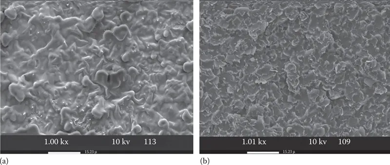
Figure 1.1 Electron micrograph of external beam therapy (EBT) film (a) unexposed and (b) exposed to 2 Gy of X-rays. (Details of techniques and magnifications are also provided in the image.)
Chapter 2 of the book provides a rare historical background and characteristics of RCF. In addition, it covers RCF action and various characteristics of films. To clearly show the action of radiation on film, Figure 1.1 shows electron micrograph of the structures in unexposed and exposed film with 2-Gy X-ray radiation. The polymerization is dose and time dependent, which is discussed in Chapter 2 of this book. Please note that polymerization reduces the structure to a flatter level, thereby changing the color from semitransparent to blue or yellow color. Additional images are also provided in other chapters whenever necessary.
The unique properties of RCF coloration are used to measure the radiation dose in every aspect of radiation dosimetry. Figure 1.2 shows the growth of research evaluated from publications searched thorough Pubmed. Until the end of 2015, there have been nearly 1000 papers published, all related to radiation-related applications of various types of RCF. These data may not reflect actual growth as Pubmed does not cover basic sciences and nonmedical journals. There are several review articles that have covered the breadth of this subject [4, 5, 6].
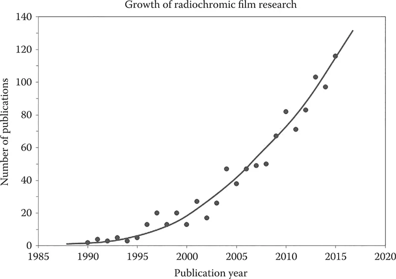
Figure 1.2 Yearly peer-reviewed publications in research journals.
The writing style is kept standard on, with introduction of modality, how RCF can be used with a summary and up-to-date references. Due to a large number of authors from various countries, uniformity of writing style was a critical factor that has been kept in mind. In addition, the presentation is made simple such that undergraduate and other students can follow on with minimum knowledge of the subject. It is hoped that this book will provide a badly needed literature for this unique topic.
REFERENCES
1. Niroomand-Rad A, Blackwell CR, Coursey BM et al. Radiochromic film dosimetry: Recommendations of AAPM radiation therapy committee task group 55. Med Phys 1998;25:2093–2115.
2. Das IJ. Radiographic film. In: Rogers DWO, Cyglar JE (Eds.) Clinical Dosimetry Measurements in Radiotherapy. Madison, WI: Medical Physics Publishing; 2009. pp. 865–890.
3. Pai S, Das IJ, Dempsey JF et al. TG-69: Radiographic film for megavoltage beam dosimetry. Med Phys 2007;34:2228–2258.
4. Devic S. Radiochromic film dosimetry: Past, present, and future. Phys Med 2011;27:122–134.
5. Devic S, Tomic N, Lewis D. Reference radiochromic film dosimetry: Review of technical aspects. Phys Med 2016;32:541–556.
6. Soares CG. New developments in radiochromic film dosimetry. Radiat Prot Dosimetry 2006;120:100–106.
2
Historical background, development, and construction of radiochromic films
2.1 Historical Development of Radiochromic Materials
2.2 Historical Development of Radiochromic Films
2.3 Historical Development and Construction of GAFchromicTM Films
2.4 Historical Development and Construction of Radiochromic Films
2.5 Historical Development and Construction of Recent Radiochromic Films
2.6 The Development and Construction of Available Radiochromic Films
2.7 Characteristics of the Currently Available Radiochromic Films
2.8 Recent Radiochromic Films for Radiology with kV Photons
2.9 Radiochromic Films for Niche Applications
2.10 Radiochromic Films Manufactured for Commercial Use
2.10.1 Radiochromic films manufactured by Far West Technology, Inc.
2.10.2 Radiochromic films produced by GEX Corporation
2.10.3 Radiochromic films produced by JP Laboratories
2.10.4 Radiochromic films produced for ultraviolet radiation exposure
2.11 Summary
References
2.1 HISTORICAL DEVELOPMENT OF RADIOCHROMIC MATERIALS
The history of development of radiochromic materials dates back to the early nineteenth century before discovery of X-ray by Roentgen (1895) and prior to the use of conventional silver halide-based radiographic films for radiographic imaging and radiation dosimetry. Radiographic films became an important tool for detecting ionizing radiation and measuring dose [1]. However, there are numerous problems associated with quantification of ionizing radiation dose using radiographic films. One problem is the energy absorption and transfer properties of radiographic films that are not similar to those of biological tissues. The radiographic films also have the disadvantages of being sensitive to room light and requiring wet chemical processing. Some of these difficulties led scientists to search and develop an alternative to radiographic films for radiation imaging and dosimetry with high spatial resolution, which did not require a special chemical processing and/or developmental procedure. The radiochromic processes always involve the direct coloration of a material by the absorption of electromagnetic radiation, without requiring latent chemical, optical, or thermal development or amplification [2, 3, 4].
In the early 1800s, a radiochromic process was first demonstrated by Joseph Niepce, a French scientist, inventor [2]. He applied a light sensitive tar-like material known as bitumen of Judea solution (an unsaturated hydrocarbon polymeric mixture) to a pewter plate, made of an alloy of tin, copper, and antimony. Niepce projected a view from a window onto a pewter plate and after about 8 h of exposure to sunlight he saw a permanent bitumen image on the plate. This image production was attributed to an unsaturated hydrocarbon polymeric mixture in the bitumen that underwent cross-linking upon irradiation [5].
By the mid-1800s, an...
Table of contents
- Cover
- Half Title
- Title Page
- Copyright Page
- Dedication
- Table of Contents
- Series preface
- Preface
- About the editor
- Contributors
- PART 1 BASICS
- PART 2 APPLICATIONS
- Index
Frequently asked questions
Yes, you can cancel anytime from the Subscription tab in your account settings on the Perlego website. Your subscription will stay active until the end of your current billing period. Learn how to cancel your subscription
No, books cannot be downloaded as external files, such as PDFs, for use outside of Perlego. However, you can download books within the Perlego app for offline reading on mobile or tablet. Learn how to download books offline
Perlego offers two plans: Essential and Complete
- Essential is ideal for learners and professionals who enjoy exploring a wide range of subjects. Access the Essential Library with 800,000+ trusted titles and best-sellers across business, personal growth, and the humanities. Includes unlimited reading time and Standard Read Aloud voice.
- Complete: Perfect for advanced learners and researchers needing full, unrestricted access. Unlock 1.5M+ books across hundreds of subjects, including academic and specialized titles. The Complete Plan also includes advanced features like Premium Read Aloud and Research Assistant.
We are an online textbook subscription service, where you can get access to an entire online library for less than the price of a single book per month. With over 1.5 million books across 990+ topics, we’ve got you covered! Learn about our mission
Look out for the read-aloud symbol on your next book to see if you can listen to it. The read-aloud tool reads text aloud for you, highlighting the text as it is being read. You can pause it, speed it up and slow it down. Learn more about Read Aloud
Yes! You can use the Perlego app on both iOS and Android devices to read anytime, anywhere — even offline. Perfect for commutes or when you’re on the go.
Please note we cannot support devices running on iOS 13 and Android 7 or earlier. Learn more about using the app
Please note we cannot support devices running on iOS 13 and Android 7 or earlier. Learn more about using the app
Yes, you can access Radiochromic Film by Indra J. Das in PDF and/or ePUB format, as well as other popular books in Medicine & Diagnostics Imaging. We have over 1.5 million books available in our catalogue for you to explore.