- 782 pages
- English
- ePUB (mobile friendly)
- Available on iOS & Android
eBook - ePub
Advanced Optical Instruments and Techniques
About this book
Advanced Optical Instruments and Techniques includes twenty-three chapters providing processes, methods, and procedures of cutting-edge optics engineering design and instrumentation. Topics include biomedical instrumentation and basic and advanced interferometry. Optical metrology is discussed, including point and full-field methods. Active and adaptive optics, holography, radiometry, the human eye, and visible light are covered as well as materials, including photonics, nanophotonics, anisotropic materials, and metamaterials.
Tools to learn more effectively

Saving Books

Keyword Search

Annotating Text

Listen to it instead
Information
1
Optics of Biomedical Instrumentation
1.1Wide-Field Microscopy
Optical Layout • Resolution
1.2Fluorescence Microscope
Introduction to Fluorescence Process • Fluorescence Imaging Systems
1.3Confocal Microscopy
Principle • Components • Types of Confocal Microscopes
1.4Optical Sectioning Structured Illumination Microscopy (OS-SIM)
Principle • Optical Sectioning Strength • Optical Sectioning Algorithm • Problem of Speed and Solution
1.5Super-Resolution Structured Illumination Microscopy (SR-SIM)
Principle • SR-SIM Instrumentation • Reconstruction Algorithm • Nonlinear SIM • Combining OS-SIM and SR-SIM
1.6Endoscopy
Introduction • Basic Optics for Endoscopes • Objective Lenses • Relay Lenses
References
Shaun Pacheco Zhenyue Chen, and Rongguang Liang
1.1Wide-Field Microscopy
The goal of a microscope is to produce a magnified image of a microscopic sample without degrading the image quality. Microscopes have become an essential tool for many biomedical applications. They are used in investigating biological processes, diagnosing diseases, and quantitatively measuring biological processes in vitro and in vivo. This section introduces the key components in an optical microscope and how diffraction-limited resolution is defined.
1.1.1Optical Layout
A typical optical layout for a microscope with an infinity corrected objective is shown in Figure 1.1. The object plane is at the front focal plane of the objective, and the output for an infinity-corrected objective is a collimated beam for every object point. A tube lens is used to form an intermediate image, which can be directly imaged onto an electronic sensor or observed by the human eye through the eyepiece. The exit pupil of the objective lens is typically set at the rear focal plane to make the objective object space telecentric. In a telecentric system, the chief rays are parallel to the optical axis and the system magnification is constant even if the object is displaced from the focal plane. Microscope objectives are well corrected for aberrations, and thus produce diffraction-limited imaging.
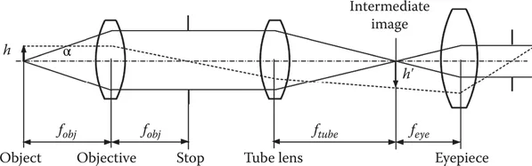
FIGURE 1.1A typical microscope design for an infinity-corrected objective.
Two important properties of a microscope are the numerical aperture (NA) and magnification. NA of the microscope objective is defined as
(1.1)
where n is the refraction index of the medium between the front lens of the objective and the object, α is half acceptance angle of the objective. The magnification of the objective is defined as
(1.2)
where ftube is the focal length of the tube lens and fobj is the focal length of the objective. The total magnification of the microscope with an eyepiece is the product of the magnification of the objective and the magnification of the eyepiece
(1.3)
The magnification of the eyepiece is approximately
(1.4)
where feye is the focal length of the eyepiece.
1.1.2Resolution
The complex exit pupil of the objective is defined as
(1.5)
where A is the amplitude function and W is the aberration function of the objective at the exit pupil for wavelength λ. Diffraction from the exit pupil to the plane of focus yields the impulse response
(1.6)
where di is the distance from the exit pupil to the image plane, and xi and yi are the spatial coordinates at the image plane. The impulse response is the Fourier transform of the complex pupil function.
For an aberration free objective with a circular aperture, the pupil function P(x, y) is unity, the point spread function (PSF) is:
(1.7)
where ri is the radial coordinate in image space, R is the radius of the exit pupil, and J1 is the Bessel function of the first kind. The normalized irradiance distribution of the diffraction-limited PSF is shown in Figure 1.2. The first zero of the Airy pattern is at a radial distance
(1.8)
Rayleigh’s criterion is often used as a measure of the resolution in microscopy. Rayleigh’s criterion states that two incoherent point sources are barely resolvable when the center of one falls exactly on the null of the second, as shown in Figure 1.3. This corresponds to a dip between the two peaks of approximately 74% of the maximum. All sources closer than Rayleigh’s criterion cannot be resolved, and are assumed to come from the same point source.
For incoherent illumination, the intensity of the image is given by
(1.9)
where * is the convolution operator, |h|2 is the incoherent PSF of the optical system and Ig is the image as predicted by geometrical optics. The resulting image is a blurring of the geometrical optics image by the point spread function.
Since an optical system can be modeled as a linear shift invariant system, the imaging equation can be written in the frequency domain. The imaging equation in frequency space is
(1.10)
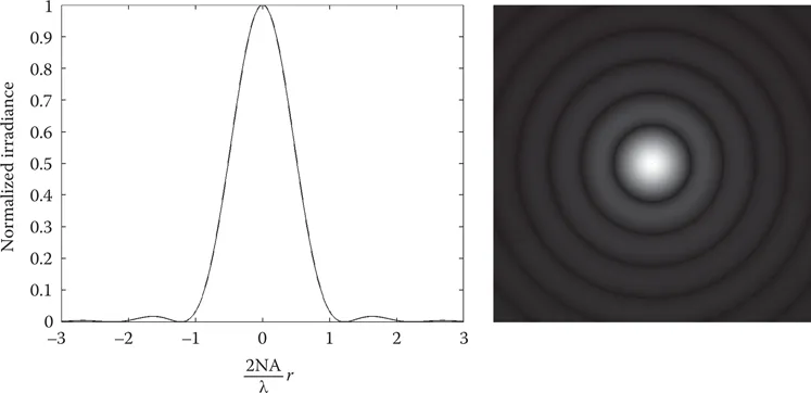
FIGURE 1.2The lateral and 2D profile of the diffraction-limited point spread function.
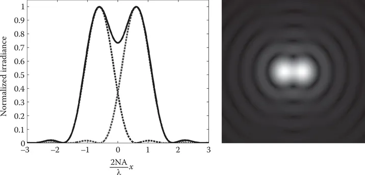
FIGURE 1.3The lateral and 2D profile of Rayleigh’s criterion.
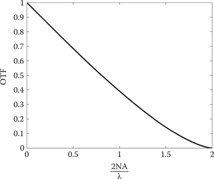
FIGURE 1.4Diffraction-limited OTF.
where kx and ky are the spatial frequency coordinates, Gi is the normalized Fourier transform of the image, Gg is the normalized Fourier transform of the image predicted by geometrical optics, and OTF is the optical transfer function of the imaging system. OTF is the normalized Fourier transform of the PSF:
(1.11)
Some important properties of the OTF are
1.OTF(0,0) = 1
2.OTF(kx,ky) = OTF*(-kx,-ky)
3.|OTF(kx,ky)| ≤ |OTF(0,0)| = 1
The OTF for a diffraction-limited system is shown in Figure 1.4; it has a cutoff frequency at 2NA/λ. Objects with a higher spatial frequency cannot be resolved, unless super-resolution techniques are used.
1.2Fluorescence Microscope
Fluorescence microscope plays a major role in the fields of cell and molecular biology, due to its intrinsic selectivity that can provide high contrast between objects of interest and background. This is important in biomedical imaging, since biological structures of interest can be fluorescently labeled to more easily study biological phenomenon. Over the past several decades, different microscope designs have appeared with the aim of increasing image contrast, penetration depth, and spatial resolution. This section discusses the principle of fluorescence microscope and related instrumental techniques.
1.2.1Introduction to Fluorescence Process
When illuminated with light in a suitable spectrum, s...
Table of contents
- Cover
- Half Title
- Series Page
- Title Page
- Copyright Page
- Table of Contents
- Preface
- Contributors
- 1 Optics of Biomedical Instrumentation
- 2 Wavefront Slope Measurements in Optical Testing
- 3 Basic Interferometers
- 4 Modern Fringe Pattern Analysis in Interferometry
- 5 Optical Methods in Metrology: Point Methods
- 6 Optical Metrology of Diffuse Objects: Full-Field Methods
- 7 Active and Adaptive Optics
- 8 Holography
- 9 Fourier Optics and Image Processing
- 10 Light-Sensitive Materials: Silver Halide Emulsions, Photoresist, and Photopolymers
- 11 Electro-Optical and Acousto-Optical Devices
- 12 Radiometry
- 13 Color and Colorimetry
- 14 The Human Eye and Its Aberrations
- 15 Incoherent Light Sources
- 16 Lasers
- 17 Spatial and Spectral Filters
- 18 Optical Fibers and Accessories
- 19 Isotropic Amorphous Optical Materials
- 20 Anisotropic Materials
- 21 Optical Fabrication
- Index
Frequently asked questions
Yes, you can cancel anytime from the Subscription tab in your account settings on the Perlego website. Your subscription will stay active until the end of your current billing period. Learn how to cancel your subscription
No, books cannot be downloaded as external files, such as PDFs, for use outside of Perlego. However, you can download books within the Perlego app for offline reading on mobile or tablet. Learn how to download books offline
Perlego offers two plans: Essential and Complete
- Essential is ideal for learners and professionals who enjoy exploring a wide range of subjects. Access the Essential Library with 800,000+ trusted titles and best-sellers across business, personal growth, and the humanities. Includes unlimited reading time and Standard Read Aloud voice.
- Complete: Perfect for advanced learners and researchers needing full, unrestricted access. Unlock 1.4M+ books across hundreds of subjects, including academic and specialized titles. The Complete Plan also includes advanced features like Premium Read Aloud and Research Assistant.
We are an online textbook subscription service, where you can get access to an entire online library for less than the price of a single book per month. With over 1 million books across 990+ topics, we’ve got you covered! Learn about our mission
Look out for the read-aloud symbol on your next book to see if you can listen to it. The read-aloud tool reads text aloud for you, highlighting the text as it is being read. You can pause it, speed it up and slow it down. Learn more about Read Aloud
Yes! You can use the Perlego app on both iOS and Android devices to read anytime, anywhere — even offline. Perfect for commutes or when you’re on the go.
Please note we cannot support devices running on iOS 13 and Android 7 or earlier. Learn more about using the app
Please note we cannot support devices running on iOS 13 and Android 7 or earlier. Learn more about using the app
Yes, you can access Advanced Optical Instruments and Techniques by Daniel Malacara Hernández in PDF and/or ePUB format, as well as other popular books in Technology & Engineering & Electrical Engineering & Telecommunications. We have over one million books available in our catalogue for you to explore.