Imaging in Photodynamic Therapy
eBook - ePub

Imaging in Photodynamic Therapy

  1. 479 pages
  2. English
  3. ePUB (mobile friendly)
  4. Available on iOS & Android
eBook - ePub

Imaging in Photodynamic Therapy

Book details
Book preview
Table of contents
Citations

About This Book

This book covers the broad field of cellular, molecular, preclinical, and clinical imaging either associated with or combined with photodynamic therapy (PDT). It showcases how this approach is used clinically for cancer, infections, and diseases characterized by unwanted tissue such as atherosclerosis or blindness. Because the photosensitizers are also fluorescent, the book also addresses various imaging systems such as confocal microscopy and small animal imaging systems, and highlights how they have been used to follow and optimize treatment, and to answer important mechanistic questions. Chapters also discuss how imaging has made important contributions to clinical outcomes in skin, bladder, and brain cancers, as well as in the development of theranostic agents for detection and treatment of disease. This book provides a resource for physicians and research scientists in cell biology, microscopy, optics, molecular imaging, oncology, and drug discovery.

Frequently asked questions

Simply head over to the account section in settings and click on “Cancel Subscription” - it’s as simple as that. After you cancel, your membership will stay active for the remainder of the time you’ve paid for. Learn more here.
At the moment all of our mobile-responsive ePub books are available to download via the app. Most of our PDFs are also available to download and we're working on making the final remaining ones downloadable now. Learn more here.
Both plans give you full access to the library and all of Perlego’s features. The only differences are the price and subscription period: With the annual plan you’ll save around 30% compared to 12 months on the monthly plan.
We are an online textbook subscription service, where you can get access to an entire online library for less than the price of a single book per month. With over 1 million books across 1000+ topics, we’ve got you covered! Learn more here.
Look out for the read-aloud symbol on your next book to see if you can listen to it. The read-aloud tool reads text aloud for you, highlighting the text as it is being read. You can pause it, speed it up and slow it down. Learn more here.
Yes, you can access Imaging in Photodynamic Therapy by Michael R. Hamblin,Yingying Huang in PDF and/or ePUB format, as well as other popular books in Physical Sciences & Physics. We have over one million books available in our catalogue for you to explore.

Information

Publisher
CRC Press
Year
2017
ISBN
9781315278155
Edition
1

PART 1

INTRODUCTION

1 Looking out the optical window: Physical principles and instrumentation of imaging in photodynamic therapy
Hui Liu and Jonathan P. Celli
2 Photochemistry and photophysics of PDT and photosensitizers
Marcin Ptaszek

1
Looking out the optical window

Physical principles and instrumentation of imaging in photodynamic therapy
HUI LIU AND JONATHAN P. CELLI
1.1 Introduction
1.2 Physical principles: Light propagation and interactions with matter
1.2.1 Light as wave and particle
1.2.2 Phase and coherence
1.2.3 Light propagation
1.2.3.1 Reflection
1.2.3.2 Refraction
1.2.3.3 Diffraction
1.2.4 Light–matter interactions and the optical window
1.2.4.1 Scattering
1.2.4.2 Absorption
1.2.4.3 Attenuation models and tissue optics
1.3 Physical processes following light absorption: Imaging and therapeutic applications
1.3.1 Radiationless dissipation Heat
1.3.2 Fluorescence
1.3.3 Intersystem crossing Photodynamic therapy
1.3.4 Nonlinear optical processes
1.4 Instrumentation and technologies for imaging and PDT
1.4.1 Light sources
1.4.1.1 Lasers
1.4.1.2 Light-emitting diodes
1.4.1.3 Lamps
1.4.2 Endoscopes
1.4.3 Photodetectors and cameras
1.4.3.1 Cameras
1.4.4 Image processing
1.5 Conclusion, perspectives, and emerging directions
References

1.1 INTRODUCTION

As a light-based treatment modality, photodynamic therapy (PDT) is inherently conducive to integration with optical imaging. The central principle of PDT is to leverage photochemistry that occurs following activation of a photosensitizing chemical [photosensitizer (PS)] using a light source of the appropriate wavelength and mode of delivery to achieve destruction of target tissues (Dougherty et al. 1998). Importantly, some degree of specificity is achieved as PSs have been almost universally observed to exhibit quasi-selective accumulation in neoplastic tissues, going back to the early observations of Policard, who studied accumulation of hematoporphyrin in rat sarcomas (Policard 1924). Since clinical PS should have little or no dark toxicity, an additional degree of selectivity is afforded by restriction of light to the target tissue. This basic photodynamic process, the fundamentals of which are discussed more extensively throughout this volume, has been developed and adapted for treatment of numerous cancer and noncancer pathologies at diverse anatomical sites using appropriate chemical modifications of the PS and innovative light delivery strategies. Importantly, the same photosensitizing agents employed in PDT for targeted tissue destruction also have a finite probability to undergo a radiative transition back to the ground state following light absorption. In other words, PS can act as both therapeutic agents and diagnostic fluorophores. Therefore, upon illumination, longer wavelength fluorescence emission is generated from the malignant tissues in which the PS accumulates, thus marking the tumor location and margins otherwise difficult to visualize. This process has been extensively leveraged to confirm PS uptake and localization and to guide surgical resection as discussed at length in the literature and reviewed elsewhere (Celli et al. 2010).
In this chapter, we review the basic physical principles underlying PDT-related optical imaging and underscore points where these basic principles have particularly important implications for PDT and imaging and/or its key enabling technologies. This discussion will include a brief review of the fundamental nature of light itself, how it interacts with matter as both a particle and a wave, and how these interactions manifest in the propagation of light through tissue to allow therapy and imaging. Also of central importance to PDT and its associated imaging applications is the probabilistic nature of quantum mechanics and the allowability of transitions between quantum states, which ultimately determine excited state lifetimes. Finally, technological developments in light sources and light detectors will be briefly discussed in the context of their role in enabling PDT and associated optical imaging applications.

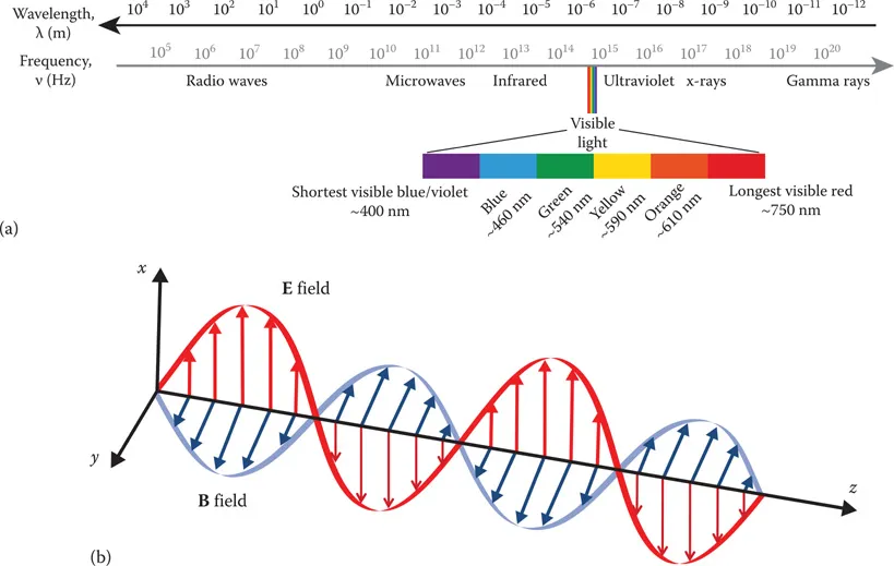
1.2 PHYSICAL PRINCIPLES: LIGHT PROPAGATION AND INTERACTIONS WITH MATTER

1.2.1 LIGHT AS WAVE AND PARTICLE

When we talk about light in this chapter, we are referring to the narrow slice of the spectrum of electromagnetic (EM) radiation that is visible to the human eye (Figure 1.1a). EM radiation, which includes radio waves, microwaves, infrared, visible light, ultraviolet, and x-ray and gamma radiation, is produced when charged particles accelerate, generating electric (E) and magnetic fields (B) propagating through space in a manner that satisfies the set of equations set down by James Clerk Maxwell (1865). For this to be true, the electric and magnetic fields are necessarily sinusoidal in space and time, and mutually perpendicular to the direction of propagation (Figure 1.1b). The descriptive categories of EM waves stated earlier are defined purely on the basis of their frequency, ν, or wavelength, λ, where λν = c. The frequency of a wave is its oscillation rate, typically reported in the SI units of Hertz (Hz), oscillations per second. The wavelength of an EM wave is its propagation distance in a vacuum during a full oscillation cycle (between two adjacent crests or troughs). In a vacuum, all EM radiation propagates at the speed of light, c, approximately 3.00 × 10⁸ m/s, though its speed is different in other media (such as biological tissue). However, although the propagation speed of EM radiation is medium dependent, it maintains the same frequency or wavelength. For the example of an EM wave propagating in the z-direction through a vacuum (empty space), this description can be written more succinctly as
Images
Figure 1.1 Electromagnetic radiation and visible light. (a) The spectrum of electromagnetic (EM) radiation. (b) The electrical field (red) and magnetic field (blue) are mutually perpendicular to the direction of propagation.
E(z,t)=E0cos(2πλ(zct))x^B(z,t)=E0ccos(2πc(zct))y^
where x^ and y^ are the unit vectors in the x- and y-directions, respectively. The energy carried by EM radiation is reported by the Poynting vector, the vector cross-product of its electric and magnetic fields, with SI units of Watts per meter squared (W/m2). Notwithstanding this universality, however, the physical processes that are relevant to the interaction of EM radiation with matter are indeed highly dependent on wavelength. Hence, our review of the light–matter interaction in the following section will be limited to the discussion of processes that are energetically allowed and relevant for light of visible and near-infrared wavelengths used in PDT and optical imaging, approximately 400–750 nm (Figure 1.1a). For example, this chapter does not deal with effects such as Compton scattering or other important processes more relevant to the interaction of ionizing EM radiation with matter. For a more complete discussion of the basic principles of electrodynamics, the curious reader is referred to any number of texts on this subject, such as the classic by J.D. Jackson (1999).
Yet simultaneously, light can be described as photons, particles that carry discrete packets of energy, E = , where h = 6.626 × 10⁻³⁴ J · s, is Planck’s constant (Einstein 1905). Photons are massless particles and have no electronic charge, but do carry momentum, p = h/λ, such that interactions with other particles are governed by the conservation of momentum. This well-known duality, in which light can be described as both a particle and as a wave, is left over from historic debates on the quantum nature of light and matter in the early twentieth century. Although the wave description is used to describe certain phenomena (coherence, interference, diffraction, etc.) and the particle description is needed for other processes (quantized transitions, photoelectric effect, etc.), both descriptions must always coexist. Indeed, even a single photon can be modeled by wave propagation and, conversely, massive particles such as electrons can also be described as waves, having a de Broglie wavelength, λ = h/p. Here too, in this chapter, we will go back and forth between both descriptions as we discuss the physics of PDT and optical imaging.

1.2.2 PHASE AND COHERENCE

Phase, φ, is a parameter in any periodical sinusoidal function that specifies where in its oscillation cycle it is at t = 0:
y(t)=sin(ωt+φ)
In general, EM waves are out of phase, with a difference in φ from 0 to 2π radians (0°–360°). Only when Δφ = 0 or 2π are the waves in phase. ...

Table of contents

  1. Cover
  2. Half Title
  3. Title Page
  4. Copyright Page
  5. Dedication Page
  6. Table of Contents
  7. Series preface
  8. Preface
  9. Acknowledgment
  10. Editors
  11. Contributors
  12. PART 1 INTRODUCTION
  13. PART 2 IN VITRO MICROSCOPY FOR PHOTOSENSITIZER LOCALIZATION IN CELLS
  14. PART 3 IN VITRO MICROSCOPY OF CELL DAMAGE AND DEATH PROCESSES AFTER PDT
  15. PART 4 THERANOSTIC AGENTS AND NANOTECHNOLOGY
  16. PART 5 SMALL ANIMAL IMAGING
  17. PART 6 CLINICAL IMAGING
  18. Index