- 350 pages
- English
- ePUB (mobile friendly)
- Available on iOS & Android
eBook - ePub
Quantitative Microbeam Analysis
About this book
Quantitative Microbeam Analysis provides a comprehensive introduction to the field of quantitative microbeam analysis (MQA). MQA is a technique used to analyze subatomic quantities of materials blasted from a surface by a laser or particle beam, providing information on the structure and composition of the material. Contributed to by international experts, the book is unique in the breadth of microbeam analytical techniques covered. For each technique, it develops the theoretical background, discusses practical details relating to choice of equipment, and describes the current advances. The book highlights developments relating to Auger electron spectroscopy in scanning electron microscopes and transmission electron microscopes and advances in surface analytical imaging and accelerated ion beam-surface interactions.
Tools to learn more effectively

Saving Books

Keyword Search

Annotating Text

Listen to it instead
Information
Topic
Physical SciencesSubtopic
Atomic & Molecular PhysicsQuantification in AES and XPS
M. P. Seah
National Physical Laboratory
Teddington, Middlesex
Teddington, Middlesex
1 Introduction
This volume covers a broad range of microbeam analytical methods and so we shall consider, here, some very general background. We shall then consider quantification using different methods and different levels of complexity. Many problems are adequately solved using very simple relationships whilst others require much more detailed analysis. Many of the aspects discussed in this chapter are now being installed by manufacturers into commercial software and the basic structures of expert systems are already being countenanced. Thus, in a few years time it is to be hoped that analysts can concentrate on surface science and applied surface science rather than working through many of the details presented here. However, if they do not understand these basics they will not be able to take effective advantage of what the software systems have to offer.
1.1 The basic principles of AES and XPS
In both Auger electron spectroscopy (AES) and X-ray photoelectron spectroscopy (XPS) we use an incident radiation which strikes the surface under study. This surface emits low energy electrons, the energy spectrum of which exhibits lines characteristic of the atoms present at the solid surface. By measuring the spectrum with an electron spectrometer in ultra-high vacuum we obtain an analysis. In AES the incident radiation is usually an electron beam in the energy range 2 to 25 keV with a beam current in the range 1 na to 1μA. This flexibility allows both insulators and conductors to be studied with rather high sensitivity or, as discussed by Prutton in this volume, at high spatial resolution. At the current time the best spatial resolution observed is 5 nm (Janssen and Venables 1978). The incident electron ejects core electrons from atoms in the surface region and then these core holes are usually filled by electrons from higher levels in the same atom with the quantum of energy release being taken by a third electron which is ejected. In terms of the shell structure shown in Figure 1, the energy of the liberated electron, the Auger electron, is approximately EA where
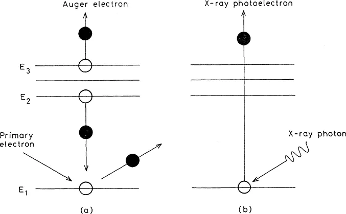
Figure 1. Schematic representation of (a) the Auger process and (b) photoelectron creation showing the atomic core levels.
Figure 2 shows an Auger electron spectrum with the lines associated with several elements marked. Figure 2a shows a spectrum in the direct mode and Figure 2b the traditional differential mode used to enhance the visibility of the peaks.
In XPS the radiation is usually of monochromatic X-rays from A1 or Mg anodes. These give sharp lines with a width below 1 eV and in the useful energy range 1250 to 1500 eV. In some instruments the Bremsstrahlung background and weak satellite peaks are removed from the A1 radiation by using a monochromator. In the past this has not been popular because considerable intensity was lost. However, in today’s instruments, this is no longer the case...
Table of contents
- Cover
- Half Title
- Title Page
- Copyright Page
- Table of Contents
- Quantification in AES and XPS
- Surface Analytical Imaging
- Electronic Structure and Electron Spectroscopy
- Auger Electron Spectroscopy in the STEM
- Electron Energy-Loss Spectroscopy—EELS
- Light Element Microanalysis and Imaging
- A Comparison of Quantification Methods and Analytical Techniques
- Data Analysis and Processing
- Microscopy and Microanalysis of Insulating Materials
- Electron Specimen Interactions
- Electron Probe X-ray Microanalysis
- Energy Dispersive X-Ray Analysis (EDX) in the TEM/STEM
- Analysis and Imaging by Proton-Induced X-Ray Emission (PIXE)
- Ion-Beam Analytical Techniques—Rutherford Backscattering, Elastic Recoil and Nuclear Reaction Analysis
- Quantitative Analysis of Solids by SIMS and SNMS
- Static SIMS
- Applications of Surface, Interface and Thin Film Analysis in an Industrial Research Laboratory
- Ion-Induced Auger Electron Emission From Solids
- Resonance Ionisation Mass Spectrometry (RIMS)
- Appendix: a list of Acronyms
- Contributed papers by students
- List of Participants
- Index
Frequently asked questions
Yes, you can cancel anytime from the Subscription tab in your account settings on the Perlego website. Your subscription will stay active until the end of your current billing period. Learn how to cancel your subscription
No, books cannot be downloaded as external files, such as PDFs, for use outside of Perlego. However, you can download books within the Perlego app for offline reading on mobile or tablet. Learn how to download books offline
Perlego offers two plans: Essential and Complete
- Essential is ideal for learners and professionals who enjoy exploring a wide range of subjects. Access the Essential Library with 800,000+ trusted titles and best-sellers across business, personal growth, and the humanities. Includes unlimited reading time and Standard Read Aloud voice.
- Complete: Perfect for advanced learners and researchers needing full, unrestricted access. Unlock 1.4M+ books across hundreds of subjects, including academic and specialized titles. The Complete Plan also includes advanced features like Premium Read Aloud and Research Assistant.
We are an online textbook subscription service, where you can get access to an entire online library for less than the price of a single book per month. With over 1 million books across 990+ topics, we’ve got you covered! Learn about our mission
Look out for the read-aloud symbol on your next book to see if you can listen to it. The read-aloud tool reads text aloud for you, highlighting the text as it is being read. You can pause it, speed it up and slow it down. Learn more about Read Aloud
Yes! You can use the Perlego app on both iOS and Android devices to read anytime, anywhere — even offline. Perfect for commutes or when you’re on the go.
Please note we cannot support devices running on iOS 13 and Android 7 or earlier. Learn more about using the app
Please note we cannot support devices running on iOS 13 and Android 7 or earlier. Learn more about using the app
Yes, you can access Quantitative Microbeam Analysis by A.G Fitzgerald,B.E Storey,D.J Fabian in PDF and/or ePUB format, as well as other popular books in Physical Sciences & Atomic & Molecular Physics. We have over one million books available in our catalogue for you to explore.