- 234 pages
- English
- ePUB (mobile friendly)
- Available on iOS & Android
eBook - ePub
Yersinia Enterocolitica
About this book
First published in 1981. Comprehensive overview of existing knowledge of Yersina enterocolitica, including laboratory models, clinical observations, and associated dieases.
Tools to learn more effectively

Saving Books

Keyword Search

Annotating Text

Listen to it instead
Information
Chapter 1
CLASSIFICATION OF YERSINIA ENTEROCOLITJCA
TABLE OF CONTENTS
I. | Introduction |
II. | Biochemical Characterization of Y. enterocolitica and Y. enterocolitica-Like Organisms |
III. | Guanine Plus Cytosine Content |
IV. | DNA Relatedness in Y. enterocolitica and Y. enterocolitica-Like Organisms |
V. | Classification |
References | |
I. INTRODUCTION
The genus Yersinia was proposed by Van Loghem26 in honor of A. J. E. Yersin to accommodate Yersinia pestis and Y. pseudotuberculosis which were previously classified in the genus Pasteurella. Thal25 first proposed that Yersinia be placed in the family Enterobacteriaceae. The Gram-negative, fermentative organism Y. enterocolitica was first reported by Schleifstein and Coleman22 who called their isolates Bacterium enterocoliticum. Hassig et al.14 were the first to isolate Y. enterocolitica from humans in Europe. Their isolates were named Pasteurella pseudotuberculosis rodentium. Daniels and Goudzwaard9 referred to similiar isolates as Pasteurella X.
Frederiksen13 examined 55 of these strains, including isolates from Schleifstein and Coleman, Hassig et al., and Daniels and Goudzwaard. Biochemical reactions were similar in all of the strains. Their antibiotic susceptibility patterns were also similar. Three different O antigens were present, none of which reacted with Y. pseudotuberculosis. Frederikson concluded: “The characteristics of this group of bacteria are sufficiently distinct to separate them from Y. pseudotuberculosis. Yet they resemble Y. pseudotuberculosis sufficiently to justify their classification in the genus Yersinia (within the family Enterobacteriaceae as a separate species: Y. enterocolitica.”
II. BIOCHEMICAL CHARACTERIZATION OF Y. ENTEROCOLITICA AND Y. ENTEROCOLITICA-LIKE ORGANISMS
Y. enterocolitica strains, unlike Y. pseudotuberculosis, show considerable biochemical variability. The biochemical profiles of Y. enterocolitica and Y. enterocolitica-like strains are quite dependent upon the temperature of incubation. Results obtained at 22°C to 28°C are often considerably different from those obtained at 36°C± FC.2,4,8,16,21,23 Furthermore, strains from different sources and/or geographical origins often have different biochemical reactions. It was, therefore, difficult to develop a biochemical definition and biochemical boundaries for Y. enterocolitica. Biochemical reactions obtained for Y. enterocolitica in several laboratories are given in Table 1.
Most strains identified as Y. enterocolitica are characterized and separated from Y. pseudotuberculosis by the reactions given in Table 2. Y. enterocolitica strains differ in reactions for lecithinase (lipase), indole, xylose, esculin, and salicin. These reactions became the basis for several biotyping schemes (Table 3). Several other biogroups of Y. enterocolitica-like strains were encountered. These included metabolically inactive strains isolated from hares.19 The hare strains are trehalose-negative and are often sucrose-negative. Niléhn21 and Wauters28 included them as biogroup 5 of Y. enterocolitica (Table 3). Stevens and Mair24 did a numerical taxonomic study of Y. enterocolitica and considered the trehalose-negative strains to be members of a separate species. A second sucrose-negative group was described by Fredericksen.13 These organisms were trehalose-positive.
Two groups of rhamnose-positive Y. enterocolitica-like strains have been described.1,4,5,6,17,28 One rhamnose-positive group is also positive in reactions for melibiose, raffinose, Simmons’ citrate, and α-methyl-D-glucoside. These strains are substantially more active at 22°C. At 37°C these reactions, except for melibiose, may be delayed or negative. The second rhamnose-positive group apparently differs from typical Y. enterocolitica only in its ability to ferment rhamnose. Botzler et al.6 described a sucrose-negative, rhamnose-positive group of Y. enterocolitica-like strains.
In addition to these Y. enterocolitica-like groups, there are a number of strains with various reactions atypical for Y. enterocolitica. These include lactose-positive, lactose and raffinose-positive, urease-negative, methyl red-negative, and D-mannose-negative strains.
Table 1
BIOCHEMICAL REACTIONS OF YERSINIA ENTEROCOLITICA
BIOCHEMICAL REACTIONS OF YERSINIA ENTEROCOLITICA
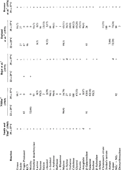
Note: + = 90% or more positive; – = less than 10% positive; v = 10 to 89.9% positive; ( ) = delayed reaction, positive in 3 days or longer. The following tests done in one or more of the laboratories cited above were 100% negative: H2S, Simmons’ citrate, Koser’s citrate, gelatin, lysine decarboxylase, arginine dihydrolase, phenylalanine deaminase, glucose (gas), dulcitol, adonitol, malonate, mucate, alginate, o-arabinos...
Table of contents
- Cover
- Title Page
- Copyright Page
- Dedication
- Table of Contents
- Chapter 1 Classification of Yersinia enterocolitica
- Chapter 2 Isolation Techniques for Yersinia enterocolitica
- Chapter 3 Yersinia enterocolitica: An Approach to Laboratory Identification with Reference to Deoxyribonucleic Acid Hybridization Groups and Biochemical Characteristics
- Chapter 4 Microbiological Aspects of Yersinia pseudotuberculosis
- Chapter 5 Antigens of Yersinia enterocolitica
- Chapter 6 Antibiotic Resistance in Yersinia enterocolitica
- Chapter 7 Human Yersinia enterocolitica Infection: Laboratory Models
- Chapter 8 Yersinia enterocolitica: Clinical Observations
- Chapter 9 Yersinia enterocolitica Gastroenteritis in Children and Their Families
- Chapter 10 Yersinia enteritis and Crohn’s Disease
- Chapter 11 Arthritis Associated with Yersinia enterocolitica Infection
- Chapter 12 Erythema Nodosum Associated with Infection with Yersinia enterocolitica
- Chapter 13 The Occurrence of Antibodies to Yersinia enterocolitica in Thyroid Diseases
- Chapter 14 Zoonotic Yersinia enterocolitica Infection: Host Range, Clinical Manifestations, and Transmission Between Animals and Man
- Chapter 15 The Occurrence of Yersinia enterocolitica in Foods
- Chapter 16 Epidemiological Aspects of Yersinia enterocolitica in New York State with Emphasis on a Recent Food-Borne Outbreak
- Chapter 17 Yersinia enterocolitica Infections in Canada
- Chapter 18 Yersinia enterocolitica in South Africa
- Chapter 19 Epidemiologic Aspects of Yersiniosis in Japan
- Index
Frequently asked questions
Yes, you can cancel anytime from the Subscription tab in your account settings on the Perlego website. Your subscription will stay active until the end of your current billing period. Learn how to cancel your subscription
No, books cannot be downloaded as external files, such as PDFs, for use outside of Perlego. However, you can download books within the Perlego app for offline reading on mobile or tablet. Learn how to download books offline
Perlego offers two plans: Essential and Complete
- Essential is ideal for learners and professionals who enjoy exploring a wide range of subjects. Access the Essential Library with 800,000+ trusted titles and best-sellers across business, personal growth, and the humanities. Includes unlimited reading time and Standard Read Aloud voice.
- Complete: Perfect for advanced learners and researchers needing full, unrestricted access. Unlock 1.4M+ books across hundreds of subjects, including academic and specialized titles. The Complete Plan also includes advanced features like Premium Read Aloud and Research Assistant.
We are an online textbook subscription service, where you can get access to an entire online library for less than the price of a single book per month. With over 1 million books across 990+ topics, we’ve got you covered! Learn about our mission
Look out for the read-aloud symbol on your next book to see if you can listen to it. The read-aloud tool reads text aloud for you, highlighting the text as it is being read. You can pause it, speed it up and slow it down. Learn more about Read Aloud
Yes! You can use the Perlego app on both iOS and Android devices to read anytime, anywhere — even offline. Perfect for commutes or when you’re on the go.
Please note we cannot support devices running on iOS 13 and Android 7 or earlier. Learn more about using the app
Please note we cannot support devices running on iOS 13 and Android 7 or earlier. Learn more about using the app
Yes, you can access Yersinia Enterocolitica by Edward J. Bottone in PDF and/or ePUB format, as well as other popular books in Biological Sciences & Biology. We have over one million books available in our catalogue for you to explore.