The contents of this book are focused on the use of chemical modification to study the properties of proteins in solution. Particular emphasis has been placed on the practical laboratory aspects of this approach to the study of the relationship between structure and function in the complex class of biological heteropolymers. As a result, little emphasis is given to the individual consideration of the functional consequence of chemical modification.
Frequently asked questions
Simply head over to the account section in settings and click on “Cancel Subscription” - it’s as simple as that. After you cancel, your membership will stay active for the remainder of the time you’ve paid for. Learn more here.
At the moment all of our mobile-responsive ePub books are available to download via the app. Most of our PDFs are also available to download and we're working on making the final remaining ones downloadable now. Learn more here.
Both plans give you full access to the library and all of Perlego’s features. The only differences are the price and subscription period: With the annual plan you’ll save around 30% compared to 12 months on the monthly plan.
We are an online textbook subscription service, where you can get access to an entire online library for less than the price of a single book per month. With over 1 million books across 1000+ topics, we’ve got you covered! Learn more here.
Look out for the read-aloud symbol on your next book to see if you can listen to it. The read-aloud tool reads text aloud for you, highlighting the text as it is being read. You can pause it, speed it up and slow it down. Learn more here.
Yes, you can access Chemical Reagents for Protein Modification by Roger L. Lundblad in PDF and/or ePUB format, as well as other popular books in Medicine & Alternative & Complementary Medicine. We have over one million books available in our catalogue for you to explore.
Until approximately 10 years ago the specific chemical modification of arginine was relatively difficult to achieve. The high pKa of the guanidine functional group (pKa ≃ 12 to 13) necessitated fairly drastic reaction conditions (pH ≽ 12) to generate an effective nucleophile. Most proteins are not stable to extreme alkaline pH. The modification of arginyl residues was however possible and the early efforts in this area have been previously reviewed.1
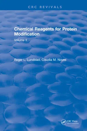
It is reasonable to suggest that the recent advances in the study of the function of arginine residues in proteins stem from the work of Takahashi2 on the use of phenylglyoxal as a reagent for the specific modification of arginine although observations on the use of 2,3-butanedione3 and glyoxal4 appeared at approximately the same time. The greatly increased interest in the elucidation of functional arginyl residues probably arises from the suggestion of Riordan and co-workers5 that arginyl residues function as “general” anion recognition sites in proteins. Patthy and Thész6 extended this concept by suggesting that the pKa of arginyl residues at anion binding sites (Figure 1) is lower than that of other arginine residues which would explain the specificity of the dicarbonyl residues which will be discussed below.
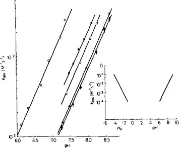
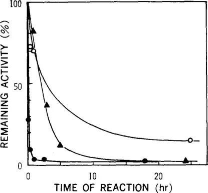
This chapter will primarily consider the reaction of arginyl residues in proteins with three different reagents; phenylglyoxal, 2,3-butanedione, and 1,2-cyclohexanedione since the vast majority of reports during the past decade have used these reagents. It is noted that several other reagents have been used for the modification of arginine. The modification of arginyl residues with ninhydrin occurs under relatively mild conditions (pH 8.0, 25°C, 0.1 M N-ethylmorpholine acetate, pH 8.0) as described by Takahashi.7 The modification of pancreatic ribonuclease A or ribonuclease T by ninhydrin is shown in Figure 2. The reaction proceeds quite rapidly at pH 8.0 with the modification of both arginyl and lysyl residues. Reducing the pH to 5.5 (0.1 M sodium acetate, pH 5.5) reduced the rate of inactivation but did not increase the specificity of the modification. The UV spectra of ribonuclease T1 before and after modification with ninhydrin are presented in Figure 3. Takahashi7 achieved specificity of modification by first modifying available lysyl residues with a reagent such as methylmaleic anhydride (citraconic anhydride) which can subsequently be removed under conditions where the arginine derivative is stable (pH 3.6). The arginine derivative is unstable under basic conditions (1% piperidine, ambient temperature, 34 hr) and arginine was regenerated. Under the conditions commonly used for the preparation of protein samples for amino acid analysis (6 Ν HCl 110°C, 24 hr), the ninhydrin-arginine derivative was destroyed with the partial regeneration of free arginine. The structure of the ninhydrin-arginine derivative (Figure 4) is similar to that proposed for the α,α’-dicarbonyl compounds such as phenylglyoxal2 or 1,2-cyclohexanedione.8 At the same time a report by Chaplin9 appeared proposing the use of ninhydrin for the reversible modification of arginine residues in proteins. The study suggested that at pH 9.1(0.1 M sodium phosphate), 37°C, the rate of reaction at arginine residues is approximately 100-fold more rapid than at lysine residues but reaction at cysteinyl residues is approximately 100-fold more rapid than at arginine. The extent of reaction was determined by measuring the decrease in the absorbance of ninhydrin at 232 nm (⋴ = 3.4 × 10⁴M−1 cm−1). As noted by Takahashi,7 the ninhydrin-arginine derivative is unstable under alkaline conditions and can be used for the reversible modification of arginine residues. The fluorescence properties of the reaction product between ninhydrin and guanidino compounds such as arginine have provided the basis for the use of ninhydrin for the detection of guanidine compounds in biological fluids (plasma) following separation by high performance liquid chromatography.10
Figure 1 Dependence of the reaction of arginine with dicarbonyl compounds on pH. Shown are second-order rate constants for the reaction of phenylglyoxal (
), hydroxypyruvaldehyde (
), glyoxal (
), and 1, 2-cyclohexanedione (
) with free arginine at 25°C at the indicated pH (the buffers were 0.1 M sodium phosphate for pH 6 to 8; 0.1 M triethanolamine-HCl for pH 7 to 9 and in HCl solutions (H„-4-0). Also included are the rate constants for the reaction of 1,2-cyclohexanedione with aldolase determined in 0.1 M triethanolamine-HCl buffers at 25°C (
). The inset shows the second-order rate constants for the arginine-glyoxal reaction over a wider pH range. (From Patthy, L. and Thész, J., Eur. J. Biochem., 105, 387, 1980. With permission.)
Figure 2 Rates of inactivation of ribonucleases A and T1 by ninhydrin. The reaction was performed at 25°C in the dark either at pH 8.0 in 0.1 M N-ethylmorpholine acetate, or at pH 5.5 in 0.1 M sodium acetate, at a protein concentration of 0.5% and a ninhydrin concentration of 1.5%. pH 8.0: ●, ribonuclease A; ▲. ribonuclease T1. pH 5.5: ○, ribonuclease A. (From Takahashi, K., J. Biochem., 80, 1173, 1976. With permission.)
The modification of arginyl residues with glyoxal has also been proposed.11 Specificity of reaction is a problem with reaction also at primary amine groups and sulfhydryl groups. For example, reaction of glyoxal with bovine serum albumin at pH 9.0 resulted in modification of greater than 80% of the arginine residues with approximately 30% modification of lysine residues.11 Glass and Pelzig12 have examined the reversible modification of arginyl residues with glyoxal in some detail. Several products are formed from the reaction of glyoxal and arginine at alkaline pH. One of these derivatives is markedly stable in strong acid(12 M HCl) at ambient temperature but rapidly is degraded to form free arginine in the presence of O-phenylenediamine (0.16 M) at pH 8.1 to 8.3. More alkaline conditions resulted in more rapid decomposition of the glyoxal-arginine derivative and ninhydrin-positive compounds other than arginine were formed. Reaction of arginine with glyoxal in borate buffer also yields the product described above. The same research group has reported on the reversible modification of arginine residues with camphorquinone-10-sulfonic acid and derivatives such as camphorquinone-10-sulfonylnorleucine.13 The synthesis of the parent compounds and various derivatives is reported. The sulfonic acid function provides a basis for the attachment of a “tag” such as norleucine which cajn be used for determining the extent of modification.14 Reaction with arginine occurs in 0.2 M sodium borate, pH 9.0. Under these conditions, reaction of camphorquinone-sulfonic acid with an amino acid analysis standard showed a greater than 90% loss of arginine and a 25% loss of cystine. Loss of cystine was not observed in the proteins studied (soybean trypsin inhibitor, ribonuclease S-peptide). The arginine derivative is stable for 24 hr in trifluoroacetic acid and under other mild acid conditions. The derivative is stable to 0.5 M hydroxylamine, pH 7.0, conditions under which the cyclohexanedione derivative of arginine decomposes8 but arginine is regenerated in 0.2 M o-phenylenediamine, pH 8.5 (approximately 75% after 4 hr; complete after 16 hr).
Figure 3 Changes in the UV absorption spectrum of ribonuclease T1 on reaction with ninhydrin. The spectra were measured in 0.01 M ammonium acetate. ——: (a) native ezyme; (b) n...
Table of contents
Cover
Title Page
Copyright Page
Contents
Preface
Chapter 1 The Modification of Arginine
Chapter 2 Chemical Modification of Tryptophan
Chapter 3 The Modification of Tyrosine
Chapter 4 The Modification of Carboxyl Groups
Chapter 5 The Chemical Cross-Linking of Peptide Chains