Bioimaging: Imaging by Light and Electromagnetics in Medicine and Biology explores new horizons in biomedical imaging and sensing technologies, from the molecular level to the human brain. It explores the most up-to-date information on new medical imaging techniques, such as the detection and imaging of cancer and brain diseases.
This book also provides new tools for brain research and cognitive neurosciences based on new imaging techniques. Edited by Professor Shoogo Ueno, who has been leading the field of biomedical imaging for 40 years, it is an ideal reference book for graduate and undergraduate students and researchers in medicine and medical physics who are looking for an authoritative treatise on this expanding discipline of imaging and sensing in medicine and biology.
Features:
Provides step-by-step explanations of biochemical and physical principles in biomedical imaging
Covers state-of-the art equipment and cutting-edge methodologies used in biomedical imaging
Serves a broad spectrum of readers due to the interdisciplinary topic and approach
Shoogo Ueno, Ph.D, is a professor emeritus of the University of Tokyo, Tokyo, Japan. His research interests include biomedical imaging and bioelectromagnetics, particularly in brain mapping and neuroimaging, transcranial magnetic stimulation (TMS), and magnetic resonance imaging (MRI). He was the President of the Bioelectromagnetics Society, BEMS (2003-2004) and the Chairman of the Commission K on Electromagnetics in Biology and Medicine of the International Union of Radio Science, URSI (2000-2003). He was named the IEEE Magnetics Society Distinguished Lecturer during 2010 and received the d'Arsonval Medal from the Bioelectromagnetics Society in 2010.
Trusted by 375,005 students
Access to over 1.5 million titles for a fair monthly price.
1.2 Light and Electromagnetic Fields in Medicine and Biology
1.3 Advances in Biomedical Imaging and Stimulation
1.3.1 Computed Tomography
1.3.2 Magnetic Resonance Imaging
1.3.3 Magnetoencephalography
1.3.4 Transcranial Magnetic Stimulation
1.3.5 Magnetic Particle Imaging
1.3.6 Near-Infrared Spectroscopic Imaging
1.4 Advances in Molecular and Cellular Imaging
1.4.1 Green Fluorescent Protein
1.4.2 Optical Fluorescence and Cancer Therapy
1.4.3 Optogenetics and Studies of Neuronal Circuit Dynamics in the Brain
1.4.4 Raman Scattering and Coherent Raman Scattering Microscopy
1.4.5 Molecular Imaging Based on Magnetic Resonance Imaging
1.4.6 Magnetic Orientation of Living Systems and Biogenic Micromirrors
1.5 Summary
References
1.1 Introduction
Imaging techniques have been rapidly developing and expanding in a variety of fields in medicine and biology. This chapter gives an overview of a range of imaging techniques from molecular and cellular levels to the human brain, focusing on the imaging techniques using light and electromagnetics. We start with a brief history of the discovery of electromagnetic fields and review the imaging techniques using light, electromagnetic fields, or electromagnetic techniques in medicine and biology.
The overview includes green fluorescent protein (GFP) and its applications to bioimaging, molecular imaging of viable cancer cells, optogenetics and studies of neuronal circuit dynamics in the brain, molecular vibrational imaging by coherent Raman scattering, principles and applications of magnetic resonance imaging (MRI) such as functional MRI (fMRI), diffusion MRI of the human nervous system, and chemical exchange saturation transfer (CEST) and amide proton transfer (APT) imaging. Other important and new technologies include magnetic particle imaging (MPI) using magnetic nanoparticles and magnetic sensors, usage of the reporter gene for molecular MRI, magnetic control of the biogenic micromirror, and brain imaging by near-infrared spectroscopy (NIRS) and static magnetic fields. Transcranial magnetic stimulation (TMS) is also reviewed to discuss the functional mapping and imaging of the human brain associated with fMRI, magnetoencephalography (MEG), electroencephalography (EEG), and systems neuroscience based on optogenetics.
Historically, computed tomography (CT) and positron emission tomography (PET), as well as ultrasonic imaging systems, have been introduced in the medical world as biomedical imaging tools, and these imaging tools have changed the medical world dramatically in modern medicine. In this chapter, we do not describe ultrasonic imaging, but we focus our discussion on recent advances in electromagnetic-based and light-based biomedical imaging and stimulation techniques, which has brought a significant impact in medicine and biology in recent decades.
1.2 Light and Electromagnetic Fields in Medicine and Biology
Electromagnetic fields or electromagnetic waves were theoretically predicted by James Clerk Maxwell (1831–1879) in the UK in 1864, and the existence of electromagnetic waves was experimentally verified by Heinrich Rudolf Hertz (1857–1894) in Germany in 1888. Guglielmo Marches Marconi (1874–1937) in Italy applied electromagnetic waves for telecommunication over the English Channel between France and England in 1899. As Maxwell pointed out, light is one of the electromagnetic waves in a specific frequency band.
Maxwell derived the so-called four fundamental electromagnetic equations, and he obtained an equation to show the propagation velocity v of electromagnetic waves in free space or in vacuum space as follows.
(1.1)
where ε0 is the dielectric constant or permittivity and μ0 is the magnetic permeability in vacuum space.
If we use ε0 = 1/(36π) × 10−9 F/m and μ0 = 4π × 10−7 H/m,
we obtain
(1.3)
These values coincide with the light velocity c = 2.99792458 × 10⁸ m/s.
Here, the relationship between frequency and wavelength is described for further discussion.
Since the electromagnetic waves propagate at the light velocity in vacuum space, multiplication of wavelength λ and frequency f of electromagnetic waves are equal to the light velocity c as shown by
(1.4)
Therefore, the wavelength λ is given by
(1.5)
The wavelength of 300 Hz electromagnetic fields is 1000 km.
The wavelength of 300 kHz (300 × 103 Hz) electromagnetic fields is 1 km
The wavelength of 300 MHz (300 × 106 Hz) electromagnetic fields is 1 m
The wavelength of 300 GHz (300 × 109 Hz) electromagnetic fields is 1 mm
The wavelength of 300 THz (300 × 1012 Hz) electromagnetic fields is 1 μm
The wavelength of 300 PHz (300 × 1015 Hz) electromagnetic fields is 1 nm
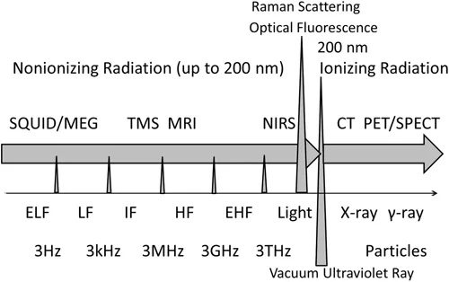
Figure 1.1 shows the classification of electromagnetic fields used in telecommunication and medical applications. Medical devices and biomedical imaging systems used at different frequency bands are marked along the frequency axis.
FIGURE 1.1 Classification of electromagnetic fields. Medical devices and biomedical imaging systems used at different frequency bands are marked along the frequency axis. The electromagnetic fields or electromagnetic waves are legally handled up to 3 THz or 3000 GHz. Electromagnetic fields lower than a frequency at the vacuum ultraviolet ray of its wavelength around 200 nm are called non-ionizing radiation. The electromagnetic fields higher than a frequency at the vacuum ultraviolet ray are called ionizing radiation. The electromagnetic fields are classified from DC to higher frequencies by ELF (extremely low frequency), LF (low frequency), IF (intermediate frequency), HF (high frequency), and EHF (extremely high frequency) electromagnetic fields. Visible light is in the frequency range from red (780 nm) to violet (380 nm). X-ray and γ-ray are in the ionizing radiation range. In non-ionizing radiation or non-ionizing electromagnetic fields, SQUID (superconducting quantum interference device) and MEG (magnetoencephalography) are used at ELF; MPI (magnetic particle imaging) is used at LF and IF; TMS (transcranial magnetic stimulation) uses pulsed magnetic fields at IF; MRI (magnetic resonance imaging) uses its resonant frequencies at HF; and NIRS (near-infrared spectroscopy) uses a near-infrared (NIR) ray. Raman scattering and optical fluorescence are used at the visible light band. In contrast, CT (computed tomography) uses X-rays, and PET (positron emission tomography) and SPECT (single-photon emission computed tomography) use γ-rays.
The usage of electromagnetic fields is legally law-handled up to 3 THz or 3000 GHz. The electromagnetic fields below a point of the vacuum ultraviolet ray at around 200 nm in wavelength are called non-ionizing radiation or non-ionizing electromagnetic fields. In contrast, the electromagnetic fields higher than a point at the vacuum ultraviolet ray at around 200 nm in wavelength are called ionizing radiation or ionizing electromagnetic fields. Exposure to ionizing radiation causes ionization of molecules in cells and living tissues, which may result in undesirable effects on living systems when the intensity of irradiation exceeds a threshold level. Radiation therapy for cancer diseases use ionizing radiation.
In non-ionizing electromagnetic fields, frequency bands are classified as follows:
Extremely low frequency (ELF) electromagnetic fields (DC ~ 3 kHz)
Low frequency (LF) electromagnetic fields (3 kHz ~ 10 kHz)
Intermediate frequency (IF) electromagnetic fields (10 kHz ~ 10 MHz)
High frequency (HF) electromagnetic fields (10 MHz ~ 6 GHz)
Extremely high frequency (EHF) electromagnetic fields (6 GHz ~ 3 THz)
In light bands, the near-infrared (NIR) band, the visible light band, and ultraviolet bands are classified in promotionally increasing frequency or in decreasing wavelength. In visible light band, from the wavelength of 780 nm (red) to the wavelength of 380 nm (violet), seven-color spectra exist like a rainbow; red, orange, yellow, green, blue, indigo, and violet.
In ionizing electromagnetic fields, the ionizing radiation is classified by X-ray, γ-ray, and the areas beyond X-ray and γ-ray where the waves act as heavy particles.
As shown in Figure 1.1, the MEG and superconducting quantum interference device (SQUID) systems are used at ELF; MPI is used at LF and IF; TMS for the stimulation of the human brain uses pulsed magnetic fields at IF; MRI uses its resonant frequencies at HF; and NIRS uses NIR rays. Raman scattering and optical fluorescence for molecular and cellular imaging are used at the visible light band. In contrast, CT uses X-rays, and PET and single-photon emission computed tomography (SPECT) use γ-rays.
MEG, MPI, TMS, and MRI are used in non-ionizing...
Table of contents
Cover
Half-Title
Title
Copyright
Contents
Preface
Acknowledgments
Editor
List of Contributors
Chapter 1 Introduction
Chapter 2 Molecular Imaging of Viable Cancer Cells
Chapter 3 Molecular Vibrational Imaging by Coherent Raman Scattering
Chapter 4 Magnetic Resonance Imaging: Principles and Applications
Chapter 5 Chemical Exchange Saturation Transfer and Amide Proton Transfer Imaging
Chapter 6 Diffusion Magnetic Resonance Imaging in the Central Nervous System
Chapter 7 Magnetic Particle Imaging
Chapter 8 Sensing of Magnetic Nanoparticles for Sentinel Lymph Nodes Biopsy
Chapter 9 Optimizing Reporter Gene Expression for Molecular Magnetic Resonance Imaging: Lessons from the Magnetosome
Chapter 10 Magnetic Control of Biogenic Micro-Mirror
Chapter 11 Non-Invasive Techniques in Brain Activity Measurement Using Light or Static Magnetic Fields Passing Through the Brain
Index
Frequently asked questions
Yes, you can cancel anytime from the Subscription tab in your account settings on the Perlego website. Your subscription will stay active until the end of your current billing period. Learn how to cancel your subscription
No, books cannot be downloaded as external files, such as PDFs, for use outside of Perlego. However, you can download books within the Perlego app for offline reading on mobile or tablet. Learn how to download books offline
Perlego offers two plans: Essential and Complete
Essential is ideal for learners and professionals who enjoy exploring a wide range of subjects. Access the Essential Library with 800,000+ trusted titles and best-sellers across business, personal growth, and the humanities. Includes unlimited reading time and Standard Read Aloud voice.
Complete: Perfect for advanced learners and researchers needing full, unrestricted access. Unlock 1.5M+ books across hundreds of subjects, including academic and specialized titles. The Complete Plan also includes advanced features like Premium Read Aloud and Research Assistant.
Both plans are available with monthly, semester, or annual billing cycles.
We are an online textbook subscription service, where you can get access to an entire online library for less than the price of a single book per month. With over 1.5 million books across 990+ topics, we’ve got you covered! Learn about our mission
Look out for the read-aloud symbol on your next book to see if you can listen to it. The read-aloud tool reads text aloud for you, highlighting the text as it is being read. You can pause it, speed it up and slow it down. Learn more about Read Aloud
Yes! You can use the Perlego app on both iOS and Android devices to read anytime, anywhere — even offline. Perfect for commutes or when you’re on the go. Please note we cannot support devices running on iOS 13 and Android 7 or earlier. Learn more about using the app
Yes, you can access Bioimaging by Shoogo Ueno in PDF and/or ePUB format, as well as other popular books in Medicine & Medical Theory, Practice & Reference. We have over 1.5 million books available in our catalogue for you to explore.