![]()
Chapter 1
Introduction to Magnetic Resonance Imaging
1.1 Introduction
Magnetic resonance imaging, commonly known as MRI, is a powerful non-invasive imaging technique that has played and will continue to play an important role in the medical community. In clinical practice, it can assist physicians in both diagnosis and presurgical planning with limited risk to the patient. In laboratory research, it can help neurologists and other biological scientists to discover novel basic anatomical structures and physiological principles. Unlike some other imaging techniques like x-ray computed tomography (CT), MRI does not require exposure of the subject to ionizing radiation and hence is considered safe. It also provides more information than other imaging modalities since MR signals are sensitive to several tissue parameters.
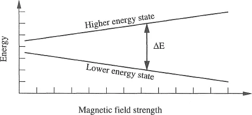
MRI belongs to a larger group of techniques which are based on the phenomenon of nuclear magnetic resonance (NMR). This phenomenon was discovered in bulk materials by Bloch and Purcell in 1946. As we will see later, certain atomic nuclei, when placed in a static magnetic field, will assume one of the two states: one has a higher energy level and the other has a lower energy level (Fig. 1.1). The energy difference between the two states is linearly proportional to the strength of the applied magnetic field. This is known as the Zeeman effect. In thermal equilibrium, the number of nuclei in the higher energy state is slightly less than the number of nuclei in the lower energy state. A nucleus in the higher energy state can fall to the lower energy state by emitting a photon with energy equal to the energy difference between the two states. A nucleus in the lower energy state can jump to the higher energy state by absorbing a photon with energy matching the energy difference between the two states. Therefore, when the nuclei in the applied magnetic field are irradiated by such photons, which are actually electromagnetic fields of certain frequency generated by a radiofrequency (RF) probe, some nuclei in the lower energy state will absorb the photons and jump to the higher energy state. This destroys the thermal equilibrium. Immediately after the irradiation of photons, the excess nuclei in the higher energy state will return to the lower energy state to recover the equilibrium, emitting photons or electromagnetic fields which can be detected by an RF probe. Since the frequency of the emitted electromagnetic signals is determined by the energy difference of the two states of the nuclei and the decay of the signals in time depends on the molecular environment of the nuclei, the NMR signals received by an RF probe can be analyzed to study the properties of the nuclei and their environment.
Figure 1.1. Splitting of energy level caused by the application of a static magnetic field.
For many years, NMR was primarily used for spectroscopic analysis before Lauterbur (1973) proposed using it for imaging purposes. The basic principle of using NMR for imaging is simple. Since the energy difference between the two states of certain nuclei in an external field depends on the strength of the external field, this energy difference at each point in the object to be imaged can be made different by varying the magnetic field from point to point. As a result, the energy of the photons and, consequently, the frequency of the electromagnetic fields absorbed or emitted by the nuclei are also different from point to point. After signals from all nuclei are received, their frequency may be used to determine spatial information about the nuclei. This simple phenomenon provides a basic foundation for all NMR imaging although the actual imaging methods are more complicated. The number of imaging techniques has blossomed since Lauterbur’s pioneering work. Technological developments have also increased the quality of NMR images. For example, the advent of superconducting magnets has made possible higher signal-to-noise ratio (SNR) and higher image resolution than were possible with resistive or permanent magnets. Modern computers have made possible whole volume imaging techniques which offer increased SNR and decreased image acquisition times. So-called “artifacts,” which are the distortions of the image due to unwanted effects such as motion, can be controlled using complex RF pulse sequences. Advances in coil technology have improved image quality and made techniques such as surface and microscopic imaging possible.
In this chapter, we first de...